The Genomic Standards Consortium
Genome sequence of th
(DSM 24565
T), a member of the marin
rich in extrachromosomal elements
Thomas Riedel1, Hazuki Teshima2, Jörn Petersen3, Anne Fiebig3, Karen Davenport2, Hajnalka Daligault2, Tracy Erkkila2, Wei Gu2, Christine Munk2, Yan Xu2, Amy Chen4, Amrita Pati5, Natalia Ivanova5, Lynne A. Goodwin2,5, Patrick Chain2, John C. Detter2,5, Manfred Rohde1, Sabine Gronow3, Nikos C. Kyrpides5, Tanja Woyke5, Markus Göker3*, Thorsten Brinkhoff6, Hans-Peter Klenk3
1 HZI – Helmholtz Centre for Infection Research, Braunschweig, Germany
2 Los Alamos National Laboratory, Bioscience Division, Los Alamos, New Mexico, USA 3 Leibniz Institute DSMZ - German Collection of Microorganisms and Cell Cultures,
Braunschweig, Germany
4 Biological Data Management and Technology Center, Lawrence Berkeley National Laboratory, Berkeley, California, USA
5 DOE Joint Genome Institute, Walnut Creek, California, USA
6 Institute for Chemistry and Biology of the Marine Environment (ICBM), Oldenburg, Germany
*Corresponding author: Markus Göker ([email protected])
Keywords: marine, biofilm, ovoid-shaped, halotolerant, heterotrophic, quorum sensing, plasmid, thiosulfate oxidation, carbon monoxide utilization
et al. 2008 is a member of the genomically well characterized
clade are metabolically versatile and involved in carbon fixation and biogeochemical processes. They form a physiologically heterogeneous group, found predominantly in coastal or polar waters, especially in symbiosis with algae, in microbial mats, in sediments or associated with invertebrates. Here we de-scribe the features ofT together with the permanent-draft genome sequence
and annotation. The 5,344,253 bp long genome consists of one chromosome and an unusually high number of seven extrachromosomal elements and contains 5,129 protein-coding and 89 RNA genes. It was sequenced as part of the DOE Joint Genome Institute Community Sequencing Program 2010 and of the activities of the Transregional Collaborative Research Centre 51 funded by the German Research Foundation (DFG).
Introduction
Strain R-26159T (= DSM 24565T = LMG 24366T =
CCUG 55860T) is the type strain of the species
currently with a validly published name in the
ge-nus
The genus
spread
rine habitats [4]. Strain R-26159T was isolated from
a marine electroactive biofilm grown on a stainless-steel cathode, which was exposed to natural sea-water at the ISMAR-CNR Marine Station within the
harbor of Genova (Italy) [1]. The genus
was named after Thomas Leisinger for his work on the bacterial methyl halide metabolism [2]; the
species epithet aquimarina refers to the Neolatin
adjective marinus, from the sea, from seawater.
PubMed records do not currently indicate any fol-low-up research with strain R-26159T after the
ini-tial description
Here we present a summary classification and a set
of features forT, together
with the description of the genomic sequencing and annotation.
Classification and features of the organism
16S rRNA analysis
A representative genomic 16S rRNA gene sequence
oT was compared using
(HSPs) from the best 250 hits) with the most recent release of the Greengenes database [7] and the rel-ative frequencies of taxa and keywords (reduced to their stem [8]) were determined, weighted by BLAST scores. The most frequently occurring
gene-ra were
four hits to sequences from other members of the genus, the average identity within HSPs was 99.4%, whereas the average coverage by HSPs was 99.3%. Among all other species, the one yielding the
high-est score wa
(NR_025637), which corresponded to an identity of 99.2% and an HSP coverage of 100.0%. (Note that the Greengenes database uses the INSDC (= EMBL/NCBI/DDBJ) annotation, which is not an au-thoritative source for nomenclature or classifica-tion.) The highest-scoring environmental sequence
was FJ202534 (Greengenes short name 'and White Plague Disease-Induced Changes Caribbean Coral
Montastraea faveolata kept aquarium 23 days clone
SGUS1024'), which showed an identity of 97.8% and an HSP coverage of 100.0%. The most fre-quently occurring keywords within the labels of all environmental samples which yielded hits were 'coral' (4.7%), 'caribbean' (3.8%), 'faveolata' (3.5%), 'chang' (3.4%) and 'white' (3.3%) (117 hits in total). Environmental samples which yielded hits of a higher score than the highest scoring species were not found, which might indicate that the spe-cies is rarely found in the environment.
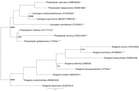
Figure 1 shows the phylogenetic neighborhood o
quences of the four identical 16S rRNA gene copies in the genome do not differ from the previously published 16S rRNA gene sequence AM900415.
Figure 1. Phylogenetic tree highlighting the position o
other species within the genus
tree was inferred from 1,383 aligned characters [9,10] of the 16S rRNA gene sequence under the max-imum likelihood (ML) criterion [11]. Rooting was done initially using the midpoint method [12] and then checked for its agreement with the current classification (Table 1). The branches are scaled in terms of the expected number of substitutions per site. Numbers adjacent to the branches are support values from 1,000 ML bootstrap replicates [13] (left) and from 1,000 maximum-parsimony bootstrap replicates [14] (right) if larger than 60%. Lineages with type strain genome sequencing projects regis-tered in GOLD [15] are labeled with one asterisk, those also listed as 'Complete and Published' with
Morphology and physiology
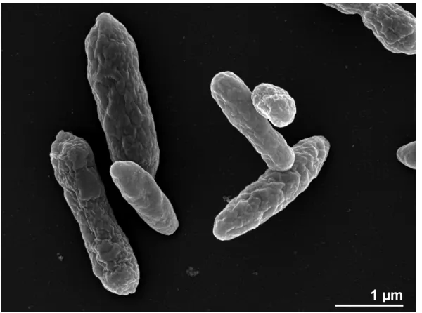
Cells of strain R-26159T are Gram-negative, ovoid
(1 × 1.4 µm) and contain a single polar flagellum (not visible in Figure 2), which is used for motility.
Poly-β-hydroxybutyrate is present in inclusion
bodies. Colonies are dark beige-pink in color, round and 1–2 mm in diameter after 3 days incu-bation on marine agar (MA). Growth occurs after 2 days incubation at 20 °C on MA, but not on Reasoner’ 2A agar (R2A), Nutrient agar (NA), Trypticase-Soy agar (TSA) or Peptone-Yeast Ex-tract-Glucose agar (PYG). The temperature range for growth is 4–37°C whereas no growth occurs at 40°C or higher. The salinity range for growth is 1– 7% NaCl. The pH range for growth is 5.5–9.0 with an optimum between 6.5–8. Growth occurs on betaine (1 mM) as a sole carbon source, but not on L-methionine (10 mM). Cells are catalase- and ox-idase-positive. Degradation of gelatin is weakly positive but cells do not degrade tyrosine, DNA, starch, casein, chitin, aesculin or Tween 80. The strain shows leucine arylamidase activity; weak alkaline phosphatase, esterase lipase (C8) and naphthol-AS-BI phosphohydrolase activities. No activity is detected for esterase (C4), valine arylamidase, acid phosphatase, α-galactosidase, β
-glucuronidase, α-glucosidase, β-glucosidase, N
-acetyl-β-glucosaminidase, α-mannosidase, lipase
(C14), cystine arylamidase, trypsin, α
-chymotrypsin, arginine dihydrolase, urease or α
-fucosidase. Nitrate is not reduced to nitrite or ni-trogen. Indole is not produced and glucose is not fermented. Cells do not assimilate D-glucose,
L-arabinose, D-mannose, D-mannitol, N
-acetylglucosamine, maltose, potassium gluconate, capric acid, adipic acid, malate, citrate or phenylacetic acid. Cells are susceptible to cefoxitin (30 mg), erythromycin (15 mg), tetracycline (30 mg) and streptomycin (25 mg), but resistant to vancomycin (30 mg), trimethoprim (1.25 mg), clindamycin (2 mg) and gentamicin (30 mg) (all data from [1]).
The utilization of carbon compounds
T grown at 20°C was also
determined for this study using Generation-III microplates in an OmniLog phenotyping device (BIOLOG Inc., Hayward, CA, USA). The microplates were inoculated at 28°C with a cell suspension at a cell density of 95-96% turbidity and dye IF-A. Fur-ther additives were vitamin, micronutrient and sea-salt solutions. The exported measurement da-ta were further analyzed with the opm package for R [31,32], using its functionality for statistically estimating parameters from the respiration curves and translating them into negative, ambig-uous, and positive reactions. The strain was stud-ied in two independent biological replicates, and reactions with a different behavior between the two repetitions were regarded as ambiguous. At 28°C the strain reacted poorly, with positive reac-tions only for 1% NaCl, 4% NaCl and lithium chlo-ride. This is in accordance with the comparatively low median of the temperature range of the strain [1].
Chemotaxonomy
The principal cellular fatty acids of strain
R-26159T are mono-unsaturated straight chain
ac-ids: C18:1 ω7c (71.6%), C14:1 iso E (11.6%), C14:1 2-OH
(4.2%), C16:0 2-OH (4.2%), C16:0 (3.5%), an unknown
fatty acid of equivalent chain-length 11.799
(2.7%), C12:0 3-OH (2.1%) as well as C10:0 3-OH
(2.0%) [28]. Remaining fatty acids were detected in very small fractions only (<1.0%) [1]. The same predominant fatty acids were also found in other
members of the
[2,28,33].
Table 1. Classification and general features oT according to the MIGS recommendations [20].
MIGS ID Property Term Evidence code
Domai TAS [21]
Phylum TAS [22]
Cla TAS [23,24]
Classification Orde TAS [24,25]
Family TAS [24,26]
Genu TAS [27,28]
Species TAS [1]
MIGS-7 Subspecific genetic lineage (strain) R-26159T TAS [1] MIGS-12 Reference for biomaterial Vandecandelaere et al. 2008 TAS [1]
Current classification
Gram stain Negative TAS [1]
Cell shape Ovoid-shaped TAS [1]
Motility Motile TAS [1]
Sporulation Not reported
Temperature range Mesophile (4– 37°C) TAS [1]
Optimum temperature 20°C NAS
Salinity Halophile, 1-7% NaCl (w/v) TAS [1]
MIGS-22 Relationship to oxygen aerobic TAS [1]
Carbon source Yeast extract, peptone, betaine TAS [1]
MIGS-6 Habitat Seawater, biofilm TAS [1]
MIGS-6.2 pH 6.5 – 8.0 TAS [1]
MIGS-15 Biotic relationship Free living TAS [1]
Biosafety level 1 TAS [29]
MIGS-23.1 Isolation Marine biofilm on stainless steel cathode TAS [1] MIGS-4 Geographic location ISMAR-CNR Marine Station, Genoa harbor, Italy TAS [1]
MIGS-4.1 Latitude +44.41 TAS [1]
MIGS-4.2 Longitude +8.92 TAS [1]
MIGS-4.3 Depth Not reported
Genome sequencing and annotation
Genome project history
This organism was selected for sequencing on the
basis of the DOE Joint Genome Institute Community
Sequencing Program 2010, CSP 441: “Whole
ge-nome type strain sequences of the genera
of highly physiologically diverse organisms”. The genome project is deposited in the GenomesOnLine Database [15] and the complete genome sequence was submitted to GenBank. Sequencing, finishing and annotation were performed by the DOE Joint Genome Institute (JGI). A summary of the project information is shown in Table 2.
Growth conditions and DNA isolation
A culture ofT was grown
in the DSMZ medium 514 (BACTO Marine Broth) [34] at 20°C. Genomic DNA was isolated from 0.5-1 g of cell paste using Jetflex Genomic DNA Purifi-cation Kit (GENOMED 600100) following the standard protocol provided by the manufacturer but modified by the use of additional 20 µl pro-teinase K and 40 minute incubation. DNA is avail-able through the DNA Bank Network [35].
Genome sequencing and assembly
The draft genome was generated using Illumina data [36]. For this genome, we constructed and se-quenced an Illumina short-insert paired-end li-brary with an average insert size of 270 bp which generated 13,668,574 reads and an Illumina long-insert paired-end library with an average long-insert size of 8047.58 +/- 2682.23 bp which generated
11,512,166 reads totaling 3,777 Mbp of Illumina data (Feng Chen, unpublished). All general aspects of library construction and sequencing can be found at the JGI web site [37]. The initial draft as-sembly contained 64 contigs in 18 scaffold(s). The initial draft data was assembled with Allpaths [38] and the consensus was computationally shredded into 10 kbp overlapping fake reads (shreds). The Illumina draft data was also assembled with Velvet [39], and the consensus sequences were computa-tionally shredded into 1.5 kbp overlapping fake reads (shreds). The Illumina draft data was assem-bled again with Velvet using the shreds from the first Velvet assembly to guide the next assembly. The consensus from the second Velvet assembly was shredded into 1.5 kbp overlapping fake reads. The fake reads from the Allpaths assembly and both Velvet assemblies and a subset of the Illumina CLIP paired-end reads were assembled using paral-lel phrap (High Performance Software, LLC) [40]. Possible mis-assemblies were corrected with man-ual editing in Consed [37,39,40]. Gap closure was accomplished using repeat resolution software (Wei Gu, unpublished), and sequencing of bridging PCR fragments with PacBio (Cliff Han, unpublished) technologies. A total of 57 PCR PacBio consensus sequences were completed to close gaps and to raise the quality of the final sequence. The final as-sembly is based on 3,777 Mbp of Illumina draft da-ta, which provides an average 699 × coverage of the genome.
Table 2. Genome sequencing project information
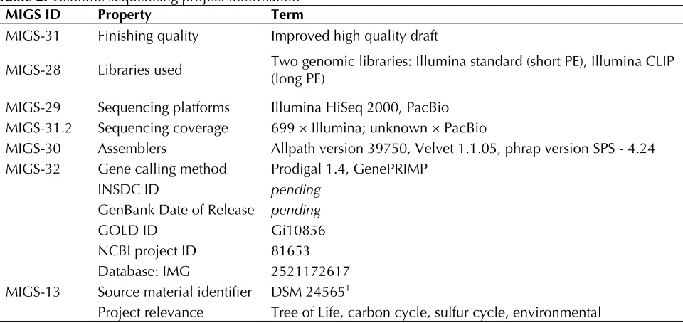
MIGS ID Property Term
MIGS-31 Finishing quality Improved high quality draft
MIGS-28 Libraries used Two genomic libraries: Illumina standard (short PE), Illumina CLIP (long PE) MIGS-29 Sequencing platforms Illumina HiSeq 2000, PacBio
MIGS-31.2 Sequencing coverage 699 × Illumina; unknown × PacBio
MIGS-30 Assemblers Allpath version 39750, Velvet 1.1.05, phrap version SPS - 4.24 MIGS-32 Gene calling method Prodigal 1.4, GenePRIMP
INSDC ID pending
GenBank Date of Release pending
GOLD ID Gi10856
NCBI project ID 81653 Database: IMG 2521172617 MIGS-13 Source material identifier DSM 24565T
Genome annotation
Genes were identified using Prodigal [41] as part of the JGI genome annotation pipeline [42], fol-lowed by a round of manual curation using the JGI GenePRIMP pipeline [43]. The predicted CDSs were translated and used to search the National Center for Biotechnology Information (NCBI) nonredundant database, UniProt, TIGR-Fam, Pfam, PRIAM, KEGG, COG, and InterPro databases. Addi-tional gene prediction analysis and funcAddi-tional an-notation was performed within the Integrated Mi-crobial Genomes - Expert Review (IMG-ER) plat-form [44].
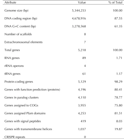
Genome properties
The genome statistics are provided in Table 3 and Figure 3. The genome consists of a 4.25 Mbp chromosome and seven extrachromosomal ele-ments of 6.2 to 248.9 kbp length with a G+C con-tent of 61.4%. Of the 5,218 genes predicted, 5,129 were protein-coding genes, and 89 RNAs. The ma-jority of the protein-coding genes (80.4%) were assigned a putative function while the remaining ones were annotated as hypothetical proteins. The distribution of genes into COGs functional catego-ries is presented in Table 4.
Table 3. Genome Statistics
Attribute Value % of Total
Genome size (bp) 5,344,253 100.00
DNA coding region (bp) 4,678,916 87.55
DNA G+C content (bp) 3,278,568 61.35
Number of scaffolds 8
Extrachromosomal elements 7
Total genes 5,218 100.00
RNA genes 89 1.71
rRNA operons 4
tRNA genes 61 1.17
Protein-coding genes 5,129 98.29
Genes with function prediction (proteins) 4,196 80.41
Genes in paralog clusters 4,110 78.77
Genes assigned to COGs 3,955 75.80
Genes assigned Pfam domains 4,253 81.51
Genes with signal peptides 419 8.03
Genes with transmembrane helices 1,037 19.87
Figure 3. Graphical map of the chromosome. From bottom to the top: Genes on forward strand (colored by COG categories), Genes on reverse strand (colored by COG categories), RNA genes (tRNAs green, rRNAs red, other RNAs black), GC content (black), GC skew (purple/olive).
Table 4. Number of genes associated with the general COG functional categories Code value %age Description
J 175 4.0 Translation, ribosomal structure and biogenesis A 1 0.0 RNA processing and modification
K 365 8.4 Transcription
L 296 6.8 Replication, recombination and repair B 2 0.1 Chromatin structure and dynamics
D 43 1.0 Cell cycle control, cell division, chromosome partitioning Y 0 0.0 Nuclear structure
V 53 1.2 Defense mechanisms
T 182 4.2 Signal transduction mechanisms
M 221 5.1 Cell wall/membrane/envelope biogenesis N 55 1.3 Cell motility
Z 2 0.1 Cytoskeleton
W 0 0.0 Extracellular structures
U 75 1.7 Intracellular trafficking and secretion, and vesicular transport O 157 3.6 Posttranslational modification, protein turnover, chaperones C 273 6.3 Energy production and conversion
G 168 3.9 Carbohydrate transport and metabolism E 550 12.6 Amino acid transport and metabolism F 95 2.2 Nucleotide transport and metabolism H 188 4.3 Coenzyme transport and metabolism
I 172 4.0 Lipid transport and metabolism
P 220 5.1 Inorganic ion transport and metabolism
Q 152 3.5 Secondary metabolites biosynthesis, transport and catabolism R 513 11.8 General function prediction only
Insights into the genome
Genome sequencing of
24565T reveals the presence of seven plasmids
with sizes between 6 kb and 249 kb (Table 5). The circular conformation of the chromosome and the two smallest extrachromosomal elements has been experimentally validated. The six larger plasmids contain characteristic replication mod-ules [45] of the RepABC-, DnaA-like, RepA- and RepB-type comprising a replicase as well as the
parAB partitioning operon [46]. The respective
replicases that mediate the initiation of replication are designated according to the established plas-mid classification scheme [47]. The different numbering of e.g. the replicases RepC-8, RepC-13 and RepC-14 from RepABC-type plasmids corre-sponds to specific plasmid compatibility groups that are required for a stable coexistence of the replicons within the same cell [48]. The cryptic 6 kb plasmid pAqui_G6 contains a solitary RepA-II type replicase without a partitioning module, but replicon maintenance in the daughter cells is probably ensured by its postsegregational killing system (PSK) consisting of a typical operon with two small genes encoding a stable toxin and an unstable antitoxin [49]. PSK systems are also lo-cated on pAqui_C182 and pAqui_F126 (Tab. 6). The locus tags of all replicases, plasmid stability
modules and the large virB4 and virD4 genes of
type IV secretion systems are presented in Table 6. A characteristic T4SS comprising the relaxase VirD2 and the coupling protein VirD4 as well as
the complete virB gene cluster for the
transmembrane channel is located on the chromo-some [50]. Its functional role is unclear, but very closely related T4SS are detected on plasmids of
e.g. T [51],
T and
T [52].
Fur-thermore, the largest plasmid pAqui_A249 con-tains the complete F factor conjugation transfer
(tra) region with 20 genes (Aqui_4678 to
Aqui_4697). It exhibits only weak homology with the typical type IV secretion system of the
chromosomal counterpart, but it resembles the F
sex factor of
for bacterial conjugation [53].
The 243 kb RepABC-13 type plasmid pAqui_B243 is predominated by seven ABC-transporters. Even more conspicuous is the presence of a couple of pentose phosphate pathway genes including an
operon of genes of the Entner-Doudoroff pathway (Aqui_4914 to Aqui_4917; EC 1.1.1.49; EC 4.2.1.12; EC 4.1.2.14; EC 5.3.1.9) that is generally used in
Roseobacters to convert D-fructose-6-phosphate
to D-glyceraldehyde-3-phosphate [54]. The exclu-sive missing gene within this operon is the chro-mosome encoded 6-phosphogluconolactonase (Aqui_2983; EC 3.1.1.31). The presence of a glyco-lytic 6-phosphofructokinase (PFK; Aqui_4950; EC 2.7.1.11) is a genetic novelty in this group of ma-rine bacteria, because the current opinion was
that “the typical pfk gene is absent from all
se-quenced hence probably catabolized via the Entner– Doudoroff pathway and not via classical glycolysis”
[55]. However, the putative functionality of the Embden-Meyerhoff-Parnas pathway (glycolysis) has to be validated e.g. via pulse-chase experi-ments with 13C labeled glucose [56]. Finally, the plasmid pAqui_B243 contains the phosphoenolpyruvate synthase (Aqui_4951; EC 2.7.1.11) that is required together with the chro-mosomal phosphoenolpyruvate carboxylase
(Aqui_0364; EC 4.1.1.31) for prokaryotic CO2
fixa-tion and the formafixa-tion of oxaloacetate from py-ruvate.
The 148 kb RepB-I type plasmid pAqui_D148 con-tains a complete rhamnose operon [50] and many genes that are required for polysaccharide biosyn-thesis. This extrachromosomal replicon also har-bors two siderophore synthetase genes (Aqui_4320; Aqui_4321), two outer membrane receptors for Fe-transport (Aqui_4319;
Aqui_4360) and genes of a putative ABC-type Fe3+
siderophore transport system (Aqui_4361 to Aqui_4364).
The 140 kb RepA-I type plasmid pAqui_E140 is largely predominated by glycosyltransferases, polysaccharide biosynthesis as well as cell-wall biogenesis genes, and it contains an operon for GDP-mannose metabolism (Aqui_5058 to Aqui_5055).
The 126 kb DnaA-like I replicon pAqui_F126 con-tains a large type VI secretion system (T6SS) with a size of about 30 kb. The role of this export sys-tem that has been first described in the context of bacterial pathogenesis, but recent findings indi-cate a more general physiological role in defense against eukaryotic cells and other bacteria in the environment [57]. Homologous T6S systems are
methylohalidivorans DSM 14336T (pMeth_A285)
and T
(pCaer_C109) as well as the RepC-8 type plasmid
of DSM23529T
(pDaep_A276).
Genome analysis of strai
24565T revealed further the presence of genes
encoding LuxI as well as LuxR homologues, which
are involved in quorum sensing (QS), an already known feature of several members of the
cation system used by many bacterial species to coordinate special behaviors based on bacterial population density [58]. Whereas two genes en-code a N-acyl-L-homoserine lactone synthase
(LuxI, Aqui_0074, Aqui_4264), some genes were
identified to encode LuxR homologues (response
and transcriptional regulators, e.g., Aqui_0075 and Aqui_3114).
Furthermore, several genes forming a putative operon are involved in the oxidation of (e.g., Aqui_3422 to Aqui_3426) indicating the oxidation of thiosulfate into sulfate to produce energy.
Addi-tionally genes for carbon monoxide utilization (Aqui_2391 and Aqui_2392, Aqui_2518, Aqui_2520, Aqui_3522, Aqui_5216 and Aqui_5217) were observed.
Interestingly, also a gene encoding a sensor of blue light using FAD (BLUF, Aqui_2375) was de-tected, indicating possible blue-light depending signal transduction.
As indicated by the 16S rRNA gene sequence anal-ysis (Figure 1), the classification of some
be reconsidered. We conducted a preliminary phylogenomic analysis with GGDC [59-61] applied
to the genome ofT and
the draft genomes of the type strains of the other
shown in Table 7 indicate that the DNA-DNA
hy-bridization (DDH) similarities calculated in silico
of
quence analysis. Thus a taxonomic revision of
Table 5. General genomic features of the chromosome and extrachromosomal replicons from
T.
Replicon Scaffold Replicase Length (bp) GC (%) Topology No. Genes#
Chromosome 1 DnaA 4,250,010 61 circular 4245
pAqui_A249 2 RepC-14 248,908 59 linear* 238
pAqui_B243 3 RepC-13 242,809 61 linear* 231
pAqui_C182 4 RepC-8 182,150 63 linear* 159
pAqui_D148 5 RepB-I 148,175 63 linear* 121
pAqui_E140 6 RepA-I 140,244 62 linear* 109
pAqui_F126 7 DnaA-like I 125,793 62 circular 105
pAqui_G6 8 RepA-II 6,164 58 circular 10
Table 6. Integrated Microbial Genome (IMG) locus tags oT genes for the initia-tion of replicainitia-tion, toxin/antitoxin modules and two representatives of type IV secreinitia-tion systems (T4SS) that are required for conjugation. The locus tags are accentuated in blue1,2,3.
Replication Initiation Plasmid Stability Type IV Secretion Replicon Replicase Locus Tag Toxin Antitoxin VirB4 VirD4
Chromosome DnaA Aqui_0952 - - Aqui _3705 Aqui _3720³
pAqui_A249 RepC-14 Aqui_4671 - - Aqui _4685² Aqui _4598³
pAqui_B243 RepC-13 Aqui_4931 - - - -
pAqui_C182 RepC-8 Aqui_5105 Aqui_5145 Aqui_5144 - -
pAqui_D148 RepB-I Aqui_4343 - - - -
pAqui_E140 RepA-I Aqui_4076 - - - -
pAqui_F126 DnaA-like I Aqui_4459 Aqui_4464 Aqui_4465
pAqui_G6 RepA-II¹ Aqui_5224 Aqui_5228 Aqui_5229 - -
¹solitary replicase without partitioning module; ²traC gene of F factor conjugation system; 3presence of adja-cent DNA relaxase VirD2.
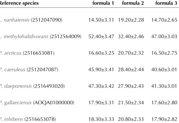
Table 7. DDH similarities betweenT and the other
†
Reference species formula 1 formula 2 formula 3
14.50±3.11 19.20±2.28 14.70±2.65
16.60±3.25 20.70±2.32 16.50±2.75
45.90±3.41 28.40±2.44 40.60±3.01
47.30±3.42 27.90±2.43 41.30±3.01
17.90±3.31 21.50±2.34 17.60±2.80
18.30±3.33 20.80±2.33 17.90±2.82
†DDH similarities were calculated in silico with the GGDC server version 2.0 [57]. The standard deviations indicate the inherent uncertainty in estimating DDH values from intergenomic distances based on models derived from empiri-cal test data sets (which are always limited in size); see [56] for details. The dis-tance formulas are explained in [56]. The numbers in parentheses are IMG object
IDs (GenBank accession number in the case o
Acknowledgements
The authors would like to gratefully acknowledge the assistance of Iliana Schröder for growing L. aquamarina
cultures and Evelyne-Marie Brambilla for DNA extrac-tion and quality control (both at the DSMZ). The work conducted by the U.S. Department of Energy Joint Ge-nome Institute was supported by the Office of Science of the U.S. Department of Energy under contract No.
DE-AC02-05CH11231; the work conducted by the
members of th
ed by the German Research Foundation (DFG) Transregio-SFB 51. We also thank the European Com-mission which supported phenotyping via the Microme project 222886 within the Framework 7 program.
References
1. Vandecandelaere I, Segaert E, Mollica A, Faimali
M, Vandamme P.
isolated form a marine electroactive biofilm, and emended descriptions of
Int J
Syst Evol Microbiol 2008; 58:2788-2793
2. Schaefer JK, Goodwin KD, McDonald IR, Murrell
JC, Oremland RS
gen. nov., sp. nov., a marine methylotroph that grows on methyl bromide. Int J Syst Evol
Microbiol 2002; 52:851-859
3. Sun F, Wang B, Liu X, Lai Q, Du Y, Li G, Luo J,
Shao Z
from marine sediment. Int J Syst Evol Microbiol
2010; 60:275-280
4. Buchan A, González JM, Moran MA. Overview of
the marineAppl Environ
Microbiol 2005; 71:5665-5677
5. Altschul SF, Gish W, Miller W, Myers EW, Lipman DJ. Basic local alignment search tool. J
Mol Biol 1990; 215:403-410
6. Korf I, Yandell M, Bedell J. BLAST, O'Reilly, Se-bastopol, 2003.
7. DeSantis TZ, Hugenholtz P, Larsen N, Rojas M, Brodie EL, Keller K, Huber T, Dalevi D, Hu P, Andersen GL. Greengenes, a Chimera-Checked 16S rRNA Gene Database and Workbench Com-patible with ARB. Appl Environ Microbiol 2006; 72:5069-5072
8. Porter MF. An algorithm for suffix stripping. Pro-gram: electronic library and information systems
1980; 14:130-137.
9. Lee C, Grasso C, Sharlow MF. Multiple sequence alignment using partial order graphs.
Bioinformat-ics 2002; 18:452-464
10. Castresana J. Selection of conserved blocks from multiple alignments for their use in phylogenetic analysis. Mol Biol Evol 2000; 17:540-552
11. Stamatakis A, Hoover P, Rougemont J. A rapid bootstrap algorithm for the RAxML web-servers.
Syst Biol 2008; 57:758-771
12. Hess PN, De Moraes Russo CA. An empirical test of the midpoint rooting method. Biol J Linn Soc Lond 2007; 92:669-674.
13. Pattengale ND, Alipour M, Bininda-Emonds ORP, Moret BME, Stamatakis A. How Many Bootstrap Replicates Are Necessary? Lect Notes Comput Sci
2009; 5541:184-200.
14. Swofford DL. PAUP*: Phylogenetic Analysis Us-ing Parsimony (*and Other Methods), Version 4.0 b10. Sinauer Associates, Sunderland, 2002. 15. Pagani I, Liolios K, Jansson J, Chen IM, Smirnova
T, Nosrat B, Markowitz VM, Kyrpides NC. The GenomesOnLine Database (GOLD) v.4: status of genomic and metagenomic projects and their as-sociated metadata. Nucleic Acids Res 2012; 40:D571-D579
16. Ruiz-Ponte C, Cilia V, Lambert C, Nicolas JL.
sp. nov., a marine
bacte-rium isolated from rearings and collectors of the scallop Pecten maximus.Int J Syst Bacteriol 1998; 48:537-542
17. Moran MA, Buchan A, González JM, Heidelbarg JF, Witman WB, Kiene JR, Henriksen JR, King GM, Belas R, Fuqua C, et al. Genome sequence
marine environment. Nature 2004; 432:910-913
18. Beyersmann PG, Chertkov O, Petersen J, Fiebig A, Chen A, Pati A, Ivanova N, Lapidus A, Goodwin LA, Chain P, et al. Genome sequence of
a surface-associated member of the marine
Stand Genomic Sci 2013; (this
issue).
19. Freese HM, Dalingault H, Petersen J, Pradella S, Davenport K, Teshima H, Chen A, Pati A, Ivanova N, Goodwin LA, et al. Genome sequence of the plasmid and phage-gene rich marine
Stand
Ge-nomic Sci 2013; (this issue).
20. Field D, Garrity G, Gray T, Morrison N, Selengut J, Sterk P, Tatusova T, Thomson N, Allen MJ, Angiuoli SV, et al. The minimum information about a genome sequence (MIGS) specification.
Nat Biotechnol 2008; 26:541-547
21. Woese CR, Kandler O, Weelis ML. Towards a natural system of organisms. Proposal for the
do-mainsProc Natl Acad Sci
USA 1990; 87:4576-4579
22. Garrity GM, Bell JA, Lilburn T. Phylum XIV.
In: Brenner DJ, Krieg
NR, Stanley JT, Garrity GM (eds), Bergey’s Manu-al of Sytematic Bacteriology, second edition. Vol.
2 (The
2005, p. 1.
23. Garrity GM, Bell JA, Lilburn T. Class I.
In: Brenner DJ,
Krieg NR, Stanley JT, Garrity GM (eds), Bergey’s Manual of Sytematic Bacteriology, second
edi-tion. Vol. 2 (The
Al-pha-, Beta-, Delta-, a
Springer, New York, 2005, p. 1.
24. Validation list No. 107. List of new names and new combinations previously effectively, but not validly, published. Int J Syst Evol Microbiol 2006; 56:1-6
25. Garrity GM, Bell JA, Lilburn T. Order III.
In: Brenner DJ, Krieg
NR, Staley JT, Garrity GM (eds), Bergey’s Manual of Systematic Bacteriology, second edition. vol. 2
(TheAlpha-, Beta-,
Delta-, a
York, 2005, p. 161.
26. Garrity GM, Bell JA, Lilburn T. Family I.
In: Brenner DJ, Krieg
NR, Staley JT, Garrity GM (eds), Bergey’s Manual of Systematic Bacteriology, second edition. vol. 2
(TheAlpha-, Beta-,
Delta-, a
York, 2005, p. 161.
27. Schaefer JK, Goodwin KD, McDonald IR, Murrell JC, Oremland RS gen. nov., sp. nov., a marine methylotroph that grows on methyl bromide. Int J Syst Evol
Microbiol 2002; 52:851-859
28. Martens T, Heidorn T, Pukall R, Simon M, Tindall BJ, Brinkhoff T. Reclassification o
et al. 1998 as
description of
classification o
1995) Uchino et al. 1999 as gen. nov., comb. nov., and emended descriptions
of the gener
Int J Syst Evol Microbiol 2006;
56:1293-1304
29. BAuA. Classification o
risk groups. TRBA 2010; 466:93.
30. Ashburner M, Ball CA, Blake JA, Botstein D, But-ler H, Cherry JM, Davis AP, Dolinski K, Dwight SS, Eppig JT, et al. Gene ontology: tool for the unification of biology. The Gene Ontology Con-sortium. Nat Genet 2000; 25:25-29
31. Vaas LAI, Sikorski J, Michael V, Göker M, Klenk HP. Visualization and curve-parameter estimation strategies for efficient exploration of phenotype microarray kinetics. PLoS ONE 2012; 7:e34846
32. Vaas LAI, Sikorski J, Hofer B, Fiebig A, Buddruhs N, Klenk HP, Göker M. opm: An R package for analyzing OmniLog Phenotype Microarray data.
Bioinformatics 2013; 29:1823-1824
33. Yoon JH, Kang SJ, Lee SY, Oh TK
the Yellow Sea in Korea. Int J Syst Evol Microbiol
2007; 57:856-861
http://www.dmsz.de/catalogues/cataloque- microorganisms/culture-technology/list-of-media-for-microorganisms.html.
35. Gemeinholzer B, Dröge G, Zetzsche H,
Haszprunar G, Klenk HP, Güntsch A, Berendsohn WG, Wägele JW. The DNA Bank Network: the start from a German initiative. Biopreserv Biobank
2011; 9:51-55.
36. Bennett S. Solexa Ltd. Pharmacogenomics 2004; 5:433-438
37. The DOE Joint Genome Institute. www.jgi.doe.gov
38. Butler J, MacCallum I, Kleber M, Shlyakhter IA, Belmonte MK, Lander ES, Nusbaum C, Jaffe DB. ALLPATHS: de novo assembly of whole-genome shotgun microreads. Genome Res 2008; 18
:810-820
39. Zerbino DR, Birney E. Velvet: algorithms for de novo short read assembly using de Bruijn graphs.
Genome Res 2008; 18:821-829
40. Phrap and Phred for Windows. MacOS, Linux, and Unix. http://www.phrap.com
41. Hyatt D, Chen GL, LoCascio PF, Land ML, Lar-imer FW, Hauser LJ. Prodigal: prokaryotic gene recognition and translation initiation site identifi-cation. BMC Bioinformatics 2010; 11:119
42. Mavromatis K, Ivanova NN, Chen IM, Szeto E, Markowitz VM, Kyrpides NC. The DOE-JGI Standard operating procedure for the annotations of microbial genomes. Stand Genomic Sci 2009;
1:63-67
43. Pati A, Ivanova NN, Mikhailova N, Ovchinnikova G, Hooper SD, Lykidis A, Kyrpides NC.
GenePRIMP: a gene prediction improvement pipeline for prokaryotic genomes. Nat Methods
2010; 7:455-457
44. Markowitz VM, Ivanova NN, Chen IMA, Chu K, Kyrpides NC. IMG ER: a system for microbial ge-nome annotation expert review and curation.
Bio-informatics 2009; 25:2271-2278
45. del Solar G, Giraldo R, Ruiz-Echevarria MJ, Espi-nosa M, Diaz-Orejes R. Replication and control of circular bacterial plasmids. Microbiol Mol Biol Rev 1998; 62:434-464
46. Petersen J, Brinkmann H, Berger M, Brinkhoff T, Päuker O, Pradella S. Origin and evolution of a novel DnaA-like plasmid replication type in
Mol Biol Evol 2011; 28
:1229-1240
47. Petersen J. Phylogeny and compatibility: plasmid classification in the genomics era. Arch Microbiol
2011; 193:313-321
48. Petersen J, Brinkmann H, Pradella S. Diversity and evolution of repABC type plasmids in
Environ Microbiol 2009;
11:2627-2638
49. Zielenkiewicz U, Ceglowski P. Mechanisms of plasmid stable maintenance with special focus on plasmid addiction systems. Acta Biochim Pol
2001; 48:1003-1023
50. Giraud MF, Naismith JH. The rhamnose pathway.
Curr Opin Struct Biol 2000; 10:687-696
51. Wagner-Döbler I, Ballhausen B, Berger M, Brinkhoff T, Buchholz I, Bunk B, Cypionka H, Daniel R, Drepper D, Gerdts G, et al. The com-plete genome sequence of the algal symbiont life in the sea. ISME J 2010; 4:61-77
52. Petersen J, Frank O, Göker M, Pradella S. Extrachromosomal, extraordinary and essential-the plasmids of essential-theAppl Microbiol Biotechnol 2013; 97:2805-2815
53. Lawley TD, Klimke WA, Gubbins MJ, Frost LS. F factor conjugation is a true type IV secretion sys-tem. FEMS Microbiol Lett 2003; 224:1-15
54. Zech H, Thole S, Schreiber K, Kalhöfer D, Voget S, Brinkhoff T, Simon M, Schomburg D, Rabus R. Growth phase-dependent global protein and
me-tabolite profiles o
strain DSM 17395, a member of the marine
Proteomics 2009; 9
:3677-3697
Microbiol 2012; 14:2661-2672
56. Fürch T, Preusse M, Tomasch J, Zech H, Wagner-Döbler I, Rabus R, Wittmann C. Metabolic fluxes in the central carbon metabolism of
BMC Microbiol 2009; 9:209
57. Schwarz S, Hood RD, Mougous JD. What is type VI secretion doing in all those bugs? Trends
Microbiol 2010; 18:531-537
58. Wagner-Döbler I, Thiel V, Eberl L, Allgaier M, Bodor A, Meyer S, Ebner S, Hennig A, Pukall R, Schulz S. Discovery of complex mixtures of novel long-chain quorum sensing signals in free-living and host-associated marine alphaproteobacteria.
ChemBioChem 2005; 6:2195-2206
59. Auch AF, Von Jan M, Klenk HP, Göker M. Digital DNA-DNA hybridization for microbial species delineation by means of genome-to-genome se-quence comparison. Stand Genomic Sci 2010; 2:117-134
60. Auch AF, Klenk HP, Göker M. Standard operating procedure for calculating genome-to-genome dis-tances based on high-scoring segment pairs.
Stand Genomic Sci 2010; 2:142-148
61. Meier-Kolthoff JP, Auch AF, Klenk HP, Göker M. Genome sequence-based species delimitation with confidence intervals and improved distance functions. BMC Bioinformatics 2013; 14:60
![Table 1. Classification and general features of L. aquimarina DSM 24565T according to the MIGS recommendations [20]](https://thumb-us.123doks.com/thumbv2/123dok_us/698579.2067750/4.612.57.558.181.679/table-classification-general-features-aquimarina-according-migs-recommendations.webp)