http://www.sciencepublishinggroup.com/j/ijmi doi: 10.11648/j.ijmi.20190701.12
ISSN: 2330-8303 (Print); ISSN: 2330-832X (Online)
Color Doppler Evaluation of Ureteral Jets in Patients with
Ureteral Stones
Sameh Ahmad Khodair
1, Mohamad Marzouk Abdallah
21
Radiology Department, Faculty of Medicine, Tanta University, Tanta, Egypt
2Urology Department, Faculty of Medicine, Minoufia University, Minofia, Egypt
Email address:
To cite this article:
Sameh Ahmad Khodair, Mohamad Marzouk Abdallah. Color Doppler Evaluation of Ureteral Jets in Patients with Ureteral Stones. International Journal of Medical Imaging. Vol. 7, No. 1, 2019, pp. 11-17. doi: 10.11648/j.ijmi.20190701.12
Received: December 20, 2018; Accepted: January 21, 2019; Published: February 13, 2019
Abstract:
Objective: To evaluate the characteristics of ureteral jets in patients with ureteral stones by color Doppler ultrasonography. Method: 49 patients (patient group) with unilateral ureteral stones proved by non contrast spiral CT and another 40 healthy volunteers (control group) who have no any previous urinary complaint were examined for their ureteral jets by color Doppler ultrasonography for ureteral jet number per 5 minutes as well as its peak velocity (cm/s). Statistical analysis was done by using Social Sciences computer program (SPSS) version 17, p-values less than or equal to 0.05 were considered statistically significant difference. Results: There was a significant statistical difference between the healthy volunteers and the patients with ureteral stones (p < 0.001). However, there was no statistical difference between the healthy volunteers on both sides and also the healthy volunteers and the non – obstructed side of the patients with ureteral stones (p > 0.05). So the healthy side could be used as a reference to the diseased side according to our results. Conclusion: The color Doppler evaluation of the ureteral jets (its number and velocity) is a valid method of evaluating ureteral calcular obstruction and could be a first line of management to detected ureteric obstruction especially in patients with acute flank pain which may be controversial for other causes of acute abdomen in the emergency department.Keywords:
Color Doppler, Ureter, Uretral Jets, Urinary Stones1. Introduction
Acute flank pain is a common complaint of patients who are examined in emergency departments. It represent lifetime risk 12% in men and 6% in women [1] with ureteral stones as the cause in a large number of patients. Patients typically present with intermittent pain radiating to right or left side with or without hematuria. Unfortunately, the clinical findings are nonspecific, and can mimic other conditions such as; appendicitis, tubo-ovarian abscess, pelvic inflammatory disease, inflammatory bowel disorders, and pyelonephritis. Ultrasonography and CT have become important imaging modalities in the evaluation of patients with flank pain. [2] Ultrasonography is frequently requested as the initial imaging study for the evaluation of acute abdomen. Color Doppler ultrasound evaluation of ureteral jets is a useful tool beside the grey-scale B - mode in the diagnosis of ureteral obstruction. [3] A variety of Doppler
spectral analysis can enable detection not only qualitative but also quantitative determination of the degree of ureteral obstruction in many patients with unilateral ureteral stone. [6]
2. Material & Methods
This study had been performed on selected patients whom underwent ultrasonography and color Doppler examination by the same ultrasonographic physician examiner followed by examination with non – contrast spiral CT urinary tract at the same day. These patients were referred to the radiology department at our institution for investigation of acute flank pain. The research protocol was approved by the research committee of our institution before starting the study. A routine signed patient consent form was obtained to use the patient's data in research work. All the ultrasonographic and non – contrast spiral CT data were recorded in an electronic archiving system which allow reviewing the whole collected patients data and archived images.
The inclusion criteria were patients suffering from acute flank pain submitted for ultrasonographic examination and color Doppler evaluation of the ureteral jet and proved by non contrast spiral CT to have a unilateral ureteral stone. The exclusion criteria were patients with bilateral ureteral stones, patients with previous ureteral operation, patients with impaired renal function, patients with impaired cardiac output and patients proved by non contrast CT to have no ureteral stones on both sides.
A total 49 patients were included in this study; they were presented with acute flank pain. In addition a total 40 volunteers were included; all of them had no previous history of urinary tract problem, they were submitted only to ultrasonographic examination and color Doppler evaluation of the ureteral jet in the same manner of patients group and were considered as a control healthy group.
Ultrasonographic examination was performed by using a commercially available color Doppler scanner (Voluson 730 Pro; GE, Milwaukee, WI) with a 3.0–5.0 MHz convex probe. Patients were instructed to drink a liter of water half an hour before the ultrasound examination to be well hydrated. The patients were first examined by B-mode ultrasonography of the kidneys to detect renal, ureteral or bladder stones and degree of renal hydronephrosis when present and then color Doppler examination of the urinary bladder at the region of the trigone was applied for continues five minutes. The scanning of the urinary bladder was done in the transverse plan where the two vesico-ureteric orifices seen at the same time, and the patient in a supine position. The color box was adjusted to include the both vesico-ureteric orifices. The color scale, pulse repetition frequency (PRF) was adjusted as needed, and flow toward the transducer was assigned a red color. Color gain was set just below the level at which noise was seen. A wide sample gate was applied to cover the ureteral jet at the vesico-ureteric orifice. The PRF was adjusted at 1.4 - 1.8 KHz. Pulsed waves Doppler was applied to measure the peak velocity obtained from each ureteral jet.
The number of jets per 5 minutes was recorded from each orifice. All the perceived waveform from the ureteral jet from the obstructed side and from the healthy side in patient groups as well as from both sides of the control group were a continuous (venous – like) waves. The peak velocity of each jet wave was measured from each orifice and average was calculated for each side.
All the 49 patients were submitted for non contrast CT scanning after finishing the ultrasonography examination. The non – contrast spiral CT examination started from the upper pole of the kidneys to the base of the urinary bladder with a 5-mm collimation, 120 kv, 200mAs, and a reconstruction at 2 mm intervals. No oral or intravenous contrast was administered. The images were analyzed at a workstation which had a reconstruction processing.
Statistical analysis was based on comparison of the number of ureteral jets and its velocity in the patient group (49 patient) between the obstructed and non obstructed side. Also between the right and left side in the healthy control group (40 healthy individual) as well as the healthy side in the patients group and the right and the left side in the healthy group.
Statistical analysis was performed with the Statistical Package for Social Sciences computer program version 17.0 (SPSS). The Paired t - test was used to analyze differences between the number of ureteral jets and its velocity in the healthy side and diseased side in patient group. It is also used to analyze the difference between the number of ureteral jets and its velocity on the right side and the left side in the control group. Analysis of the ureteral jet numbers and its velocity of the healthy side of patients group and the right side and left side of the control group was investigated using the ANOVA test (F - test). p-values less than or equal to 0.05 indicated a statistically significant difference.
3. Results
On Color Doppler study of the ureteral jets of the 40 selected healthy asymptomatic volunteers (Control group), the mean average of jet number in the right side was 16.1±4.0 jets/ per 5 minutes while the left side was 16.2 ±3.9 jets/ per 5 minutes (P value 0.712) that statically non significant. The mean average of jet flow velocity in the right side was 40.3±7.0cm/s while the left side was 40.3±6.7cm/s
(P value 0.654) that statically non significant (table 2). In comparison between the ureteric jets number and velocity of the healthy side of the patient group and the right and left side of the control group it was statistically non significant as the F – test was 1.675 (P value 0.191) and 0.172 (P value 0.842) for the jet number and jet velocity respectively (table 3).
Table 1. Comparison of number of jets/5 min and its velocity between diseased side and healthy side in patient group.
Side Number of jets/ 5 min range mean ± SD P value Jet velocity cm/s range mean ± SD P value
Healthy side n= 49 10 – 20 14.5 ±2.9
0.000 30 – 60 cm/s 39.7±6.8 0.000
Diseased side n = 49 0 – 8 2.89±1.6 5 – 14 cm/s 8.7±2.2
Table 2. Comparison of number of jets/5 min and its velocity between Right & Left side of control group.
Side Number of jets/ 5 min range mean± SD P value Jet velocity cm/s range mean± SD P value
Right side n= 40 10 – 25 16.1±4.0
0.712 30 – 65 40.3±7.0 0.754
Left side n = 40 11 – 25 16.2±3.9 33 – 65 40.3±6.7
Table 3. Comparison of number of jets/5 min and its velocity in healthy side of patient group and Right & Left side of control group.
Side Number Number of jets/ 5 min Mean ± SD P value
F- test Jet velocitycm/s range Mean ± SD
P value F- test
Healthy side patient group 49 10 – 20 14.5 ±2.9
0.191
30 – 60 cm/s 39.7±6.8
0.842
Right side control group 40 10 – 25 16.1±4.0 30 – 65 40.3±7.0
Left side control group 40 11 – 25 16.2±3.9 33 – 65 40.3±6.7
Figure 2. Color Doppler sonography of the right ureteric jet of a healthy subject with the sample box of the pulsed Doppler is adjusted at the right vesico-ureteric orifice (peak velocity = 35.8 cm/s).
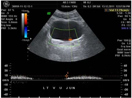
Figure 4. Color Doppler sonography of the left ureteric jet of another patient proved by non contrast spiral CT to have a left ureteric stone (peak velocity = 16.8 cm/s).
4. Discussion
Acute abdomen pain account for more than half of all surgical procedures performed in the emergency unit. Prompt and accurate diagnosis is essential for the proper management of such acute conditions. [7] Imaging techniques leads to accurate insight in the diagnosis of acute abdomen, and specifies cases to a more rapid diagnosis. [8] The different imaging modalities to be performed for the patients with acute abdominal pain includes; conventional abdomen and pelvis radiography, ultrasonography and computed tomography (CT) which are the most often used modalities in this setting. Choosing of the initial imaging technique will depend upon the localization and nature of pain as well as the probability of the professional diagnosis. [9] Conventional radiography of the abdomen is an effective technique in diagnosis of urinary stones, regarding its availability, its economic costs especially in the developing countries, that will remain widely used, however its accuracy is limited by body size, patients preparation, especially with the presence of intestinal gases as well as its inability to diagnose the radiolucent stones. Non contrast spiral computed tomography has a high sensitivity (96%) and specificity (100%) for the detection of both radiopaque and radiolucent stones, but it is still limited by costs and in certain populations such as pregnant women and young children due to radiation exposure. [10] Ultrasonography is considered as imaging modality of choice in acute abdomen as a first line of imaging especially when renal colic is the professional diagnosis [11] As hydronephrosis of the kidneys and ureteric obstruction are closely associated, but upper urinary tract dilatation can occur without significant obstruction and vice versa. [12] Dubbins, P. A., et al, stated that during the real-time ultrasound examinations of the urinary bladder, that jets of urine were noted by B- mode to enter the bladder during the filling phase as the jets appeared as motion in the fluid from the vesico-ureteric orifice at irregular intervals. [5] Some of color Doppler artifacts could be detected in variant pelvic and abdomenal conditions and could be usefully used in different clinical applications, the ureteric jets could be evaluated by color Doppler examination at the vesico-ureteric orifice as a color flow going into the lumen of the urinary bladder and emitting from the vesico-ureteric orifices [13] In their study using color Doppler to evaluate the ureteric jets in children patients with hydronephrosis as well as patients with acute renal colic, Cvitkovic Kuzmic et al, [14] found that ureteric jets were completely absent in eight of nine children patients (89%) with acute renal colic. In our study we found also absent ureteric jet but this was recorded in only 10 patients (20%) from the side with ureteric stone presented in acute stage. This difference may be due to the age group of our study was adult and regarding to Vivian Yee-fong [15], who found a statistically difference between child group and adult group in the number and velocity of the ureteric jets, however the site and size of the calculi may be
another factors.
In our study, statistically significant difference were found between the diseased ureter having a stone and the other healthy ureter (p value less than 0.001). This is in agree with Burge, H. J., et al. [6] who found in their study that the side with ureteric stone showed abnormal pattern of ureteric jets in both low grade and high grade obstruction, however they depended upon the flow pattern (direction and appearance) and not the velocity as in our study.
In our study, we found no statistical significant difference between the right side and the left side of the healthy control group and this is in agreement with Vivian Yee-fong [15], who found in their study on a large group - to evaluate the ureteric jet patterns and factors affecting it regarding age, sex and others - that, the studied adult group, there were no statically significant difference between right and left side of ureteric jets in healthy asymptomatic subjects. Also they stated that the mean average number of normal jets per 5 minutes was 8 jets and the mean velocity was 57 – 63 cm/s, these mean values are near our study mean values regarding the number and velocities. In our study, no statistical significant difference between the healthy side of the patient group and the control healthy group regarding the number and velocity of ureteric jets and this stated that in unilateral renal colic the healthy side could be taken as a reference for the diseased side
5. Conclusion
The color Doppler evaluation of the ureteral jets (its number and velocity) is a valid method of evaluating ureteral calcular obstruction and could be a first line of management to detected ureteric obstruction especially in patients with acute flank pain which may be controversial for other causes of acute abdomen in the emergency department.
6. Recommendation
We advise for a study with larger group than this study as – for our knowledge - this is the first study use the flow velocity of urine flow and so need studies with larger patients group.
References
[1] Matthew Bulttitude, a. J. R., Management of renal colic. www.bmj.com/content/345/bmj.e5499. 2012. August.
[2] Douglas H. Sheafor, Barbara S. Hertzberg, Kelly S. Freed, et al., Nonenhanced helical CT and US in the emergency evaluation of patients with renal colic prospective comparison. Radiology, 2000. 217 (3): p. 792-7.
[4] Shannon C. Campbell, Jeanne A. Cullinan, and Deborah J. Rubens, Slow Flow or No Flow? Color and Power Doppler US Pitfalls in the Abdomen and Pelvis. RadioGraphics, 2004. 24: p. 497-506.
[5] P A Dubbins, A B Kurtz, J Darby, and, B B Goldberg., Ureteric jet effect: the echographic appearance of urine entering the bladder. A means of identifying the bladder trigone and assessing ureteral function. Radiology, 1981. 140 (2): p. 513-5.
[6] H J Burge, W D Middleton, B L McClennan, and C F Hildebolt. Ureteral jets in healthy subjects and in patients with unilateral ureteral calculi: comparison with color Doppler US. Radiology, 1991. 180 (2): p. 437-42.
[7] Ajay Singh, Raman Danrad, Peter F. Hahn, Michael A. Blake, Peter R. Mueller, and Robert A. Novelline. MR imaging of the acute abdomen and pelvis: acute appendicitis and beyond. RadioGraphics, 2007. 27 (5): p. 1419-31.
[8] Keeman, J. N., [New diagnostic imaging technology often offers no advantage in the differential diagnosis of acute abdomen]. Ned Tijdschr Geneeskd, 1999. 143 (45): p. 2225-9.
[9] Excoffier, S., P. A. Poletti, and H. Brandstatter, [Acute abdominal pain of the upper abdomen: which imaging to choose?]. Rev Med Suisse. 2013. 9 (399): p. 1710, 1712-4.
[10] Sorensen M. D., Harper JD, His RS, et al., B-mode ultrasound versus color Doppler twinkling artifact in detecting kidney stones. J Endourol. 2013. 27 (2): p. 149-53.
[11] Mishra DS, Magu S, Sharma N, Rattan KN, Tewari AD, and Rohilla S. Imaging in acute abdomen. Indian J Pediatr, 2003. 70 (1): p. 15-9.
[12] de Bessa J Jr, Dénes FT, Chammas MC, Cerri L, et al., Diagnostic accuracy of color Doppler sonographic study of the ureteric jets in evaluation of hydronephrosis. J Pediatr Urol, 2008. 4 (2): p. 113-7.
[13] Drs. Michael Hirsch S, Tamara Palavecino B, and Boris León R. Color doppler twinkling artifact: A misunderstood and useful sign. Revista Chilena de Radiología 2011. 17 (N2): p. 82-84.
[14] Cvitković Kuzmić A, Brkljacić B, Rados M, and Galesić K., Doppler visualization of ureteric jets in unilateral hydronephrosis in children and adolescents. Eur J Radiol, 2001. 39 (3): p. 209-14.