ISSN 0015–5659 journals.viamedica.pl
Address for correspondence: Dr. M. Farimaz, Department of Anatomy, Faculty of Medicine, Hacettepe University, Ankara, 06100, Turkey, tel: +90 312 3052357, fax: +90 312 3107169, e-mail: [email protected]
The morphometry of the cavernous part
of the internal carotid artery
M. Farımaz
1, H.H. Çelik
1, K.M. Ergun
1, A. Akgöz
2, B. Urfalı
31Department of Anatomy, Faculty of Medicine, Hacettepe University, Ankara, Turkey 2Department of Radiology, Faculty of Medicine, Hacettepe University, Ankara, Turkey 3Department of Neurosurgery, Faculty of Medicine, Mustafa Kemal University, Hatay, Turkey
[Received: 8 March 2018; Accepted: 23 April 2018]
Background: In the study, the morphometric evaluation of internal carotid artery (ICA) was performed in order to show the differences between the age groups and genders.
Materials and methods: In the study, descriptive measurements of intercarotid distance on the computed tomography of 173 (88male [M], 85 female [F]) pa-tients and the intercavernous distance on magnetic resonance images (MRIs) of 49 (19 M, 30 F) individuals were reviewed.
Results: Intercarotid distance was found to be close to the border of statistical significance and for results of the comparative measurements that were per-formed in the study; no significant sex-associated difference was observed for the distance between the gender and midpoint of the sella turcica and medial margin of the right ICA. Compared to gender, the distance between the base of the sella turcica and the base of the left ICA is found to be closed to of statistical significance. A statistically significant difference was obtained for the distance between the midpoint of sella turcica and medial margin of the left ICA and for the distance between the base of the sella turcica and the base of right ICA. Although it is observed that there is a weak correlation between the age and the distance between midpoint of the sella turcica and medial margin of the right ICA, statistically there is a significant difference between them.
Conclusions: Obtained results, planning of surgical interventions are supportive and guiding in terms of prevention of damage of to ICA in three dimensional thinking and operations. (Folia Morphol 2019; 78, 1: 54–62)
Key words: internal carotid artery, cavernous sinus, morphometry, transsphenoidal surgery
INTRODUCTION
The internal carotid artery (ICA) is one of the main arteries of the central nervous system. The cavernous part of this artery is important clinically in transsphe-noidal surgical procedures. Transsphetranssphe-noidal surgical procedures minimise the risk of complications and prevent bleeding of the vessel [12]. Transsphenoidal
surgery and its modifications are important while
taking biopsies from sellar and parasellar lesions
[3, 8, 10, 15, 21, 24]. Bleeding of the ICA in the sellar region may be important for mortality and morbidities. Therefore, prior to surgical procedures it
is clinically important to know the transverse distance between the right and left internal carotid arteries, which are found on both sides of the sella turcica [12].
The aim of this study is the morphometric analysis of the cavernous part of the ICA in an anatomically
-formed according to age groups and sex. We believe
that this analysis will have a significant impact on
planning surgical interventions in the region.
MATERIALS AND METHODS
Patient population
The morphometric analysis was performed in two groups. The first group consisted of patients who underwent brain computed tomography (CT) an -giography examination in May, June and July 2015 in the Radiology Department of Hacettepe Medical
Faculty. The second group consisted of patients who underwent three-dimensional (3D) contrast magnetic
resonance (MR) examinations in 2015 in same
depart-ment. The 201 patients who underwent CT angiog
-raphy were between 40 and 71 years old. However, 28 of them were not included in the study because
of presence of stents, stenosis, occlusion, plaques or
technical reasons. The patients who underwent 3D contrast MR examination were between 21 and 84
years of age. This group included 50 patients, but
one of the patients was excluded due to the presence of a meningioma. Ethical approval for the study was
obtained from the ethics committee of our institution (December 16, 2015, GO 15/777-18).
CT examination protocol
Computed tomography angiographies of the
patients were performed using dual CT, which has a double tube system and 64 sections (Somatom Defi -nition; Siemens Erlangen, Germany). The MR images
(MRI) were taken in a 1.5 Tesla (Signa HDxT, GE Health -care, USA) instrument. The acquisition details are as
follows: section thickness was 1 mm, NEX 1, matrix
256 × 256, TR-10 ms, TE-4.2 ms, T1-450, FA-15, sequence 3D T1 FSPGR.
Image evaluation and analysis
The measurements were taken in sagittal, coronal
and axial sections of CT angiographies and 3D con-trast MRI. In the sagittal axis, the deepest part of the
sella turcica was chosen as a reference point for the
measurements. The measured sections of the MRI
were taken post contrast in axial and sagittal planes and were in 3D. In these images, the morphometric analysis was done in a plane vertical to the sagittal base and coronal reformats were obtained. The dis
-tances measured in the CT angiographies were as follows: in the first sagittal axis obtained, the deep
-est section of the sella turcica was found. This axis
was used in every measured parameter. In the axial
sections, the intercarotid distance (the nearest point of the medial borders of the internal carotid arteries)
was measured. In the coronal sections, the mid-point
of the sella turcica (determined by measuring the
distance between the carotid sulci on each side and dividing this value by two) and the distance between
the medial borders of the internal carotid arteries
were measured. In the coronal sections, the distance between the base of the sella turcica and the bases
of the internal carotid arteries in the cavernous sinus
was calculated.
In the 3D contrast MRI, the distance between the medial borders of the cavernous sinus was measured. The measurements were taken by obtaining the cor -onal reformats in a plane that is vertical to the base of the sella turcica.
Statistical analysis
The obtained data were analysed statistically us
-ing the SPSS 22 software. The correlations between the different parameters were determined with the
Pearson test. For the comparison of the parameters
between two groups Student’s t test was used. Be
-forehand the usage of Student’s t test, a normality was assessed for the statistical analysis. In all the sta
-tistical analyses, a p value under 0.05 was considered statistically significant.
RESULTS
In the study, the intercarotid distance was meas -ured on CT scans of 173 patients (88 males [M],
85 females [F]) and the intercavernous distance was
measured in the MRI of 49 patients (19 M, 30 F).
Additionally, comparative measurements were done
on CT brain scans. These comparative measurements
included the distance between the medial borders of
the right and left internal carotid arteries and the
mid-point of the sella turcica and the distance between
the base of sella turcica and the bases of the right and left internal carotid arteries (Table 1).
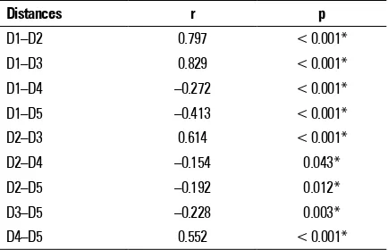
The obtained data showed that there was a strong correlation between the intercarotid distance and the distance between the mid-point of the sella turcica
and the medial borders of the right and left
inter-nal carotid arteries (p < 0.001). There was a weak correlation between the intercarotid distance and the distance between the base of sella turcica and base of the right ICA. However, in this group, statis
the distances (p < 0.001). Although, there was only a mild degree of correlation between the intercarotid distance and the distance between the base of the
sella turcica and the base of the left ICA (p < 0.001),
the measured distances showed statistically signifi -cant differences in this group. Additionally, an inverse
correlation was detected between the intercarotid distance and the distance between the base of the
sella turcica and the bases of the right and the left
internal carotid arteries. There was a mild degree of correlation between the mid-point of the sella turcica and the medial border of the right ICA and between
the mid-point of the sella turcica and the medial border of the left ICA (p < 0.001). In this group, the
measured distances showed statistically significant differences. There were weak correlations between
the mid-point of the sella turcica and the medial
bor-der of the right ICA and between the base of the sella
turcica and bases of the right and the left internal
ca-rotid arteries. However, in these groups, the measured distances showed statistically significant differences. There were mild degree of correlations between the
distance of the base of the sella turcica and the base
of the right ICA and the distance between the base
of the sella turcica and the left ICA (p < 0.001). There
were statistically significant differences among the
distances in these groups (Table 2).
Additionally, the types of sphenoidal sinuses were
also examined in the study. Of the 173 cases, 86.7% had sellar type, 10.98% had presellar type, 2.31%
had conchal type sinuses. There was no statistically significant difference between the types of sphenoidal
sinuses and measured distances (p > 0.05).
The mean value of the intercarotid distance was
13.6 ± 2.8 mm in both sexes (Fig. 1). In females, the
mean value was 14.1 ± 2.8 mm and in males, it was
13.0 ± 2.8 mm (Table 3). The mean value of the
in-tercarotid distance was 16.5 ± 4.3 mm in both sexes (Figs. 2, 3). In females, the mean value was 15.8 ± 3.8 mm, and in males, it was 17.1 ± 4.7 mm (Table 1). The mean value of the intercavernous distance was
13.6 ± 2.8 mm of both sexes (Fig. 1). In the statistical
analysis for differences between females and males, the intercarotid distance was found to be close to the significance level (p = 0.051) and the intercavernous distance was found to be non-significant (p > 0.05). Additionally, the intercarotid distance did not show any correlation with age and the intercavernous distance showed a negative correlation with age (r = –0.079).
Table 1. Statistical evaluation results according to age and gender groups of measured distances
D1 D2 D3 D4 D5
Gender (n = 173)
Female (n = 85) 15.8 ± 38 9 ± 2.3 8.8 ± 1.8 0.2 ± 1.7 0 ± 2
Male (n = 88) 17.1 ± 4.7 9.6 ± 2.5 9.7 ± 2.7 –0.6 ± 2.10 0 ± 2
P 0.051* 0.111 0.014* 0.006* 0.07
Age groups
≤ 40 (n = 26) 15 ± 3.9 8.3 ± 2 8.7 ± 1.9 –0.5 ± 1.8 0 ± 2
41–50 (n = 29) 16.3 ± 4.7 9 ± 2.7 9.1 ± 3.1 –0.7 ± 1.8 0 ± 2
51–60 (n = 40) 17.2 ± 4.1 9.4 ± 2.2 9.7 ± 2.2 –0.5 ± 17 0 ± 2
61–70 (n = 35) 17.1 ± 4.7 9.6 ± 2.5 9.63 ± 2.2 0.4 ± 2.2 0 ± 2
≥ 71 (n = 43) 16.4 ± 4.2 9.7 ± 2.3 9.0 ± 2.2 0 ± 2.1 0 ± 2
Data is shown as mean ± standard deviation. D1 — intercarotid distance (mm), D2 — in coronal sections, the distance between the mid-point of sella turcica and medial border of right internal carotid arteries coursing in cavernous sinus (mm), D3 — in coronal sections, the distance between the mid-point of the sella turcica and the medial border of the left internal carotid artery coursing in the cavernous sinus (mm), D4 — in coronal sections, the distance between the base of the sella turcica and the base of the right internal carotid artery coursing in cavernous sinus (mm), D5 — in coronal sections, the distance between the base of the sella turcica and the base of the left internal carotid artery coursing in cavernous sinus (mm); p > 0.05 — statistically not significant difference; *p < 0.05 — statistically significant difference
Table 2. Correlation analysis test results
Distances r p
D1–D2 0.797 < 0.001*
D1–D3 0.829 < 0.001*
D1–D4 –0.272 < 0.001*
D1–D5 –0.413 < 0.001*
D2–D3 0.614 < 0.001*
D2–D4 –0.154 0.043*
D2–D5 –0.192 0.012*
D3–D5 –0.228 0.003*
D4–D5 0.552 < 0.001*
The mean value of the distance between the
mid-point of the sella turcica and the right medial
bor-der of the ICA coursing in the cavernous sinus was 9.3 ± 2.4 mm. In females, it was 9.0 ± 2.3 mm, and in males, it was 9.6 ± 2.5 mm. The mean value of the distance in between the mid-point of the sella turcica and the left internal carotid artery’s medial border coursing in the cavernous sinus was 9.3 ± 2.3 mm. In females, it was 8.8 ± 1.8 mm and, in males, it was
9.7 ± 2.7 mm (Fig. 4).
The mean value of the distance between the base of the sella turcica and the base of the right ICA was –0.2 ± 2 mm. In females, it was 0.2 ± 1.7 mm, and in males, it was –0.6 ± 2.1 mm. The mean value of the distance between the base of the sella turcica and base of the left ICA was 0 ± 2 mm (Fig. 5). In both sexes, the mean value was found to be 0 ± 2 mm. In the measurement of the distance between the base of
the sella turcica and base of the right and left internal carotid arteries coursing in the cavernous sinus; the
value was recognised as (+) when the base of ICA was over the base of the sella turcica. When the base
of the ICA was under the base of the sella turcica; it was recognised as (–) (Fig. 5).
In the statistical analysis of the comparative
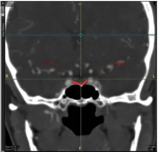
meas-urements between males and females, the distance Figure 1. The measurement of intercavernous distance (magnetic
resonance).
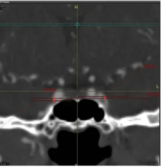
Figure 2. The measurement of intercarotid distance (computed tomography angiography).
Figure 3. The minimum intercarotid distance measurement (com-puted tomography angiography).
Figure 4. The measurement of the distance between the mid-point of the sella turcica and the medial borders of the right and left inter-nal carotid artery (computed tomography angiography).
Table 3. Intercavernous distance
Intercavernous distance Mean ± SD Min–Max p Female (n = 30) 14.1 ± 2.8 7.8/21.5
0.183
Male (n = 19) 13.0 ± 2.8 7.8/18.6
Sex (n = 49) 13.6 ± 2.8 7.8/21.5
Age 56.5 ± 12.8 24/84
between the base of the sella turcica and the base of the left ICA was found to be close to the signifi
-cance level (p = 0.07; Fig. 5). There was no statisti
-cally significant difference in the measurement of the distance between the mid-point of the sella turcica and the medial border of the right ICA. However, statistically significant differences were observed in the measurements of distances between mid-point
of the sella turcica and the medial border of the ICA,
between the mid-point of the sella turcica and the medial border of the left ICA and between the base of sella turcica and the base of right ICA (p = 0.014, p = 0.006; Figs. 4, 5). Additionally, there was a weak correlation in the distance between mid-point of the
sella turcica and the medial border of the right ICA
with age (r = 0.186).
In the statistical analysis of the sex parameter
between MR and CT groups, χ2 test was used. No sta
-tistically significant difference was observed between these two groups according to sex (p = 0.14). In the statistical analysis of the age parameter between MR and CT groups, Mann-Whitney U test was used. No sta
-tistically significant difference was observed between these two groups according to age (U = 3926.5, p = 0.43).
DISCUSSION
The intercarotid distance between the cavernous
parts of the internal carotid arteries, sellar region, cavernous sinus, and their relation to each other are important clinically to prevent arterial bleeding during surgical procedures [1, 2, 5–7, 12, 19, 21, 22, 27]. The ICA is located on the medial side of the cavernous sinus
(C4 segment) [13, 14]. As there are no cranial nerves in the medial part of the cavernous sinus, ICA is the ma-jor structure encountered in transsphenoidal surgery. In surgical procedures, after the exposure to the base of the sella turcica, the dura mater covering the
hypophysis is defined. The dura mater splits into two layers. The periosteal dura forms the first layer and
is related to the bone. After covering the base of the sella turcica, the periosteal dura courses to the lateral
side and forms the medial wall of the cavernous sinus.
Later, it continues as the base of the sinus. The dural
layer, which is adherent to the hypophysis separates
the gland from the medial side of the cavernous sinus. This layer continues as the lateral layer on the lateral side and roof of the cavernous sinus [25]. Therefore,
the medial wall of the cavernous sinus is divided into two parts. In the upper section, the medial wall of the
cavernous sinus is formed as the dural layer of the
hy-pophysis without a bony support. In the lower section, the medial wall of the cavernous sinus is formed by the
periosteal dura of the sella turcica and supported by
the bony lateral wall of the sphenoidal sinus. Destrieux
et al. [9] studied the microanatomy of the dura layers in
the sellar region. Their definitions were in accordance with Romano et al. [21], the mean distance between the medial walls of the cavernous sinus was found to be 14.9 mm (10.1/18.22, n = 20) in their study.
In all the measurements done in our study, the
sagittal axis, in which the deepest point of sella tur
-cica was defined, was used first. The deepest point
of sella turcica can best be observed and measured in CT images taken in the sagittal axis and, therefore,
this axis was used for the morphometric examination.
In studies performed on cadavers, Fujii et al. [11], (at the level of the sellar base), Abuzayed et al. [1] (at the sellar level), Perondi et al. [19] (at level of the sellar base), and Aktas et al. [2] (at level of the dorsum
sellae) measured the intercarotid distances. It was
observed that the measurements done on CT scans
were in accordance with the cadaver measurements as shown in Table 4.
Figure 5. The measurement of the distance between the base of the sella turcica and the bases of the right and left internal carotid arteries (computed tomography angiography).
Table 4. Intercarotid distance (ID) at the level of the sella turcica
Authors Mean ID [mm]
Fujii et al. (1979) 17
Abuzayed et al. (2010) 18
Perondi et al. (2013) 18
Aktas et al. (2013) 15.33
In our study, in the measurements done on the
MRI, the mean intercavernous distance was found to
be 14.1 ± 2.8 mm in females and 13.0 ± 2.8 mm in
males. There was no statistically significant difference between males and females for this measurement (p = 0.183). Additionally, the intercavernous distance showed a negative correlation with age (r = –0.079). The mean value of the intercavernous distance was
found to be 14.9 mm by Romano et al. [21], 12.7 mm
by Zada et al. [27]. However, in our study, we found
this distance to be 13.6 mm.
In this study, in the measurements done on the CT scans, the mean value of the nearest distance of
medial walls of the internal carotid arteries cours
-ing in the cavernous sinus was found to be 15.8 ±
± 3.8 mm in females and 17.1 ± 4.7 mm in males.
In surgical approaches, the medial wall of the ICA will have to be penetrated before an injury could
occur to the ICA coursing in the cavernous sinus. Therefore, if abundant venous bleeding is observed during a transsphenoidal surgery, it is likely to be secondary to the penetration of the cavernous sinus
and surgeons must be aware of the course of the ICA, which is approximately 3 mm away. The study also showed a negative correlation between age and the mean distance between the medial walls of the internal carotid arteries. All these findings are
important clinically.
In our study, the CT scan data showed that the
measurement of the intercarotid distance of the
cav-ernous part of the ICA was 16.5 mm (mean value). The intercarotid distance was measured in the ax
-ial sections of CT images. In MRI, studies found in literature measured this distance to be 16.6 mm
in Scotti et al.’s study [23], 17.8 mm in Knappe et al.’s research [16], 16.2 mm in Zada et al.’s work [27], 20.6 mm in Zhang et al.’s manuscript [28] and 19.4 mm in Sasagawa et al.’s study [22]. The mean value of the intercarotid distance in cadavers was
found to be 17 mm by Fujii et al. [11], 13.33 mm by Ozcan et al. [18], 18 mm by Abuzayed et al. [1], 18 mm by Perondi et al. [19], 15.33 mm by Aktas et al. [2], and 12.15 mm by Cebula et al. [5] (Table 5).
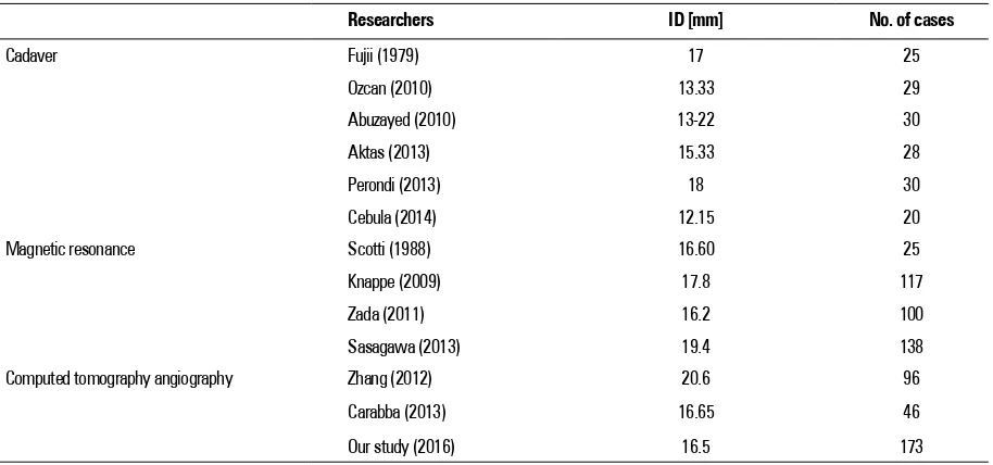
As shown in Table 5, measurement of the interca
-rotid distance was primarily done on cadavers. Simi
-larity in the measured intercarotid distances between
cadavers and living patients suggests that radiologic methods can also be used accurately to obtain these measurements. Comparatively, our study had the
highest number of patients in whom the measure
-ments were done.
Gupta [12] and Yilmazlar et al. [26] measured the
distances from three different views: anterior, medial
and posterior. According to Gupta [12], the nearest
distance between the internal carotid arteries was
found at the maximum sellar depth. Yilmazlar et al.
[26] reported a statistically significant difference in
the medial and posterior intercarotid distances in studies done on MRI and cadavers. Zhang et al. [28] and Carrabba et al. [4] measured the intercarotid dis-tances on CT scan and found them as to be 20.6 mm and 16.65 mm, respectively. The main difference
between our study and these two studies was the
large size of our patient population. Table 5. Comparison of the measured intercarotid distances (ID)
Researchers ID [mm] No. of cases
Cadaver Fujii (1979) 17 25
Ozcan (2010) 13.33 29
Abuzayed (2010) 13-22 30
Aktas (2013) 15.33 28
Perondi (2013) 18 30
Cebula (2014) 12.15 20
Magnetic resonance Scotti (1988) 16.60 25
Knappe (2009) 17.8 117
Zada (2011) 16.2 100
Sasagawa (2013) 19.4 138
Computed tomography angiography Zhang (2012) Carabba (2013) Our study (2016)
20.6 16.65 16.5
If the intercarotid distance between the cavern -ous parts of the internal carotid arteries is less than
10 mm, transsphenoidal surgery will be difficult [20].
In our study, the minimum intercarotid distance
was found to be 5.5 mm. Although, the short in -tercarotid distance does not make the operation impossible, it can only be performed by compe-tent surgeons. In literature, the mean intercarotid distance of internal carotid arteries coursing in the
cavernous sinus was found to be 12 mm (4–18 mm). However, the artery may be tortuous in the carotid
sulcus or more laterally located in the sinus [17]. In
our series, the maximum intercarotid distance was found to be 27.3 mm. However, this value does
not create a contraindication for transsphenoidal surgery [26].
The data in Table 5 show the various studies done on this subject. However, in our study, we also meas
-ured the distances between the internal carotid arter -ies and the sella turcica, in addition to the intercarotid and intercavernous distances.
In our study, the distances between the mid-point
of the sella turcica and the right and left internal
carot-id arteries were measured. These measurements were
performed in the coronal sections of CT images. On the
right side, it was 9.0 ± 2.3 mm in females and 9.6 ± ± 2.5 mm in males. This distance did not show any sta
-tistically significant sex-related difference (p = 0.111). The mean distance between mid-point of the
sella turcica and the medial border of the left ICA
coursing in the cavernous sinus was 8.8 ± 1.8 mm in females and 9.7 ± 2.7 mm in males. There was a statistically significant sex-related difference for this distance (p = 0.014). The mean distance in between the mid-point of the sella turcica and the
medial border of the right ICA coursing in the
cav-ernous sinus showed a weak correlation with age groups. These findings will be important during
surgical procedures.
Cheng et al. [7] studied 144 CT scans (75 females and 69 males). In the coronal plane, the mean distance
of the ICA to the mid-point of the sella turcica was
found to be 11.25 mm on the right and 11.06 mm on the left side. Cheng et al. [7] measured the
dis-tance between the mid-point of the posterior ver -tical segment of the ICA and the transverse line
drawn from the mid-point of the sella turcica. It was
6.41 mm on the right and 6.31 mm on the left side.
They did not observe any statistically significant dif
-ference between right and left (p > 0.05). In our
study, the distance between the mid-point of the
sella turcica and the medial borders of the right and left internal carotid arteries coursing in the cavernous
sinus was measured directly from the mid-point of the
sella turcica using an oblique coursing measurement
method. In our study, there was no statistically sig
-nificant difference in the measurement between the
mid-point of the sella turcica and the medial border of
the right ICA between males and females (p = 0.111). However, the measurement between the mid-point of
the sella turcica and the medial border of the left ICA
showed a statistically significant difference between males and females (p = 0.014).
Cheng et al. [6] studied the relationships of the optic canal and the ICA on CT scans of 200 patients
in the coronal, sagittal, and axial planes. In two of
their measurements, they used the sagittal axis, in
which the sella turcica is deepest, which is similar to our study. In these two measurements, they drew
a vertical plane to the sellar base in the axial plane
and measured the distance between the right and left internal carotid arteries. The mean value was 8.87 mm
on the right and 8.94 mm on the left side. In another
measurement, they drew a vertical line to the sellar base where the cavernous part of the posterior verti -cal segment of the ICA made the largest bulge. They
measured the distance between this line and the right
and left internal carotid arteries. The mean distance
was 11.34 mm on the right and 11.08 mm on the left side. They did not find any statistically significant difference between the right and left sides (p > 0.05). In our study, there was a statistically significant differ
-ence in the measurement of the distance between the
mid-point of the sella turcica and the medial border of
the left ICA coursing in the cavernous sinus between
males and females. The observations from our study
were different from those made by Cheng et al. [6]. In our study, the measured parameters were different and the obtained data were analysed according to
age and sex.
Another parameter measured in our study was the distance between the base of the sella turcica and the
bases of the right and left internal carotid arteries coursing in the cavernous sinus. These measurements
were performed in the coronal sections of CT images. The mean value of the distance between the base of the sella turcica and the base of the right ICA was
0.2 ± 1.7 mm in females and –0.6 ± 2.1 mm in
(p = 0.006). This distance did not show any corre
-lation with age in our study. The mean value of the distance between the base of the sella turcica and the base of the left ICA was 0 ± 2 mm in males and females. This distance was close to being statistically significant both in males and females (p = 0.07). These measurements will be important during surgical procedures while the surgeon is working with the
microscope or endoscope. During transsphenoidal
surgery, the distance between the base of the sella turcica and the base of the ICA is very important while penetrating to the intrasellar region. There were no data on this finding in literature.
Surgeons must remember the importance of very small differences in the intercarotid and intercav-ernous distances. The measurement of the distance
between the mid-point of the sella turcica and the
medial borders of the right and left internal carotid
arteries coursing in the cavernous sinus will be very
important during angulations to either the left or right sides for a surgeon to be most successful. The
distance between the base of the sella turcica and
the bases of the right and left internal carotid arteries
coursing in the cavernous sinus will be important for the angulation by the surgeon after defining the base
of the sella turcica.
CONCLUSIONS
In this study, some of the important anatomic
measurements were determined in order to allow sur
-geons to use new approaches during transsphenoidal
surgical procedures. In vivo radiologic measurements
were also performed. The data obtained by the com
-parison of these measurements will be very important during the planning of surgical procedures and while
thinking three dimensionally. With the help of these
measurements, the ICA will be better protected dur -ing surgical manipulations.
Acknowledgements
The authors would like to thanks Prof. Mustafa F. Sargon, who is the head of the Anatomy Depart -ment, Hacettepe University Faculty of Medicine for his invaluable assistance and encouragement in per-forming of this study. Thanks Prof. Mutlu Hayran, at Department of Preventive Oncology, Hacettepe
University Faculty of Medicine, for his assistance with
the statistical analysis and thanks Department of Radiology, Hacettepe University Faculty of Medicine
for radiological images which are used in this study.
REFERENCES
1. Abuzayed B, Tanriover N, Gazioglu N, et al. Endoscopic anat-omy and approaches of the cavernous sinus: cadaver study.
Surg Radiol Anat. 2010; 32(5): 499–508, doi: 10.1007/ s00276-010-0651-3, indexed in Pubmed: 20443000. 2. Aktas U, Yilmazlar S, Ugras N. Anatomical restrictions in
the transsphenoidal, transclival approach to the upper clival region: a cadaveric, anatomic study. J
Cranio-maxillofac Surg. 2013; 41(6): 457–467, doi: 10.1016/j. jcms.2012.11.011, indexed in Pubmed: 23257317. 3. Arai H, Sato K, Okuda O, et al. Transcranial
transsphenoi-dal approach for tuberculum sellae meningiomas. Acta Neurochir (Wien). 2000; 142(7): 751–6; discussion 756,
indexed in Pubmed: 10955669.
4. Carrabba G, Locatelli M, Mattei L, et al. Transphenoidal surgery in acromegalic patients: anatomical considerations and potential pitfalls. Acta Neurochir (Wien). 2013; 155(1):
125–130, doi: 10.1007/s00701-012-1527-6, indexed in
Pubmed: 23180167.
5. Cebula H, Kurbanov A, Zimmer LA, et al. Endoscopic, endonasal variability in the anatomy of the internal carotid artery. World Neurosurg. 2014; 82(6): e759–
e764, doi: 10.1016/j.wneu.2014.09.021, indexed in
Pubmed: 25238676.
6. Cheng Y, Liu M, Zhang S, et al. Optic canal (OC) and inter-nal carotid artery (ICA) in sellar region. Surg Radiol Anat.
2013; 35(9): 797–801, doi: 10.1007/s00276-013-1193-2,
indexed in Pubmed: 24005376.
7. Cheng Ye, Zhang H, Su L, et al. Anatomical study of cavernous segment of the internal carotid artery and its relationship to the structures in sella region.
J Craniofac Surg. 2013; 24(2): 622–625, doi: 10.1097/ SCS.0b013e3182801f30, indexed in Pubmed: 23524760. 8. Couldwell WT, Weiss MH, Rabb C, et al. Variations on
the standard transsphenoidal approach to the sellar
region, with emphasis on the extended approaches
and parasellar approaches: surgical experience in 105 cases. Neurosurgery. 2004; 55(3): 539–547, indexed in
Pubmed: 15335421.
9. Destrieux C, Kakou MK, Velut S, et al. Microanatomy of the hypophyseal fossa boundaries. J Neurosurg. 1998; 88(4):
743–752, doi: 10.3171/jns.1998.88.4.0743, indexed in
Pubmed: 9525722.
10. Fahlbusch R, Thapar K. New developments in pituitary
surgical techniques. Baillieres Best Pract Res Clin En-docrinol Metab. 1999; 13(3): 471–484, indexed in
Pu-bmed: 10909437.
11. Fujii K, Chambers SM, Rhoton AL. Neurovascular re-lationships of the sphenoid sinus. A microsurgical
study. J Neurosurg. 1979; 50(1): 31–39, doi: 10.3171/ jns.1979.50.1.0031, indexed in Pubmed: 758376. 12. Gupta T. An anatomical study of inter carotid distances
in the sellar region with a surgical perspective. J Morphol
Sci. 2009; 26: 23–26.
13. Hakuba A, Tanaka K, Suzuki T, et al. A combined orbi-tozygomatic infratemporal epidural and subdural ap-proach for lesions involving the entire cavernous sinus.
J Neurosurg. 1989; 71(5 Pt 1): 699–704, doi: 10.3171/ jns.1989.71.5.0699, indexed in Pubmed: 2809723. 14. Harris FS, Rhoton AL. Anatomy of the cavernous
169–180, doi: 10.3171/jns.1976.45.2.0169, indexed in
Pubmed: 939976.
15. Kitano M, Taneda M. Extended transsphenoidal approach
with submucosal posterior ethmoidectomy for parasell -ar tumors. Technical note. J Neurosurg. 2001; 94(6):
999–1004, doi: 10.3171/jns.2001.94.6.0999, indexed in
Pubmed: 11409533.
16. Knappe UJ, Jaursch-Hancke C, Schönmayr R, et al. Assessment of normal perisellar anatomy in 1.5 T
T2-weighted MRI and comparison with the anatomic criteria defining cavernous sinus invasion of pitui -tary adenomas. Cent Eur Neurosurg. 2009; 70(3):
130–136, doi: 10.1055/s-0029-1216363, indexed in
Pubmed: 19701871.
17. Lang J. Clinical Anatomy of the Head: Neurocranium, Orbit, Craniocervical Regions, 6th ed. Springer Verlag, Berlin 1983: 200–201.
18. Ozcan T, Yilmazlar S, Aker S, et al. Surgical limits in trans-nasal approach to opticocarotid region and planum sphe-noidale: an anatomic cadaveric study. World Neurosurg.
2010; 73(4): 326–333, doi: 10.1016/j.wneu.2010.01.015,
indexed in Pubmed: 20849787.
19. Perondi GE, Isolan GR, de Aguiar PH, et al. Endoscop-ic anatomy of sellar region. Pituitary. 2013; 16(2):
251–259, doi: 10.1007/s11102-012-0413-9, indexed in
Pubmed: 22847021.
20. Renn WH, Rhoton AL. Microsurgical anatomy of the sellar
region. J Neurosurg. 1975; 43(3): 288–298, doi: 10.3171/ jns.1975.43.3.0288, indexed in Pubmed: 1151464. 21. Romano A, Zuccarello M, van Loveren HR, et al.
Expand-ing the boundaries of the transsphenoidal approach: a microanatomic study. Clin Anat. 2001; 14(1): 1–9, doi: 10.1002/1098-2353(200101)14:1<1::AID-CA1000>3.0. CO;2-3, indexed in Pubmed: 11135390.
22. Sasagawa Y, Tachibana O, Doai M, et al. Internal carotid arterial shift after transsphenoidal surgery in pituitary
adenomas with cavernous sinus invasion. Pituitary. 2013; 16(4): 465–470, doi: 10.1007/s11102-013-0492-2,
in-dexed in Pubmed: 23720159.
23. Scotti G, Yu CY, Dillon WP, et al. MR imaging of cavernous sinus involvement by pituitary adenomas. AJR Am J
Roent-genol. 1988; 151(4): 799–806, doi: 10.2214/ajr.151.4.799,
indexed in Pubmed: 3262283.
24. Spencer WR, Levine JM, Couldwell WT, et al. Approach -es to the sellar and parasellar region: a retrospective comparison of the endonasal-transsphenoidal and sub-labial-transsphenoidal approaches. Otolaryngol Head
Neck Surg. 2000; 122(3): 367–369, doi: 10.1016/S0194-5998(00)70050-7, indexed in Pubmed: 10699812. 25. Umansky F, Nathan H. The lateral wall of the cavernous
sinus. With special reference to the nerves related to
it. J Neurosurg. 1982; 56(2): 228–234, doi: 10.3171/ jns.1982.56.2.0228, indexed in Pubmed: 7054432. 26. Yilmazlar S, Kocaeli H, Eyigor O, et al. Clinical importance
of the basal cavernous sinuses and cavernous carotid ar-teries relative to the pituitary gland and macroadenomas: quantitative analysis of the complete anatomy. Surg
Neu-rol. 2008; 70(2): 165–74; discussion 174, doi: 10.1016/j. surneu.2007.06.094, indexed in Pubmed: 18262607. 27. Zada G, Agarwalla PK, Mukundan S, et al. The neurosur
-gical anatomy of the sphenoid sinus and sellar floor in
endoscopic transsphenoidal surgery. J Neurosurg. 2011;
114(5): 1319–1330, doi: 10.3171/2010.11.JNS10768,
indexed in Pubmed: 21235317.
28. Zhang Y, Tian Y, Song J, et al. Internal carotid artery in endoscopic endonasal transsphenoidal surgery.