Test-retest reliability of intrinsic human brain default-mode
fMRI connectivity: a study of slice acquisition and physiological
noise correction effects
Author: Advisor:
Rocco MARCHITELLI Dr. Jorge JOVICICH
A THESIS SUBMITTED FOR THE DEGREE OF
PHILOSOPHIAE DOCTOR (PHD)
DOCTORAL SCHOOL IN COGNITIVE AND BRAIN SCIENCES
XXVIII CYCLE
Declaration! vi
Acknowledgements! vii
1.
Introduction
1
1.1. Motivations & Goals 1
1.1.1. General summary 1
1.1.2. Physiological noise artifacts 3
1.1.3. Head-motion artifacts 4
1.1.4. Thesis goals 5
1.2. Research Outline 7
1.3. Contributions 8
1.3.1. Publications 8
1.3.2. Conferences and Workshops 9
2.
Resting-State Functional Connectivity MRI
12
2.1. Discovery and properties of resting-state networks 12
2.2. Origins of resting-state fMRI signals 14
2.3. The Default Mode Network 15
2.4. Current issues in intrinsic DMN connectivity studies 17
2.4.1. Data acquisition factors that affect DMN connectivity 17
2.4.2. Image preprocessing issues 19
2.4.3. Physiological denoising issues 20
2.4.4. Motion correction issues 23
2.4.5. DMN extraction methods 25
2.5. Intrinsic DMN connectivity: a potential biomarker? 28
2.5.1. Intrinsic DMN fMRI-based biomarker 29
2.5.2. Test-retest reliability methods 31
2.6.3. Comparison of functional connectivity methods 37
2.7. Challenges addressed in this thesis 40
3.
Experiment 1: Influence of physiological denoising on multisite DMN
reliability in healthy aging
43
3.1. Introduction 43
3.2. Materials & Methods 44
3.2.1. MRI data acquisition 44
3.2.2. Data preprocessing 47
3.2.3. Retrospective physiological denoising methods 47
3.2.4. DMN extraction methods 51
3.2.5. Intra-site TRT reliability metrics 52 3.2.6. Inter-site reliability consistency metrics 52
3.2.7. Statistical measures 53
3.3. Results 54
3.3.1. Head-motion metrics 54
3.3.2. Intrinsic DMN connectivity 54
3.3.3. Intra-site TRT reliability metrics 60 3.3.4. Inter-site reliability consistency 64
3.4. Limitations 65
4.
Experiment 2: Influence of slice-order acquisition and head-motion
correction methods on the DMN reliability in healthy adults
67
4.1. Introduction 67
4.2. Materials & Methods 69
4.2.1. MRI data acquisition 69
4.2.2. Head-motion correction methods 69
4.2.3. Data preprocessing 71
4.3. Results 76
4.3.1. tSNR & head-motion metrics 76
4.3.2. Intrinsic DMN connectivity 77
4.3.3. TRT reliability metrics 79
4.4. Limitations 82
5.
Discussion
85
5.1. Main findings 85
5.2. DMN connectivity in aging: MRI-site and physiological denoising effects 88
5.3. DMN reliability in aging: MRI-site and physiological denoising effects 90
5.4. DMN connectivity: slice-order acquisition and motion correction effects 91
5.5. DMN reliability: slice-order acquisition and motion correction effects 93
5.6. Future studies 94
I herewith declare that I have produced this paper without the prohibited assistance of third parties and without making use of aids other than those specified; notions taken over directly or indirectly from other sources have been identified as such. This thesis has not previously been presented in identical or similar form to any other Italian or foreign examination board. The thesis work was conducted under the supervision of Jorge Jovicich at the University of Trento.
Trento, February 2016
I would like to thank my parents for the longstanding support provided during these years
and all the people who guided me through this important path and shared this experience with me. It
was very important for me to find professional researchers who invested their expertise in me with
passion and steadfastness, believing in my dedication to cognitive neuroimaging.
I express unlimited gratitude to my advisor, Dr. Jorge Jovicich, and the members of the
oversight committee, Dr. Uri Hasson and Dr. Olivier Collignon, for methodically supervising my
doctoral works. I herewith also thank all the other doctors of the MRI method group, Domenico
Zaca, Ludovico Minati, Paolo Ferrari and the LNIF staff for providing precious help when needed.
Going back in time, I am bound to recognize the contribution of Ben Davis who introduced me to
fMRI data analysis, de facto shaping the way to my doctoral studies.
I am grateful to all members and collaborators of the PharmaCog project, funded by the
EU-FP7 for the Innovative Medicine Initiative (grant no. 115009). Moreover, special thanks go to
Giulia Elli for resting-state data acquisition and to the CiMEC MRI laboratory for medical and
technical support. Research was supported in part by a European Research Council starting grant
(MADVIS; ERC-StG 337573) attributed to O. Collignon.
I give thanks to all the doctoral colleagues and students whom I have had the plasure to
spend my time with. This fascinating international environment allowed me to get closer to
different people and cultures from all over the world and discover their own characteristics and
peculiarities. I take this opportunity to recall all my flatmates Eduard, Ulisse, Alberto, Reshanne,
Evelyn, Moahmed, Marilina, Caterina, Matthias and all my friends Mauro, Yagmur, Dario, Betul,
Yanina, Lorilei, Delyan, Katya, Anuch, Kusala, Megan, Kristen, Ronny, Klarissa, Deniz, Vania and
Adrià. Forgive me for missing somebody and remember that each of you has contributed to my
Introduction
Motivations & Goals
General summary
Resting-state or task-free functional magnetic resonance imaging (RS-fMRI) of
the human brain is a functional neuroimaging technique that exploits the magnetic
resonance (MR) technology to measure spontaneous brain activity in-vivo using
endogenous contrasts associated with blood flow changes (Ogawa et al., 1990). fMRI
experiments are characterized by the acquisition of T1-weighted (structural) and
T2*-weighted (functional) images, the latter obtained using the
blood-oxygen-level-dependent (BOLD) contrast (Ogawa et al., 1992). This endogenous contrast is
advantageous for scientific and medical research for being non-invasive and
comparatively cheap for not requiring administration of paramagnetic external contrasts
or radioactive tracers (Ogawa et al., 1990).
The RS-fMRI methodology was introduced a couple of decades ago with the
discovery of functional co-activation patterns in sensory-motor areas even in absence of
task performance (Biswal et al., 1995; Biswal et al., 1997) and has since been developed
into well structured experimental protocols. During RS-fMRI studies, subjects are
typically instructed to stay still in the scanner, refraining from being engaged in any
cognitive task. This allows the investigation of the whole human brain intrinsic
functional organization across several neurophysiological states.
This thesis will consider functional connectivity MRI (FC-fMRI) measures of
cortical brain activity using RS-fMRI. FC-fMRI is defined as a statistical measure of
temporal dependencies of the BOLD signal (defined on T2*-weighted images) among
distinct brain regions or areas of the brain (defined on T1-weighted images).
Resting-state FC-fMRI revealed the existence of several widely distributed coactive brain areas,
1.1.
usually referred to as resting-state or intrinsic brain networks that are associated with
self-oriented cognition and other neurophysiologic and metabolic processes in the
cortical areas under consideration (Van den Heuvel & Hulshoff Pol, 2010).
In this thesis we focus on one particular intrinsic brain network, the
default-mode network (DMN) (Greicius, 2003). The DMN has recently received particular
attention from clinical neurosciences since its intrinsic FC-fMRI appears to be sensitive
to a wide range of neurodevelopmental, neuropsychiatric and neurodegerative disorders
(Anticevic et al., 2012). Therefore, considering the ease of acquisition and the technical
feasibility of RS-fMRI with non-cooperative clinical populations, intrinsic DMN
connectivity properties could be used to validate clinical and develop preclinical
biomarkers to predict and monitor disease progression (Chhatwal & Sperling, 2012;
Greicius et al., 2004; Zhou et al., 2010).
Despite the potentials of the intrinsic DMN connectivity as a disease marker,
there are several signal fluctuations of non-neural origins that confound intrinsic
FC-fMRI estimates within the DMN. The BOLD contrast is only an indirect measure of
cortical brain activity and captures several unwanted artifacts rising from human
physiology or MRI hardware system. This means that suboptimal choices in defining
RS-fMRI protocols could lead to reduced BOLD sensitivity to neural activity. Recently,
many issues related to different aspects of RS-fMRI protocol were investigated,
including acquisition issues (Biswal et al., 2010; Patriat et al., 2013), data preprocessing
(Cordes et al., 2001) and analysis (Chang et al., 2009; Fox et al., 2009; Murphy et al.,
2009; Power et al., 2012; Tzourio-Mazoyer et al., 2002; Van Dijk et al., 2012; Yeo et al.,
2011). Altogether, these observations indicate that physiological and MRI hardware
related artifacts confound FC-fMRI metrics in RS-fMRI making their interpretation
quite challenging (Power et al., 2014; Van Dijk et al., 2012; Weissenbacher et al., 2009).
This thesis focuses on the identification and elimination of two main resting-state
FC-fMRI confounds that hamper the characterization of intrinsic FC-FC-fMRI within the
default-mode system: physiological noise (intended as cardiac and respiratory related
Physiological noise artifacts
Physiological noise rising from cardiac activity and respiration can either
increase or decrease intrinsic FC-fMRI in resting-state networks leading to an
overestimation or underestimation of intrinisc DMN connectivity (Churchill et al.,
2012; Liu et al., 2006; Welvaert & Rosseel, 2012). In particular, unless monitored
during the fMRI scanning sessions and removed offline (Birn et al., 2006; Birn et al.,
2009; Lund, 2001) signals associated with human physiology unavoidably introduce
unwanted variability in resting-state FC-fMRI metrics across subjects or repeated
sessions. However, due to technical limitations or lack of MRI equipment it is often not
possible to monitor physiological signals during MR acquisitions. This is often the case
in multisite studies where MRI-compatible pulse-oxymeters and respiration belts might
not be available at each site.
In these cases, retrospective data-based correction methods can be used to
estimate and reduce the effects of physiological noise. These technical approaches
include the averaged white matter (WM) and cerebral spinal fluid (CSF) nuisance
regressions (Behzadi et al., 2007; Jo et al., 2010), Bayesian methods to track the
frequency trajectories of cardiac and respiration for removing physiological noise
(Sarkka et al., 2012) or canonical correlations analysis to identify autocorrelated
physiological noise (Churchill & Strother, 2013). Other methods exploit independent
component analysis (ICA) to remove temporal (Beall, 2010; Beall & Lowe, 2007) or
spatial components associated to physiological noise (Griffanti et al., 2014;
Salimi-Khorshidi et al., 2014).
Retrospective physiological denoising methods have their limitations.
Retrospective estimation of physiological fluctuations based on the periodicity of the
respiration or the heart beat (Glover et al., 2000; Hu et al., 1995) is limited by the
model-order selection of the Fourier transformations (Beall & Lowe, 2007). While low
orders would only minimize noise in the data (Glover et al., 2000) higher
model-orders would overfit heart beat and respiration profiles (Harvey et al., 2008) reducing
also non-noise signal variance (Beall & Lowe, 2007). On the other hand, ICA-based
separation does not reduce temporal correlations between resting-state brain networks
and parallel measures of human physiology (Beall & Lowe, 2010). This could depend
on ICA model-order dimensionality (Beall & Lowe, 2010) and highlights the need of a
specific physiological denoising strategy prior running ICA algorithms.
Therefore, it remains an open question what is an optimal physiological
correction strategy that increases DMN specificity. The contributions of cardiac and
respiratory activity on BOLD signal fluctuations as a function of age are unknown;
therefore physiological effects in young and elderly people may be different.
Furthermore, the application of resting-state FC-fMRI as a longitudinal biomarker of
dementia would require both reliability and sensitivity to longitudinal neuropathological
changes. An optimal physiological noise correction is expected to improve the stability
of DMN connectivity measures over time, thereby increasing the sensitivity of
longitudinal studies evaluating DMN connectivity changes related to normal aging,
disease progression or disease treatment.
Head-motion artifacts
Another important confounding factor in RS-fMRI comes from head
movements, even if small, which can introduce spurious but structured noise in
functional brain connectivity measures (Power et al., 2015). Movements typically
induce reduction and increase in FC-fMRI metrics between spatially distant and close
areas, respectively. Considering the main regions of the DMN, connectivity between
anterior-posterior regions is particularly vulnerable to head motion (Power et al., 2012;
Van Dijk et al., 2012). Connectivity loss induced by head-motion effects in the
anterior-posterior regions of the DMN could lead to erroneous diagnosis of pathological
conditions characterized by reduction of connectivity in the anterior regions of the
network. In fact, motion effects are particularly problematic in children (Fair et al.,
2007) and elderly (Andrews-Hanna et al., 2007) individuals who are generally
characterized by higher motion than healthy young adults. This limits motion-unbiased
cross-sectional comparisons of intrinsic DMN in development and aging.
Furthermore, longitudinal evaluations demonstrated a predisposition to motion
in young adults (Van Dijk et al., 2012). This indicates that motion is not exclusively
incidental but more broadly constitutes an individual biological trait (Van Dijk et al.,
2012; Zeng et al., 2014). It remains challenging to distinguish between “biological” and
“incidental” head-motion because no “gold standard” is available to date.
There are two general types of approaches aimed at addressing head motion
correction in MRI studies, one is based on prospective “real-time” measures of head
motion with reorientation of the imaging slices during acquisition, and the other is
based on retrospective estimation and reorientation of the data during processing of the
data after acquisition (Godenschweger et al., 2016). The study of prospective measures
is a promising active field of research, but such methods are not yet standardly
available. Here we focus on retrospective head motion correction methods, moreover in
some aspects of data acquisition.
Standard retrospective motion correction approaches implemented in FC-fMRI
studies account only for rigid head-motion effects occurring between the acquired
volumes (Jenkinson et al., 2002). However, in standard single-shot 2D echo-planar
imaging (EPI) acquisition protocols there are two acquisition choices, interleaved or
sequential acquisitions. One open question is whether there are quantitative advantages
between using one of these methods for motion sensitivity effects on RS-fMRI, in
particular it is not clear how slice-timing differences between the acquisition of adjacent
slices might influence BOLD sensitivity to head-motion and consequently intrinsic
DMN connectivity.
A separate issue relates to the implementation of the co-registrations done for the
head motion correction, which can be performed on the full 3D volume or at the 2D
slice level. Standard volumetric correction methods are therefore not sensitive to true
motion occurring during the acquisition of each single volume. Although novel methods
have been recently introduced to correct for deformable (non-rigid) motion within each
acquired volume (Beall & Lowe, 2014) more effort should be made to account for
through-plane motion effects (Godenschweger et al., 2016). These technical limitations
are potentially relevant in longitudinal study designs since motion might be associated
with reduced FC-fMRI reliability in the DMN (Guo et al., 2012).
Thesis goals
This thesis aims at evaluating, in two separate studies, strategies for
physiological noise and head motion correction in resting state brain FC-fMRI. In
particular, as a general marker of noise correction performance we use the test-retest
reproducibility of the DMN. The guiding hypothesis is that methods that improve
reproducibility should reflect more efficient corrections and thus be preferable in
longitudinal studies.
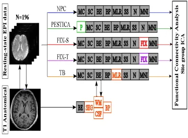
The physiological denoising study evaluated longitudinal changes in a 3T
harmonized multisite fMRI study of healthy elderly participants from the PharmaCog
Consortium (Jovicich et al., 2016). Retrospective physiological noise correction (rPNC)
methods were here implemented to investigate their influence on several DMN
reliability measures within and between 13 MRI sites. Each site involved five different
healthy elderly participants who were scanned twice at least a week apart (5 participants
per site). fMRI data analysis was performed once without rPNC and then with WM/CSF
regression, with physiological estimation by temporal ICA (PESTICA) (Beall & Lowe,
2007) and FMRIB's ICA-based Xnoiseifier (FSL-FIX) (Griffanti et al., 2014;
Salimi-Khorshidi et al., 2014). These methods differ for their data-based computational
approach to identify physiological noise fluctuations and need to be applied at different
stages of data preprocessing. As a working hypothesis, physiological denoising was in
general expected to improve DMN reliability.
The head motion study evaluated longitudinal changes in the DMN connectivity
from a 4T single-site study of 24 healthy young volunteers who were scanned twice
within a week. Within each scanning session, RS-fMRI scans were acquired once using
interleaved and then sequential slice-order acquisition methods. Furthermore, brain
volumes were corrected for motion using once rigid-body volumetric and then
slice-wise methods. The effects of these choices were then evaluated computing multiple
DMN reliability measures and investigating single regions within the DMN to assess
the existence of inter-regional effects associated with head-motion. In this case, we
expected to find slice-order acquisition effects in reliability estimates under standard
volumetric motion correction and no slice-order acquisition effect under 2D slice-based
motion correction.
Both studies used ICA to characterize the DMN using group-ICA and dual
regression procedures (Beckmann et al., 2009). This methodology proved successful at
defining consistent DMN connectivity metrics in longitudinal and clinical RS-fMRI
studies (Zuo & Xing, 2014). Automatic DMN selection procedures and other quality
Both studies considered several test-retest (TRT) reliability estimates (Vilagut,
2014) for some DMN connectivity measurements: absolute percent error between the
sessions, intraclass correlation coefficients (ICC) between sessions and multiple sites,
the Jaccard index to evaluate the degree of voxel-wise spatial pattern actiavtion overlap
between sessions.
Research Outline
After a general introduction (Chapter 2), the research in this thesis focuses on the
evaluation of physiological noise correction methods (Chapter 3) and on the evaluation
of acquisition and analyses strategies to reduce the effects of head motion (Chapter 4).
The physiological noise study (Chapter 3) revealed that retrospective
physiological denoising methods significantly affected the mean z-scores and, albeit
less markedly, the cluster-size in the DMN; in particular, FSL-FIX tended to increase
the DMN z-scores compared to others. Within-site test-retest reliability was consistent
across sites, with no differences across denoising methods. The absolute percent errors
were in the range of 5-11% for DMN z-scores and cluster-size reliability. DMN pattern
overlap was in the range 60-65%. In particular, no rPNC method showed a significant
reliability improvement compared with no physiological correction implemented.
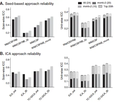
However, FSL-FIX and WM/CSF regressions showed both similar and significant
improvements of reproducibility consistency across the consortium (ICC = 0.67) for the
DMN z-scores relative to no physiological noise correction (NPC). Overall these
findings support the use of rPNC methods like WM/CSF regressions or FSL-FIX to
characterize multisite longitudinal changes of intrinsic FC-fMRI.
The head motion study (Chapter 4) revealed that mean z-scores within the DMN
are influenced by both slice-order acquisition and motion correction methods in all
DMN regions even in presence of low motion. In contrast, no combinations-of-interest
influenced or systematically improved the TRT reliability in all regions. In the DMN,
TRT reliability errors were overall below 8% and ICC were overall moderate 0.47 (C.I.
0.29-0.57), indicating longitudinally stable spatio-temporal network characteristics. The
average DMN pattern overlap was 40% (range: 14-65%). The longitudinal spatial
reproducibility was the lowest in the ACC region at 30%. Acquisition protocols and
variability in frontal DMN areas. These results support freedom of choice between the
examined acquisition protocols and head-motion correction methods in longitudinal
DMN studies.
The main thesis findings are outlined and put in perspective with the literature in
the discussion (Chapter 5). These indicate that the intrinsic DMN connectivity is
significantly sensitive to the factorts manipulated (slice-order acquisition and
preprocessing correction choices), while TRT reliability of the DMN connectivity is not,
or at least not systematically.
Contributions
The entire work presented in this thesis has been realized in the Laboratory for
the Functional Neuroimaging (Lnif) within the Interdepartmental Center for Brain/Mind
Sciences (CIMeC, University of Trento). Within the duration of the PhD activity,
contributions were given with extensive data analysis to two main projects, namely
PharmaCog, a European multisite consortium of Alzheimer research, and a resting-state
Lnif project. The work performed during the PhD resulted in published works as well as
several proceedings, in national and international conferences, both as first author or
co-author. A list of author’s publications is provided in the following.
Publications
Jovicich J, Minati L, Marizzoni M, Marchitelli R, Sala-Llonch R, Bartrés-Faz
D, Arnold J, Benninghoff J, Fiedler U, Roccatagliata L, Picco A, Nobili F, Blin O,
Bombois S, Lopes R, Bordet R, Sein J, Ranjeva JP, Didic M, Gros-Dagnac H, Payoux P,
Zoccatelli G, Alessandrini F, Beltramello A, Bargalló N, Ferretti A, Caulo M, Aiello M,
Cavaliere C, Soricelli A, Parnetti L, Tarducci R, Floridi P, Tsolaki M, Constantinidis M,
Drevelegas A, Rossini PM, Marra C, Schönknecht P, Hensch T, Hoffmann KT, Kuijer
JP, Visser PJ, Barkhof F, Frisoni GB; PharmaCog Consortium. Longitudinal
reproducibility of default-mode network connectivity in healthy elderly participants: A
multicentric resting-state fMRI study. Neuroimage. 2016 Jan 1;124(Pt A):442-54.
1.3.
Marchitelli R, Minati L, Marizzoni M, Bosch B, Bartrés-Faz D, Müller BW,
Wiltfang J, Fiedler U, Roccatagliata L, Picco A, Nobili F, Blin O, Bombois S, Lopes R,
Bordet R, Sein J, Ranjeva JP, Didic M, Gros-Dagnac H, Payoux P, Zoccatelli G,
Alessandrini F, Beltramello A, Bargalló N, Ferretti A, Caulo M, Aiello M, Cavaliere C,
Soricelli A, Parnetti L, Tarducci R, Floridi P, Tsolaki M, Constantinidis M, Drevelegas
A, Rossini PM, Marra C, Schönknecht P, Hensch T, Hoffmann KT, Kuijer JP, Visser PJ,
Barkhof F, Frisoni GB, Jovicich J; The Pharmacog Consortium. Test-Retest Reliability
of the Default Mode Network in a Multi-centric fMRI Study of Healthy Elderly: Effects
of Data-Driven Physiological Noise Correction Techniques, Human Brain Mapping,
March 2016. doi: 10.1002/hbm.23157.
Marchitelli R, Collignon O. M. C., Jovicich J; Test-Retest Reproducibility of the
intrinsic Default Mode Network: Influence of Head-Motion Correction Methods and
fMRI Slice-Order Acquisition Effects. (In preparation).
Conferences and Workshops
Marchitelli R, Jovicich J, Marizzoni M, Bosch B, Bartrés-Faz D, Bargalló N,
Roccatagliata L, Picco A, Nobili F, Gross-Dagnac H, Payoux P, Frisoni G,
Physiological Noise Correction Effects on Multisite Functional Connectivity
Reproducibility; Organization for Human Brain Mapping, Hamburg, Germany, June
8-12, 2014. (Poster presentation.)
Jovicich J, Minati L, Marchitelli R, Frisoni G, The Pharmacog Consortium,
Resting state functional connectivity in the default mode network: preliminary
evaluation of multicenter test-retest reproducibility, Biennial Conference on Resting
State/Brain Connectivity, Boston, U.S.A, September 11-13, 2014. (Poster presentation.)
Jovicich J, Marchitelli R, Marizzoni M, Sala-Llonch R, Bartrés-Faz D, Bargalló
N, Frisoni G, Influences of physiological noise correction on the functional connectivity
of the default mode network and its reproducibility using TC-GICA, 13th International
Geneva Springfield Symposium on Advances in Alzheimer Therapy, Geneva,
Switzerland March 26-29, 2014. (Poster presentation.)
Marchitelli R, Collignon O, Jovicich J. Influence of Head Motion and fMRI
Slice Acquisition on Intrinsic Default Mode Network Reliability. Organization for
Human Brain Mapping, Honolulu, Hawaii, USA, June 14-18, 2015. (Poster
presentation.)
Marchitelli R, Influence of data-driven physiological correction methods on the
longitudinal reliability and multicentric consistency of the default mode network in
healthy aging. The 6th Italian Chapter of the International Society for Magnetic
Resting-State Functional Connectivity
MRI
Discovery and properties of resting-state networks
In their pioneeristic work, Biswal et al. were the first who observed spontaneous
BOLD oscillations in the motor system during resting conditions marking de facto the
beginning of RS-fMRI (Biswal et al., 1995). Later, further investigations confirmed the
relevance of RS-fMRI as a valuable paradigm to investigate brain function (Biswal et
al., 1997; Lowe et al., 1998) and functional mapping was extended to other areas
throughout the whole human brain (Xiong et al., 1999).
These studies attempted to functionally map the entire brain using the intrinsic
FC-MRI methodology. Intrinsic FC-fMRI is defined as a statistical measure of temporal
dependencies between low-frequency BOLD signal oscillations from distinct brain
areas, that is commonly obtained using temporal correlations (Figure 2.1) (Biswal et
al., 2010; Fox & Raichle, 2007; Lowe et al., 2000). Intrinsic FC-fMRI was determinant
in the exploration of the whole-brain intrinsic architecture and led to the discovery of
many resting-state networks (Figure 2.2). These networks show resilient
spatio-temporal characteristics which allows their robust identification across individuals
(Damoiseaux et al., 2006; De Luca et al., 2006; Salvador et al., 2005). Some overlap
with sensory-motor activations detected in task-based fMRI studies (Lee et al., 2013)
which indicates that spontaneous brain activity during rest is relevant to understand
Resting-state networks are
characterized by complex and
dynamic interactivity between the
regions involved in the network as
well as with other networks (Kelly,
2008). These interactions are
however difficult to be understood
using only intrinsic FC-fMRI
methods both because correlations
are not very informative about the
directionality of interactions within
and between intrinsic brain
systems, and because non-neural
fMRI signals or some effects of
particular data analysis methods can induce temporal correlations between two given
time courses (Liao, 2009; Murphy et al., 2009). These limitations will be outlined in
more details in the next sections since they represent the main concern of our
experimental studies aimed at clarifing how to minimize the impact of non-neural
spurious correlations from intrinsic FC-fMRI metrics.
Figure 2.1. Example of intrinsic FC-fMRI. The figure shows temporal correlations of the low-frequency BOLD oscillations between three different regions. These were defined in the Left, L and Right, R Motor cortex, MOT and in the Left, L Visual cortex,VIS. High correlation between the inter-hemispheric motor corteces (top) and no correlation between ipsilateral correlations in left motor and visual cortex (bottom). from Van Dijk et al., 2010.
Origins of resting-state fMRI signals
To date, the neural underpinnings of low-frequency oscillations in the RS-fMRI
and consequently the neural sensitivity of intrinsic FC-fMRI mapping are not fully
understood (Horwitz et al., 2005; Van den Heuvel & Hulshoff Pol, 2010). As previously
mentioned, the fMRI signal is not a direct measure of neural activity but rather a
measure of blood oxygenation, therefore BOLD effects do not only raise from neural
activity, primary linked to synaptic metabolic activity, but also from random nuisance
factors such as thermal or quantum mechanical noise and from structured nuisance
components associated with signal reconstruction and distortion, non-neural
physiological processes (i.e. cardiac and respiratory activity) and head-motion.
According to these considerations, the characteristics of resting-state and
task-based fMRI signals would be alike with both advantages and disadvantages associated
with the implementation of the BOLD contrast. However, since an evoked stimulus is
missing in the resting-state, it is even more challenging to interpret and quantify the
contributions of each constituent to the resulting BOLD effect (Arthurs & Boniface,
2002). For this reason, whether spontaneous oscillations include a neural constituent has
been debated for long. Some have argued that they do not reflect neural activity but
only physiological processes related to cardiac and respiratory activity (Birn, 2012; Birn
et al., 2008; Shmueli et al., 2007). In contrast, some others have not denied the influence
of physiological aliasing on intrinsic FC-fMRI but argued that spontaneous brain
activity would also be neural in origin (Buckner & Vincent, 2007; Greicius et al., 2003;
Gusnard et al., 2001) since these spontaneous oscillations mainly occur at very low
frequencies (< 0.1 Hz) whereas physiological processes occur at higher frequencies (>
0.3 Hz) (Cordes et al., 2001; Cordes et al., 2000).
Several other reasons in favor of a neural hypothesis for the RS-fMRI signals
have been proposed. Some were already outlined in the previous sections of this work
and include the consistent identification of resting-state networks across healthy
individuals (Damoiseaux et al., 2006) as well as the spatial correspondence with
task-evoked networks in motor, visual and auditory areas which suggests that the intrinsic
functional architecture of the brain develops in regions more frequently recruited for
Finally, other reasons include the inter-hemispheric synchrony found in humans
and other mammals (Biswal & Kannurpatti, 2009) and additional evidence comes from
other imaging modalities such as electroencephalography (EEG): local field potentials
at low-frequency also show temporal synchronicity (Kenet et al., 2003; Leopold &
Logothetis, 2003; Lowe, 2012), recordings of neuronal firing are indirectly associated to
the amplitude profiles of RS-fMRI correlations (Nir et al., 2008) and simultaneously
measured fluctuations in neuronal spiking are associated to spontaneous BOLD
fluctuations (Pan et al., 2013; Shmuel & Leopold, 2008; Shmuel et al., 2002).
In summary, both neural and non-neural fMRI signals contribute to intrinsic
FC-fMRI. This highlights the need for optimizing the RS-fMRI design, developing
technical solutions or improving analytical methods to minimize the influence of
non-neural BOLD signals from intrinsic FC-fMRI measures.
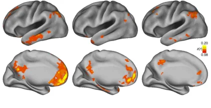
The Default Mode Network
Among all resting-state functional networks mentioned previously, the work
outlined in this thesis focuses on an intrinsic network in particular: the default mode
network (DMN). Functional coactivations associated with this network typically
encompass regions in the posterior cingulate cortex (PCC) and precuneus, medial
prefrontal cortex (MPFC), anterior cingulate cortex (ACC), inferior parietal lobule and
bilateral parietal cortex (LPC) (Buckner, 2012) (Figure 2.3). Neuroimaging research of
the DMN grows at fast pace today and is grounded on solid scientific knowledge about
its intrinsic functional dynamics and relevance for cognition (Buckner, 2012).
The DMN was observed for the first time in positron emission tomography
(PET) studies (Mazoyer et al., 2001; Shulman et al., 1997) and the idea of a
“default-mode” system was not immediately recognized until evidence in support of a plausible
self-referential processing role was put forward (Gusnard & Raichle, 2001) and
knowledge about its complex physiological dynamics was also gained from other
imaging modalities (Laufs et al., 2003). Afterwards, first descriptions of the DMN using
RS-fMRI were made using intrinsic FC-fMRI methods similar to those Biswal had used
The DMN owes its name to its main
characteristic of showing prominent
activation during the resting-state and
low-demanding passive stimulation tasks
in contrast to goal-directed cognitive
tasks (Greicius & Menon, 2004). The
DMN is a unique intrinsic brain network
with this endogeneous dynamical
dichotomy and for this reason it has been
studied extensively. These DMN
dynamics are crucial to understand its role
in cognition: since the beginning, intrinsic
FC-fMRI of the DMN was linked to
memory function (Andreasen et al., 1995; Binder et al., 1999) and in particular to the
retrieval of information from long-term memory, conscious representation of mental
imagery and thoughts (Andrews-Hanna, 2012) and cognitive operations involved in
problem solving and future planning (Buckner et al., 2008; Buckner & Carroll, 2007).
In fact, it has been shown that the exacerbated dichotomy in DMN dynamics between
attention-demanding tasks and resting-state (Fox et al., 2005) is mitigated by the
inclusion of task features related to self-referential thoughts (McGuire et al., 1996),
episodic memory (Shannon & Buckner, 2004) and meditation practice (Brewer et al.,
2011; Jao et al., 2015).
In general, evidence suggests that the DMN is not innate. There are no adult-like
patterns resembling the DMN in neonates and children aged below 4 years. Anyway,
this might result from absent distant connectivity in children, which does not exclude
the hypothesis of independent default mode function confined to single brain regions
(Fair et al., 2009). Additional evidence suggests that DMN activity tends to decrease as
a function of normal aging with substantial loss of intrinsic FC-fMRI (Andrews-Hanna
et al., 2007; Damoiseaux et al., 2008; Ferreira et al., 2015; Sambataro et al., 2010). The
most credited hypothesis is that loss of intrinsic FC-fMRI might be associated to
age-related widespread structural changes (Horn et al., 2014) that affect the network
efficiency in the transmission of neural information (Marstaller et al., 2015). Finally,
there is evidence suggesting that DMN connectivity is sensitive to circadian rhythm
gradually reducing co-activations from morning to afternoon (Blautzik et al., 2013;
Hodkinson et al., 2014).
Current issues in intrinsic DMN connectivity studies
RS-fMRI data analysis is not subjected to standardized acquisition and analysis
methods. This lessens the reproducibility of findings across studies and increases the
risk of adopting suboptimal acquisition and analysis methods (Griffanti et al., 2016)
which could bias DMN connectivity metrics. On the other side the optimization of
RS-fMRI data acquisition and analysis is challenging. It is not trivial to identify analytic
expedients which favor the characterization of the DMN and the interpretability of its
dynamics because of many issues arising from limitations in the fMRI technology and
analysis software. The most critical ones are outlined in the following.
Data acquisition factors that affect DMN connectivity
Primarily, data acquisition choices can influence the overall data quality and
determine decision making for analytic operations to be performed downstream
including data preprocessing and FC-fMRI method choices. Standard acquisition
protocols include single-shot multislice echo-planar imaging (EPI) which allows overall
moderate temporal resolution (~ 0.5 Hz) in acquired volumes. This privileges the spatial
dimension to perform data analysis and to compute FC-fMRI, unavoidably increasing
the risk of spatial overlapping between the DMN and other brain networks or, even
worse, structured noise (Birn et al., 2006). Novel acquisition sequences such as
multiband echo-planar imaging allow the acquisition of multiple slices at the same time,
improving the overal temporal resolution of fMRI (~ 1.25 Hz). This acquisition method
improves DMN sensitivity (Feinberg et al., 2010) and has been used to investigate
temporal modes of the DMN (Smith et al., 2012) but it is limited in the ability to encode
spatial information by the radio frequency coil array alone which can lead to reduction
in signal-to-noise-ratio (Feinberg et al., 2010).
2.4.
Although a wide range of sampling rates and a relatively small number of
datapoints can be used to measure sufficient BOLD activity for identifying stable
spatio-temporal DMN patterns, the standardization of these parameters is still an open
issue (Van Dijk et al., 2010; Whitlow et al., 2011). This becomes particularly
controversial when harmonizing acquisition sequences across different imaging sites,
using different MRI vendors, RF coils and fat suppression methods (Jovicich et al.,
2016).
Acquisition methods are defined upon default settings provided by MRI scanner
manufacturers which could be different across Siemens, Philips and General Electric
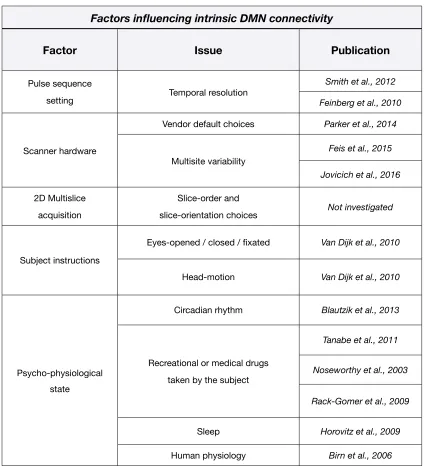
Table 2.1. Summary table of fMRI acquisition and human physiologic factors influencing the characterization of the DMN.
Factors influencing intrinsic DMN connectivity Factors influencing intrinsic DMN connectivity Factors influencing intrinsic DMN connectivity
Factor Issue Publication
Pulse sequence
setting Temporal resolution
Smith et al., 2012 Pulse sequence
setting Temporal resolution Feinberg et al., 2010
Scanner hardware
Vendor default choices Parker et al., 2014
Scanner hardware
Multisite variability
Feis et al., 2015 Scanner hardware
Multisite variability
Jovicich et al., 2016
2D Multislice acquisition
Slice-order and
slice-orientation choices Not investigated
Subject instructions
Eyes-opened / closed / fixated Van Dijk et al., 2010 Subject instructions
Head-motion Van Dijk et al., 2010
Psycho-physiological state
Circadian rhythm Blautzik et al., 2013
Psycho-physiological state
Recreational or medical drugs taken by the subject
Tanabe et al., 2011
Psycho-physiological state
Recreational or medical drugs
taken by the subject Noseworthy et al., 2003 Psycho-physiological
state
Recreational or medical drugs taken by the subject
Rack-Gomer et al., 2009 Psycho-physiological
state
Sleep Horovitz et al., 2009 Psycho-physiological
state
(GE) scanners (Jovicich et al., 2016). These differences also concern 2D slice
acquisition methods (Parker et al., 2014) which are usually acquired in an interleaved
fashion (Westbrook, 2005) with axial in-plane orientation (O'Connor, 2010). Interleaved
slice acquisition is typically chosen to allow for zero or small slice gap while avoiding
slice cross-talking effects and related magnetic saturation effects but increases BOLD
sensitivity to head motion effects, which are known to bias DMN connectivity (Van
Dijk et al., 2012). It was recently proposed that, with some slice gap to avoid slice
cross-talking effects, sequential slice acquisition methods might reduce head-motion
effects in DMN connectivity (Cheng & Puce, 2014). However, slice-order acquisition
choices remain an open issue in the field, especially because these two methods were
not directly compared on the same subjects.
As mentioned previously, recent investigations suggest that coactivations in the
default-mode system might be influenced also by psycho-physiological states and that
individual psychological characteristics should not be underestimated in RS-fMRI
designs to characterize consistent DMN connectivity metrics in a sample. In typical
designs, subjects are instructed to lay still and relaxing while in the scanner, avoiding
the engagement of particular attention towards something in particular and falling
asleep (Horovitz et al., 2009). For this reason, it is yet not fully understood whether
instructing participants to keep their eyes closed, opened or fixated on a cue could be of
help for them to relax without influencing spontaneous brain activity somehow (Van
Dijk et al., 2010; Yan et al., 2009). Moreover, considering circadian rhythm effects on
the DMN, it would be optimal to scan all individuals at the same time of the day,
particularly in longitudinal study designs as in (Meindl et al., 2010). Finally, even
though substance abuse is a common exclusion criterion in studies of healthy
individuals, the degree of coffee intake (Rack-Gomer et al., 2009), nicotine (Tanabe et
al., 2011) or food (Noseworthy et al., 2003) is usually not properly considered and
should be at least better supervised.
Image preprocessing issues
After data acquisition, 4D EPI images commonly undergo preprocessing to
enhance signal detection and prepare the data for group analysis. RS-fMRI
preprocessing is characterized by a non-standardized workflow of signal processing
operations which typically but not necessarily include the following:
- The elimination of some initial TRs to account for magnetic saturation effects. - Physiological denoising to correct for cardiac and respiratory activities. - 3D image volume realignment via spatial coregistration.
- Slice-timing correction such that all voxels represent the signal at the same time. - Spatial smoothing using a Gaussian kernel to increase temporal signal-to-noise
ratio (tSNR).
- Temporal smoothing to restrict the analysis to frequencies of interest. - Regression of motion and additional confounds.
- Mean or mode intensity normalization.
- Spatial normalization to a common space template for group analysis.
The implementation of these operations deserves caution and might be subjected
to different ordering to preserve data structure. To improve DMN signal detection,
preprocessing workflows include methods to detect and attenuate spurious BOLD
fluctuations induced by non-neural biological processes and MRI hardware noise.
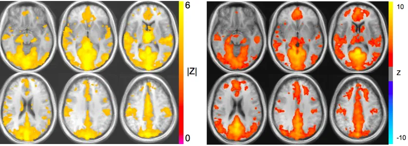
Physiological denoising issues
This is not straightforward: the success of these methods is often compromised
by the fact that neural and non-neural signals are coupled in time and overlap in space
(Figure 2.4); therefore their removal will unavoidably be associated with loss in signal
of neural origins.
Spontaneous neural BOLD oscillations in the DMN regions occur at very low
frequencies (0.01 - 0.1 Hz). In principle, these do not overlap with respiratory (0.1 - 0.5
Hz) and cardiac activity (0.6 - 1.2 Hz) even though DMN signals could also occur at
higher frequencies (Cordes et al., 2001). A bandpass filter is typically applied during
image preprocessing to remove higher frequencies; however the relatively low sampling
rate employed in standard EPI acquisitions (~ 0.5 Hz) does not allow the perfect
characterization of these physiological confounds which are aliased in the low
frequency range (< 0.1 Hz) (Cordes et al., 2001).
One partial technical solution to this issue consists of recording signals of
non-interest such as those rising from human physiology during MRI acqusition to remove
them offline by filtering them out of the BOLD timeseries (Glover et al., 2000).
MRI-compatible pulse oxymeters and respiration belts can be used to monitor cardiac and
respiratory cycles, respectively (Chia et al., 2000; Santelli et al., 2011). However, these
parallel measurements cannot often be obtained due to lack of equipment. This issue is
especially relevant in multicentric designs where limited availability in parallel
measurements across sites is often the case.
As previously mentioned in the introduction, data-based fMRI methods have
been developed to deal with this issue. These are retrospective methods that
automatically detect non-neural signal sources from the empirical data. A common
approach, the global regression (Fox et al., 2009) removes the whole-brain averaged
timecourse from the data using regression methods. This approach assumes that
physiological confounds are widespread in the entire brain and that can be consequently
catched by averaging the signal across all voxels. A methodological challenge with this
approach is that the global average also includes signal from gray matter voxels and
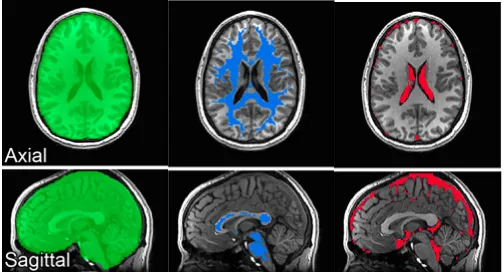
therefore unavoidably leads to remotion of signal of interest (Jo et al., 2010). One
technical solution in this case includes the adoption of probabilistic segmented
anatomical masks of WM and CSF (Figure 2.5) to avoid averaging signals from gray
matter (GM) (Weissenbacher,
2 0 0 9 ) . Th e g l o b a l s i g n a l
regression method remains
controversial for inducing
anti-c o r r e l a t i o n s i n F C - f M R I
measures in the entire brain (Fox
et al., 2009; Murphy et al., 2009)
even though they do not
negatively influence DMN
connectivity.
Other methods exploit the
ICA methodology to detect and separate signal sources associated to physiological noise
(Figure 2.6). This multivariate approach decomposes the signal into either temporal or
spatial statistically addittive independent signal sources by maximizing non-gaussianity
or minimizing mutual information between
them. Using these methods, human
physiology can be observed in the Willis
polygon, WM and the ventricles. Despite
the relatively low number of timepoints,
temporal ICA can be used to isolate
temporal modes associated with cardiac (48
- 85 bpm) and respiratory (10 - 24 bpm)
frequencies using spatio-temporal priors
(Beall & Lowe, 2007).
On the other side, spatial ICA
methods able to classify and remove
various signal artifacts according to
spatio-temporal features were also developed
(Griffanti et al., 2014; Salimi-Khorshidi et
al., 2014). Both methods share ICA
limitations and need the user supervision to
be conducted. For instance, temporal ICA limitations include the need of a user-defined
Figure 2.5. Anatomical masks adopted for cardiac and respiratory signal regressions. (green) anatomical masks of the full brain are adopted in global signal regression. Segmented WM (blue) and CSF (red) anatomical masks used to reduce loss in GM neural signals from Jo et al., 2010.
frequency range which might be difficult to be standardized across subjects. Spatial ICA
classifiers are parametric and might require training the features thus being
time-consuming. Furthermore, preprocessing workflows including ICA-based physiological
denoising must be defined with attention because slice-timing correction, spatial and
temporal smoothing might reduce ICA sensitivity (Beall & Lowe, 2007).
Motion correction issues
If not addressed properly, head motion can severely confound single-subject and
group functional DMN connectivity
(Figure 2.7). Some preprocessing
strategies known as motion correction
or volume realignment were developed
to deal with motion and are commonly
implemented in the majority of
resting-state studies. These methods consist of
coregistering all train of volumes to a
reference volume, by default the
volume in the middle of the acquisition
train. This procedure shifts all voxels in
the exact spatial location defined in the
reference volume. Complementary
motion correction approaches include
the regression of motion metrics
obtained from the coregistration process
(Satterthwaite et al., 2013) and to
censor highly motion corrupted
volumes (Power et al., 2012). Similarly to physiological noise correction, ICA-based
approaches were developed to automatically detect and correct for head-motion (Pruim
et al., 2015a; Pruim et al., 2015b; Schopf et al., 2010; Schopf et al., 2011; Wang et al.,
2012).
For several reasons, the implementation of motion correction is always
recommended even if motion is overall low. First, it has been show that even
2.4.4.
submillimetrical motion can confound DMN connectivity estimates (Power et al.,
2012). However, if motion estimated is low, ICA methods can quite efficaciously deal
with it (Figure 2.8) and therefore motion correction could be avoided to preserve data
structure (Meindl et al., 2010). Second, the
order of slice-timing and head-motion
corrections cannot be predefined but
depends on slice-order acquisition methods
and the amount of motion detected in the
data. Some methods which can perform
both corrections simultaneously have been
proposed (Roche, 2011) while other argued
that applying motion correction prior
slice-timing correction would be an optimal
solution (Jones et al., 2008) especially if
some preprocessing software and
a l g o r i t h m s r e q u i r e s l i c e - t i m i n g
information to be implemented (Beall &
Lowe, 2007). In general, relative freedom
of choice is admitted for interleaved slice
acquisition protocols. Instead, different
optimal solutions are proposed in case slices are acquired sequentially as a function of
head-motion severity (Huettel et al., 2008; Poldrack et al., 2011).
The relevance of slice-order acquisition methods for head-motion correction was
already described previously among the fMRI data acqusition issues. Regarding this
issue, volume realignment is only a part of motion correction. Volume-based motion
correction methods treat brain volumes as a rigid-body and perform inter-volume
coregistration. While the rigid-body assumption is technically wrong (Poncelet et al.,
1992), these methods do not correct for motion occurring within-volume associated
with timing differences in the acquisition of consecutive 2D slices (0 < t < TR). More
recently, advanced retrospective motion correction solutions have been proposed with
the introduction of novel slice-wise correction methods (Beall & Lowe, 2014). In
particular it has also been showed that standard volume-based methods might be quite
insensitive to motion in BOLD-weighted MRI data (Beall & Lowe, 2014). In any case,
it remains unclear how within-volume motion occurring during different slice-timing
acquisitions might influence the intrinsic DMN connectivity (Kim et al., 2008; Sladky
et al., 2011). It should be noted, however, that no methods can account for motion in its
entirety so far, with slice-based motion correction (SLOMOCO) slightly outperforming
the others.
DMN extraction methods
Following preprocessing steps, several methods have been developed to
characterize intrinsic DMN connectivity. These can be grouped into two categories here
defined as model-based or model-free methods. Model-based methods are
hypothesis-driven approaches which exploit predefined anatomical region(s)-of-interest (ROIs) to
investigate intrinsic FC-fMRI in the entire brain. They permit to gain an immediate
inspection and high interpretability about the intrinsic human brain functional
architecture. In contrast, model-free methods are exploratory and do not require the
predefinition of anatomical priors to characterize intrinsic brain networks (Beckmann et
al., 2005; Calhoun et al., 2001a). Both methods present advantages and disadvantages
and their choice strictly depends on the scientific question of the experimenters.
The simplest and most common model-based method is the seed-based
approach. This method characterizes FC-fMRI by means of univariate temporal
correlations between the averaged signal within a predefined ROI, technically referred
to as the seed, and the signal in all other voxels/ROIs throughout the brain (Fransson,
2005). Another model-based method exploits graph-theory analysis to investigate
topological properties of intrinsic brain networks. This method is a mathematical
representation of brain networks as graphs, characterizing FC-fMRI as edges or archs
between a set of predefined ROIs, technically referred to as the nodes of the graph.
Topological properties of intrinsic brain networks include clustering coefficient i.e. the
degree of local interconnectivity between nodes; the path length, i.e. the efficiency of
the neural transmission among distant nodes; hub analysis to identify those ROIs that
plays a fundamental role within the network, et cetera (Bullmore & Sporns, 2009;
Hosseini & Kesler, 2013). Both methods are used to identify the DMN by placing the
seed in the posterior cingulate cortex and the precuneus, considered the main hub of the
DMN (Fransson & Marrelec, 2008; Leech et al., 2012; Leech et al., 2011). The
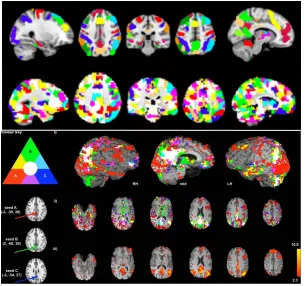
definition of the shape and size of ROIs poses serious challenges to characterize
intrinsic DMN connectivity (Figure 2.9), considering eventual parcellation scheme
(Shen et al., 2013) and, edge thresholding in graph-theory approaches (Van Wijk et al.,
2010).
Model-free methods include multivariate approaches such as ICA (Bell & Sejnowski, 1995), an exploratory method that decomposes the BOLD signal into its addittive, statistically independent, spatio-temporal sources. Therefore, ICA can
automatically identify independent spatio-temporal sources of resting-state brain
networks, including the DMN, in addition with the aforementioned artifacts associated
with head-motion and physiological noise (Kiviniemi et al., 2003; McKeown, 1997).
ICA cannot be perfomed on space and time simultaneously. For this reason, although it
is possible to identify the DMN by conducting ICA in either dimensions (Boubela et al.,
2013), the spatial dimension is preferred because of the larger number of voxels
compared with time points (Beall & Lowe, 2007).
Moreover, different ICA approaches do exist. The first approach consists of
conducting an ICA on individual EPI data. This method is usually named single-subject
ICA. An alternative approach consists of conducting an ICA on the entire group of
subjects, concatenating all the EPI volumes in time (Calhoun & Adali, 2012). Then,
methods to characterize the activation patterns in each individual include
back-reconstruction (Calhoun et al., 2001b) or dual-regression (Beckmann et al., 2009). Very
recently, it has been shown that group ICA approaches improve separability of neural
from artifactual signals improving the ultimate ICA sensitivity to FC-fMRI signals (Du
et al., 2016).
Despite its advantages, ICA also comes with some limitations. First, ICA is
non-deterministic which implies that the ultimate decomposition of the BOLD signal sources
always allows some degree of run-to-run variability. Second, ICA is also unsupervised
which implies that no predefined number of components exists and that this could be
therefore extablished using semi-quantitative methods or empirically by (re-)conducting
ICA at different dimensionality orders on the same data. Suboptimal dimensionality
order might either cause signal overlapping (erring on the side of caution) or splitting
In the former case, DMN signals will coexist with irrelevant signals in the same
component. In the latter, DMN signals will be split across several components
introducing spurious interregional variability in connectivity patterns within one single
component. Similarly, if spatio-temporal signal patterns largely overlap, ICA might fail
to separate independent signals sources. This would introduce some degree of
interdependence among components leading to the imperfect characterization of the
DMN or other resting-state networks, which will be likely to be contaminated by
non-neural sources (Beall & Lowe, 2007). Overall, these issues might lead to reduce DMN
consistency across individuals and reduce the reproducibility of intrinsic fMRI studies
(Schopf et al., 2010).
Intrinsic DMN connectivity: a potential biomarker?
Many definitions of biomarkers have been proposed over the last decades.
Fortunately they converge quite well with the general definition given by the National
Institutes of Health (NIH). According to this definition, a biological marker or
biomarker is a metric that can denote both normal biological and pathogenic processes
or pharmacological responses to therapeutic drugs (Biomarkers Definitions Working,
2001). Neuroimaging biomarkers are detailed biological features derived from
radiological images or recorded brain signals that can be used in isolation and jointly
with known biomarkers to assess or predict the presence of disease and evaluate
treatment response (Richter, 2006).
Diagnostic biomarkers can be clinical, i.e. able to assess the presence of disease,
and preclinical, i.e. able to assess signs or risk factors for health and function
deterioration (Shoji, 2012). Preclinical neuroimaging biomarkers are particularly
important in case of neurodegenerative diseases or cancer that present a symptom-free
phase. In these cases, these biomarkers might timely identify high-risk individuals so
that they receive either preventive treatments or further follow-up evaluations (Frank &
Hargreaves, 2003; Sapsford et al., 2010). This class of biomarkers include hippocampal
atrophy measured in morphometric MRI studies of Alzheimer Disease (AD) (Fraser et
al., 2015). This MRI-based biomarker can be used in conjunction with CSF analytes
that measure abnormal protein aggregates (i.e. amyloid beta, phosphorilated tau protein)
or with indicators of neural loss to distinguish between disease-modifying and
symptomatic treatment effects (Hampel et al., 2011; Saumier et al., 2009).
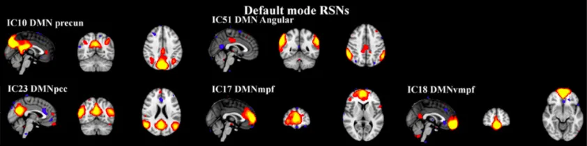
Figure 2.10. The DMN splitting phenomenon following ICA. The figure shows an example of strong DMN splitting onto 5 independent components following group ICA. From (Rytty et al., 2013).
Other relevant longitudinal biomarkers of AD are obtained from molecular
neuroimaging using PET. The Pittsburgh compound-B (PiB-PET) tracer and
Fludeoxyglucose PET (FDG-PET) represent validated biomarkers of amyloid-beta
plaques accumulation and abnormal glucose metabolism, respectively (Frisoni et al.,
2013; Habeck et al., 2012). Nevertheless, healthcare providers resist compensating for
the expensive costs of these diagnostic biomarkers for insufficient clinical evidence
about diagnosis or treatment of illness and improvement of cognitive functioning in
people affected by dementia (Frisoni & Visser, 2015).
While technique noninvasiveness, ease of acquisition and unexpensive costs are
standard criteria to qualify preclinical biomarkers (Katz, 2004a, 2004b), further
evidence from comparative and longitudinal studies involving subjects from appropriate
populations is demanded (Frisoni & Visser, 2015). In contrast, these neuroimaging
biomarkers reflect neurodegeneration and metabolic dysfunction only in symptomatic
stages of AD but not in its preclinical asymptomatic stages where structural MRI and
FDG-PET efficacy is lessened while PiB-PET becomes more challenging considering
that amyloid-beta deposition is not AD specific and may be also found in normal aging
(Jack et al., 2012; Toledo et al., 2015).
Intrinsic DMN fMRI-based biomarker
Intrinsic DMN connectivity as defined by fMRI offers attractive possibilities for
evaluation as a biomarker for some diseases. Evidence from cross-sectional studies
suggets that the intrinsic DMN connectivity is sensitive to neuropsychiatric illness and
neurodegenerative diseases showing aberrant loss in connectivity strength with regional
dysconnectivity or hyperconnectivity. Since BOLD-weighted FC-fMRI is not a direct
measure of neural activity, these alterations likely indicate metabolic dysfunction that
would cause an incomplete recruitment of DMN regions or induce additional brain
effort to safeguard network integrity and support cognition with higher computation
costs (Palop et al., 2006).
DMN-based biomarkers can be easily developed considering that the network is
consistently observable in healthy young adults (Damoiseaux et al., 2006) and that
default-mode connectivity predicts cognitive performance (Sala-Llonch et al., 2012).
The ability to characterize the DMN in young healthy adults has allowed the study of its
neurophysiological changes (Chou et al., 2012; Patriat et al., 2013; Shehzad et al., 2009)
and to characterize abnormal DMN connectivity changes in pathological conditions
(Somandepalli et al., 2015; Song et al., 2012). This is of primary importance for
studying typical and atypical development in children (Uddin et al., 2010) where
aberrant connectivity patterns undergoing atypical brain maturation trajectories were
identified (Uddin et al., 2010). These are found in the DMN that show a retarded
formation of distant connectivity in autism (ASD) until late adolescence (Washington et
al., 2014).
There is evidence supporting DMN-based biomarkers of major depression which
causes changes in anterior-posterior DMN connectivity with increased connectivity in
anterior DMN regions (Craddock et al., 2009; Greicius et al., 2007; Mulders et al.,
2015; Sheline et al., 2009). Increases in intrinsic DMN connectivity are also typically
found in case of structural white matter degeneration in multiple sclerosis (MS)
probably because of compensatory mechanisms that preserve deficitary cognition
(Hawellek et al., 2011).
Diminished FC-fMRI can instead be used as a DMN-based biomarker of normal
aging and more relevantly for mild cognitive impairment (MCI) and mild AD
(Balthazar et al., 2014; Greicius et al., 2004). DMN connectivity shrinkage in normal
elderly is a natural process associated with aging decline and gray matter atrophy which
does not necessarily indicate the presence of disease. However, evidence suggests that
DMN connectivity loss is related to increased amyloid-beta (Hedden et al., 2009;
Sheline et al., 2010) and symptoms’ severity of dementia (Hafkemeijer et al., 2012).
These findings consequently promoted the ability to track and monitor clinical
deterioration in MCI and AD patients (Binnewijzend et al., 2012; Damoiseaux, 2012)
and discriminate between AD and other forms of dementia such as fronto-temporal
dementia (FTD) (Zhou et al., 2010) or dementia with Lewy bodies (Galvin et al., 2011).
Importantly, intrinsic DMN connectivity might distinguish between MCI who would
undergo cognitive decline, hence conversion to AD, from those who would remain more
stable over time (Prestia et al., 2013; Vos et al., 2013).
Longitudinal task-based BOLD fMRI studies provided evidence in support of
DMN-based predictive biomarkers of dementia (Petrella et al., 2011) bu