143 Volume 6 Issue 3 Spring 2014
Prevalence of Osteoporosis among Thalassemia
Patients from Zafar Adult Thalassemia Clinic, Iran
Hashemieh M
1, Azarkeivan A
2* , Radfar M
3, Saneifard H
4, Hosseini-Zijoud SM
5,
Noghabaei G
6, Yaseri M
71. Imam Hossein Medical Center, Shahid Beheshti University of Medical Sciences, Tehran, Iran, 2. Research Center of Iranian blood Transfusion Organization, Thalassemia Clinic, Tehran, Iran. 3. Imam Hossein Medical Center, Shahid Beheshti University of Medical Sciences, Tehran, Iran. 4. Mofid Childrens’ Hospital, Shahid Beheshti University of Medical Sciences, Tehran, Iran. 5. Hamadan University of Medical Sciences, Hamadan, Iran.
6. Imam Hossein Medical Center, Shahid Beheshti University of Medical Sciences, Tehran, Iran. 7. Department of Epidemiology and Biostatistics, Tehran University of Medical Sciences, Tehran, Iran.
*Corresponding Author: Azarkeivan A, Email: azazarkeivan@yahoo.com
Submitted: 25-11-2013 , Accepted: 12-04-2014
Abstract
Background: The advances in treatment regimes for thalassemic patients have increased the survival among them; therefore osteoporosis has emerged as an important cause of morbidity. The aim of this study was to determine the prevalence of osteoporosis and osteopenia in patients with thalassemia from Zafar Adult Thalassemia Clinic, Tehran, Iran.
Patients and Methods: In this cross sectional investigation, we studied 239 patients with β thalassemia major and 87 patients with thalassemia intermedia with a mean age of 29±8 years. All demographic data including age, weight, height, sex, age at diagnosis, age at blood transfusion initiation, chelating agent therapy and serum ferritin level were obtained from patients’ history. Bone mineral density of the lumbar spine (L1-L4) and femoral neck was determined using dual-energy X-ray absorptiometry (DEXA).
Results: The prevalence of osteoporosis was 65.6% (214 out of 326 patients). Osteoporosis was present in 10.7%, 11% and 43.9% of patients in the lumbar spine alone (L1-L4), femoral neck alone and both places, respectively. In the rest of patients 18.7% showed osteopenia and only 15.7% were normal. Osteoporosis was more prevalent in patients with thalassemia intermedia compared to thalassemia major (p<0.001). Also higher age of patients, longer duration of transfusion and longer intervals between transfusions had a positive correlation with osteoporosis.
Conclusion: The prevalence of osteoporosis among Iranian thalassemia patients is similar to prevalence reported elsewhere. Bone Mineral density is a good index of bone status in patients with thalassemia and recommended to be done for thalassemic patients annually.
Key words: Thalassemia, bone mineral density, osteoporosis, DEXA.
Introduction
Thalassemia is a group of inherited disorders of hemoglobin synthesis, and is the most common monogenetic disease worldwide 1. In ß thalassemia, mutations of the ß globin gene lead to an imbalance in globin chain synthesis and ineffective erythropoiesis. The low number of mature red blood
cells leads to anemia and other associated health
problems 1. Patients with beta-thalassemia major are susceptible to osteopenia and osteoporosis due to several factors which interfere with bone remodeling . Delay in sexual maturation, presence of diabetes and hypothyroidism, accelerated
hemopoiesis with progressive marrow expansion and direct iron toxicity have been identified as major causes of osteoporosis in thalassemic patients 2. Reduced bone mass, skeletal pain and fractures are common causes of morbidity and disability among patients with homozygous ß thalassemia. Bone abnormalities, such as spinal deformities, scoliosis, nerve compression, spontaneous fractures, osteopenia and osteoporosis have also been described in patients with thalassemia 3.
Over the past three decades, management of patients with thalassemia has improved IJBC 2014;6(3): 143-148
ORIGINAL ARTICLE
144 IRANIAN JOURNAL OF BLOOD AND CANCER dramatically with hypertransfusion therapy, which
has nearly normalized pre-transfusion hemoglobin levels, reduced the ineffective erythropoietic process and prevented bone deformities. Iron chelation using prolonged subcutaneous infusions of desferrioxamine, has also improved the survival and diminished the endocrine complications of transfusional hemochromatosis 4,5. As treatment with transfusion programs and chelation therapy has significantly prolonged the survival in thalassemia patients, osteopenia and osteoporosis represent prominent causes of morbidity in young adults of both genders with thalassemia major or
thalassemia intermedia 6,7.
During the last decade, many studies have shown low bone mass in adult patients with
thalassemia7,8. Bone mass is the result of a balance between bone formations gained during growth and the subsequent bone loss. According to the World Health Organization, osteoporosis is a disease characterized by low bone mass and microarchitectural deterioration of bone tissue, leading to enhanced bone fragility and a consequential increase in fracture risk 9.
Thalassemic patients show a variety of bone disorders including bone pain, bone deformity, growth failure, rickets, scoliosis, spinal deformities, pathologic fracture, osteopenia and osteoporosis. In a large study, adult thalassemia major patients presented a 51% prevalence of osteoporosis and 45% prevalence of osteopenia 7.
Several sensitive techniques are available for the quantitative assessment of the degree of total bone mass. Bone mineral density (BMD) measurement by dual X-ray absorptiometry (DEXA) of the lumbar spine, femoral neck and forearm is recommended as one of the most reliable and non-invasive techniques for the assessment of bone mass 8-10. DEXA is an excellent non-invasive
choice for repeated measurements of any temporal changes of BMD because of its 1% precision rate and low radiation exposure 11. As shown in previous works by other authors in thalassemic population, the lumbar spine and femoral neck BMD is below the normal reference 3,11.
Earlier studies of bone mineral metabolism in thalassemic patients have mainly concentrated on the pediatric and adolescence age groups 12,13. As mentioned, osteoporosis and osteopenia is common even in well-treated thalassemic patients 14. There
were more than 13,000 thalassemic patients in Iran at 2007 15, but the current frequency of osteopenia and osteoporosis in various thalassemia syndromes has not been well assessed in Iran.
In the present study Data from the Zafar Adult Thalassemia Clinic, Tehran, Iran, were analyzed to determine the current prevalence of osteopenia and osteoporosis among thalassemic patients.
Materials and Methods
Patients
A total of 326 patients with beta-thalassemia attending the Zafar Adult Thalassemia Clinic in Tehran (239 thalassemia major, 87 thalassemia intermedia) from June 2012 to April 2013 were enrolled in this study. They had a mean age of 29 ± 8 years (range 12–52 years) with an average body mass index (BMI) of 20.18±2.96 (Table 1). All patients were examined by one hematologist and one endocrinologist.
For this study, beta-thalassemia major (TM) was defined as homozygous or compound heterozygous beta-thalassemia requiring eight or more transfusions in the 12 months prior to enrolling in the registry. Thalassemia intermedia (TI) was defined as beta-thalassemia receiving less than 8 units per year. All TM patients were receiving regular transfusions from the age of 2 years. History of chelation and transfusion therapy, initiation and duration of blood transfusion, pre-transfusion hemoglobin, ferritin values and desferrioxamine (DFO) dosage in the last year were obtained from the patients’ medical records. None of the patients or controls was treated with vitamin D or calcium supplements. Characteristics of study group are shown in table 1.
The study was performed according to the principles of the Declaration of Helsinki and was approved by the ethic committee of the Shahid Beheshti University of Medical Sciences, Tehran, Iran. Each subject above 18 years old or the patient’s parent or guardian for patients under 18 years old gave written consent before patient being included in this study.
Bone mineral density (BMD)
In all patients, BMD was determined using dual X-ray absorptiometry (DEXA, Lunar DPX-Plus), both at the lumbar spine (L1–L4) in A-P projection and at the femoral neck.
145 Volume 6 Issue 3 Spring 2014
The instrument was calibrated on a daily basis according to the manufacturer’s instructions. Reproducibility was calculated as a CV obtained by weekly measurements of a standard phantom
on the instrument and by repeated measurements obtained in three patients of different ages. The CV of our instrument was 0.5% with the standard phantom; and in vivo, we calculated a CV of 1.1% for
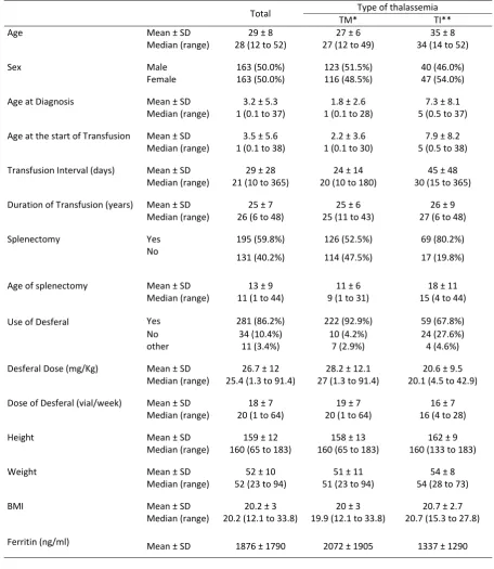
١١ Table 1: Demographic findings of patients with thalassemia major and thalassemia intermediate.
Total Type of thalassemia
TM* TI**
Age Mean ± SD 29 ± 8 27 ± 6 35 ± 8
Median (range) 28 (12 to 52) 27 (12 to 49) 34 (14 to 52)
Sex Male 163 (50.0%) 123 (51.5%) 40 (46.0%)
Female 163 (50.0%) 116 (48.5%) 47 (54.0%)
Age at Diagnosis Mean ± SD 3.2 ± 5.3 1.8 ± 2.6 7.3 ± 8.1
Median (range) 1 (0.1 to 37) 1 (0.1 to 28) 5 (0.5 to 37)
Age at the start of Transfusion Mean ± SD 3.5 ± 5.6 2.2 ± 3.6 7.9 ± 8.2 Median (range) 1 (0.1 to 38) 1 (0.1 to 30) 5 (0.5 to 38)
Transfusion Interval (days) Mean ± SD 29 ± 28 24 ± 14 45 ± 48 Median (range) 21 (10 to 365) 20 (10 to 180) 30 (15 to 365)
Duration of Transfusion (years) Mean ± SD 25 ± 7 25 ± 6 26 ± 9 Median (range) 26 (6 to 48) 25 (11 to 43) 27 (6 to 48)
Splenectomy Yes 195 (59.8%) 126 (52.5%) 69 (80.2%)
No 131 (40.2%) 114 (47.5%) 17 (19.8%)
Age of splenectomy Mean ± SD 13 ± 9 11 ± 6 18 ± 11
Median (range) 11 (1 to 44) 9 (1 to 31) 15 (4 to 44)
Use of Desferal Yes 281 (86.2%) 222 (92.9%) 59 (67.8%)
No 34 (10.4%) 10 (4.2%) 24 (27.6%)
other 11 (3.4%) 7 (2.9%) 4 (4.6%)
Desferal Dose (mg/Kg) Mean ± SD 26.7 ± 12 28.2 ± 12.1 20.6 ± 9.5 Median (range) 25.4 (1.3 to 91.4) 27 (1.3 to 91.4) 20.1 (4.5 to 42.9)
Dose of Desferal (vial/week) Mean ± SD 18 ± 7 19 ± 7 16 ± 7 Median (range) 20 (1 to 64) 20 (1 to 64) 16 (4 to 28)
Height Mean ± SD 159 ± 12 158 ± 13 162 ± 9
Median (range) 160 (65 to 183) 160 (65 to 183) 160 (133 to 183)
Weight Mean ± SD 52 ± 10 51 ± 11 54 ± 8
Median (range) 52 (23 to 94) 51 (23 to 94) 54 (28 to 73)
BMI Mean ± SD 20.2 ± 3 20 ± 3 20.7 ± 2.7
Median (range) 20.2 (12.1 to 33.8) 19.9 (12.1 to 33.8) 20.7 (15.3 to 27.8)
Ferritin (ng/ml) Mean ± SD 1876 ± 1790 2072 ± 1905 1337 ± 1290
*TM = Thalassemia Major **TI = Thalassemia Intermedia
146 IRANIAN JOURNAL OF BLOOD AND CANCER the lumbar spine and 1.5% for the neck. BMD data
were expressed as grams per square centimeter and compared with BMD values of normal subjects of the same age and sex. Osteopenia or osteoporosis was calculated according to WHO criteria, based on BMD expressed as Z-score indicating osteopenic (Z-score between –1 to –2.5 SD) and osteoporotic patients (Z-score below –2.5 SD). We considered the patients osteoporotic if they showed osteoporosis in at least one site and osteopenic if they showed osteopenia in at least one site.
Ferritin Assay
In all patients, after an overnight fasting, blood samples were obtained and serum level of ferritin was determined by ELISA method using materials provided by RAMCO (TX, USA). In all assays, the intra-assay coefficient of variation was 6% or less, and the inter-assay coefficient of variation was 15%
or less.
Statistical analysis
Data were analyzed using SPSS software, version
19 (SPSS Inc., Chicago, IL, USA). Students’ t-test was applied to compare the means. Association between variables was compared using Pearson’s Chi-Square test. Results are expressed as mean±SD. P values <0.05 were considered significant.
Result
This study included 239 patients with thalassemia major (116 women and 123 men; mean age: 13.2±5.8 years) and 87 cases with thalassemia intermedia (47 women and 40 men; mean age: 14.2±6.9 years). Basic demographics and therapeutic characteristics are presented in table 1. The prevalence of osteopenia and osteoporosis and its relation to different variables is presented in table 2. According to our data, the prevalence rate of osteoporosis, based on the WHO criteria was 65.6% (214 out of 326). In 35 patients (10.7%) only lumbar spine was osteoporotic, in 36 patients (11%) only femur was osteoporotic and in 143 (43.3%) patients both femur and lumbar spine were osteoporotic. The prevalence of osteopenia was estimated as 18.7% (61 out of 326).
١٢
Table 2: Prevalence of osteopenia and osteoporosis related to different variables.
Subject
Osteoporosis Number (%)
Osteopenia
Number (%) Number (%) Normal P
Overall 214 (65.6) 61 (18.7) 51 (15.7)
Age 31 ± 8 27 ± 6 25 ± 8 <0.001
29 (13 to 52) 27 (17 to 45) 24 (12 to 49)
Sex male 103 (63.2) 33 (20.2) 27 (16.6) .376 female 111 (68.1) 28 (17.2) 24 (14.7)
Age at Diagnosis 3.7 ± 6 2.1 ± 2.9 2.7 ± 4.1 .124 1 (0.1 to 37) 1 (0.1 to 20) 1 (0.1 to 20)
Type of thalassemia TM 143 (59.8) 54 (22.6) 42 (17.6) .001
TI 71 (81.6) 7 (8) 9 (10.3)
Transfusion interval 32 ± 34 22 ± 8 24 ± 10 .011 23 (10 to 365) 20 (10 to 60) 20 (12 to 60)
Duration of Transfusion 26 ± 7 25 ± 5 23 ± 7 .007 26 (9 to 48) 25 (14 to 38) 23 (6 to 45)
147 Volume 6 Issue 3 Spring 2014
In men, the prevalence rate of osteoporosis was 63.2% and of osteopenia was 20.2%. In women, the rate of osteoporosis was 68.1%, whereas osteopenic rate were 17.2%. There was no correlation between the sex and the prevalence of osteoporosis (p = 0.378).
In this survey there was a significantly higher rate of osteoporosis in TI patients than TM cases (p<0.001). Also higher age of patients, longer duration of transfusion and longer intervals between transfusions had a positive correlation with osteoporosis.
Discussion
Since osteoporosis is a progressive disease, prevention and early diagnosis are equally important as well as treatment of the established disease. Despite normalization of hemoglobin levels, adequate hormone replacement, and effective iron chelation therapy patients show an unbalanced bone turnover with an increased resorptive phase resulting in diminished bone mineral density 16.
Various studies have demonstrated that multiple acquired factors are involved in the pathogenesis of osteopenia/osteoporosis in thalassemia. They include the primary disease, itself causing bone marrow expansion 17, and several secondary factors, such as hormonal deficiency 7, iron overload, desferrioxamine toxicity 4, calcium, zinc and vitamin D deficiencies, hypothyroidism, hypoparathyroidism, diabetes mellitus, hypogonadism and inadequate physical activity 2. These factors mainly act by inhibiting the osteoblast activation and/or increasing the osteoclastic function, leading to bone loss and osteoporosis 17.
This study presents the current osteoporosis and osteopenia prevalence in a large series of thalassemia patients from Iran. The results of the current study indicate a high prevalence of osteoporosis among thalassemic Iranian patients (65.6%). This data is in agreement with other cross-sectional studies indicating the prevalence of osteoporosis to be 52- 96% among thalassemic patients 18-20.
According to this study patients with TM and TI had low bone mineral density. In this study, reduced BMD of the spine and femoral neck was present in about two-thirds of adult patients with
beta-thalassemia major and intermedia which is in agreement with previous works by other authors 3,10. Our longitudinal data suggest that,
despite long-term transfusions, iron chelation and hormone replacement therapy, thalassemic patients continue to lose BMD over time. It seems that underlying genetic factors play a significant role in the imbalance of bone remodeling 3.
In the current study there was no significant gender difference regarding bone mineral values. This finding is in agreement with a previous report21, but was in contrast to the findings of Jensen et al. who reported that the bone lesions in thalassemic are more frequent and more prominent in males 7.
Conclusion
The prevalence of osteoporosis among Iranian thalassemia patients is similar to prevalence reported elsewhere. Bone Mineral density is a good index of bone status in patients with thalassemia and recommended to be done for thalassemic patients annually.
References
1. Weatherall DJ, JB C. The thalassemia syndromes 4ed. Oxford: Malden, MA: Blackwell Science 2001.
2. Wonke B. Bone disease in beta-thalassaemia major. Br J Haematol. 1998;103(4):897-901.
3. Voskaridou E, Terpos E. New insights into the pathophysiology and management of osteoporosis in patients with beta thalassaemia. Br J Haematol. 2004;127(2):127-39.
4. Olivieri NF. The beta-thalassemias. N Engl J Med. 1999;341(2):99-09.
5. Calleja EM, Shen JY, Lesser M, Grady RW, New MI, Giardina PJ. Survival and morbidity in transfusion-dependent thalassemic patients on subcutaneous desferrioxamine chelation. Nearly two decades of experience. Ann N Y Acad Sci. 1998;850:469-70.
6. Vichinsky EP. The morbidity of bone disease in thalassemia. Ann N Y Acad Sci. 1998;850:344-8.
7. Jensen CE, Tuck SM, Agnew JE, Koneru S, Morris RW, Yardumian A, et al. High prevalence of low bone mass in thalassaemia major. Br J Haematol. 1998;103(4):911-5.
8. Orvieto R, Leichter I, Rachmilewitz EA, Margulies JY. Bone density, mineral content, and cortical index in patients with thalassemia major and the correlation to their bone fractures, blood transfusions, and treatment with desferrioxamine. Calcified tissue
148 IRANIAN JOURNAL OF BLOOD AND CANCER
international. 1992;50(5):397-9.
9. Assessment of fracture risk and its application to screening for postmenopausal osteoporosis. Report of a WHO Study Group. World Health Organization technical report series. 1994;843:1-129.
10. Cefalu CA. Is bone mineral density predictive of fracture risk reduction? Current medical research and opinion. 2004;20(3):341-9.
11. Angelopoulos NG, Katounda E, Rombopoulos G, Goula A, Kaltzidou V, Kaltsas D, et al. Evaluation of bone mineral density of the lumbar spine in patients with beta-thalassemia major with dual-energy x-ray absorptiometry and quantitative computed tomography: a comparison study. J Pediatr Hematol Oncol. 2006;28(2):73-8.
12. Soliman AT1, El Banna N, Abdel Fattah M, ElZalabani MM, Ansari BM. Bone mineral density in prepubertal children with beta-thalassemia: correlation with growth and hormonal data. Metabolism. 1998 May;47(5):541-8.
13. Roth C, Pekrun A, Bartz M, Jarry H, Eber S, Lakomek M, et al. Short stature and failure of pubertal development in thalassaemia major: evidence for hypothalamic neurosecretory dysfunction of growth hormone secretion and defective pituitary gonadotropin secretion. Eur J Pediatr. 1997;156(10):777-83.
14. Hajjar RR1, Kamel HK. Osteoporosis for the home care physician. Part 1: etiology and current diagnostic strategies.J Am Med Dir Assoc. 2004;5(3):192-6.
15. Abolghasemi H, Amid A, Zeinali S, Radfar MH, Eshghi P, Rahiminejad MS, et al. Thalassemia in Iran: epidemiology, prevention, and management. J Pediatr Hematol Oncol. 2007;29(4):233-8.
16. Carmina E, Di Fede G, Napoli N, Renda G, Vitale G, Lo Pinto C, et al. Hypogonadism and hormone replacement therapy on bone mass of adult women with thalassemia major. Calcif Tissue Int. 2004;74(1):68-71.
17. Mahachoklertwattana P, Chuansumrit A, Sirisriro R, Choubtum L, Sriphrapradang A, Rajatanavin R. Bone mineral density, biochemical and hormonal profiles in suboptimally treated children and adolescents with beta-thalassaemia disease. Clin Endocrinol (Oxf). 2003;58(3):273-9.
18. Bielinski BK, Darbyshire P, Mathers L, Boivin CM, Shaw NJ. Bone density in the Asian thalassaemic population: a cross-sectional review. Acta Paediatr. 2001;90(11):1262-6.
19. Saffari F, Mahyar A, Jalilolgadr S. Endocrine and
metabolic disorders in beta-thalassemiamajor patients. Caspian J Intern Med. 2012;3(3):466-72. 20. Dresner Pollack R, Rachmilewitz E, Blumenfeld A,
Idelson M, Goldfarb AW. Bone mineral metabolism in adults with beta-thalassaemia major and intermedia. Br J Haematol. 2000;111(3):902-7.
21. Anapliotou ML, Kastanias IT, Psara P, Evangelou EA, Liparaki M, Dimitriou P. The contribution of hypogonadism to the development of osteoporosis in thalassaemia major: new therapeutic approaches. Clin Endocrinol (Oxf). 1995;42(3):279-87.