C
hairmanMohaMMad Saeid RahiMinejad, Md
E
ditor-
in-C
hiEfhaSSan abolghaSeMi, Md
S
CiEntiCiE
ditorS
aMina
lavi, Md
I
ranIan
J
ournal
of
B
lood
and
C
anCer
The Official Journal of
Iranian Pediatric Hematology and Oncology Society (IPHOS)
Volume 8, Number 1, March 2016
ISSN: 2008-4595
ناریا ناکدوک ناطرس و نوخ نمجنا
Iranian Pediatric Hematology & Oncology Society
Aggarwal Bharat, India
Alebouyeh Mardawij, Iran
Arzanian Mohammad Taghi, Iran
Biondi Andrea, Italy
Cappellini Maria-Domenica, Italy
Faranoush Mohammad, Iran
Ghavamzadeh Ardeshir, Iran
Khaleghnejad Tabari Ahmad, Iran
Kowsari Farid, Iran
Najmabadi Hosein, Iran
Nakagawara Akira, Japan
Oberlin Odile, France
Pedram Mohammad, Iran
Peyvandi Flora, Italy
Ravindranath Yaddanapudi, USA
Rezvan Houri, Iran
Samiei Farhad, Iran
Schrappe Martin, Germany
Taher Ali, Lebanon
Telfer Paul, UK
Vosough Parvaneh, Iran
Wagner Hans-Peter, Switzerland
Zandian Khodamorad, Iran
EDITORIAL BOARD
“Iranian Journal of Blood and Cancer” is published by “Iranian Pediatric
Hematology and Oncology Society (IPHOS)” in collaboration with
“Iranian Blood Transfusion Organization (IBTO)”
Iranian Journal of Blood and Cancer
is Covered in IranMedex
®Editorial Office
Pediatric Hematology and Oncology Society, 1st floor, NO.63, Shahid Toosi
Street, Tohid Square, Tehran, Iran
Postal Code: 1419783311
Tel/Fax:
+98(21)66912679
Website:
www.ijbc.ir
Email:
Info@ijbc.ir
“IJBC” is approved as an “Academic Research Journal” by Medical Journal
Commissions of the “Ministry of Health” and Medical Education of Islamic
Republic of Iran”.
Abolghasemi Hassan
Aghaeipour Mahnaz
Alavi Samin
Alilou Sam
Alizadeh Shaban
Amin Kafiabad Sedigheh
Ansari Shahla
Arjmandi Rafsanjani Khadijeh
Arzanian Mohammad Taghi
Azarkeivan Azita
Bahoosh Gholamreza
Dehghani Fard Ali
Eghbali Aziz
Ehsani Mohammad Ali
Enderami Ehsan
Eshghi Peyman
Faranoush Mohammad
Farshdoosti Majid
Habibi Roudkenar Mehryar
Hadipour Dehshal Mahmoud
Haghi Saba Sadat
Hashemieh Mozhgan
Hedayati Asl Amir Abbas
Honarfar Amir
Ghasemi Fariba
Goudarzipour Kourosh
Jamshidi Khodamorad
Karimi Gharib
Karimijejad Mohammad Hassan
Kariminejad Roxana
Kaviani Saeid
Khaleghnejad Tabari Ahmad
Keikhaei Bijan
Kompany Farzad
Koochakzadeh Leili
Maghsoudlu Mahtab
Mehrvar Azim
Najmabadi Hossein
Naseripour Masood
Nazari Shiva
Rahiminejad Mohammad Saeid
Rahimzadeh Nahid
Ramyar Asghar
Roozrokh Mohsen
Saki Najmaldin
Saki Nasrin
Shamsian Bibi Shahin
Seighali Fariba
Sharifi Zohreh
Tashvighi Maryam
Reviewers
Aim and Scope
The Iranian Journal of Blood and Cancer (IJBC)
is published quarterly in print and
online and includes high quality manuscripts including basic and clinical investigations
of blood disorders and malignant diseases and covers areas such as diagnosis, treatment,
epidemiology, etiology, biology, and molecular aspects as well as clinical genetics of these
diseases editor., as they affect children, adolescents, and adults. The IJBC also includes
studies on transfusion medicine, hematopoietic stem cell transplantation, immunology,
genetics, and gene-therapy. The journal accepts original papers, systematic reviews, case
reports, brief reports and letters to the editor, and photo clinics.
The IJBC is being published since 2008 by the Iranian Pediatric Hematology and
Oncology Society (IPHOS). The contents of the journal are freely available for readers
and researchers and there is no publication or processing fee.
The IJBC has a scientific research rank and is indexed in Directory of Open Access
Journals (DOAJ), Islamic World Science Center (ISC), Index COpernicus (IC), and
Embase. It is also visible in the following databases: Magiran, IranMedex, ISC, Scientific
Information Database (SID), Cambridge Scientific Abstracts (CSA) Academic Search
Complete (ASC), Electronic Journals Library (EJB), CINAHL, GEOBASE, CABI,
Global Health, Open-J-Gate, Excerpta Medica, and Google Scholar.
All Submission should be sent online via our online submission system. For further
inquiries please email the journal directly. The IJBC benefits from editorial freedom.
Our editorial policy is consistent with the principles of
editorial independence presented
by WAME.
http://www.wame.org/resources/policies#independence
Instructions to Authors
Submission Process:
Manuscripts should be sent through the on-line submission system. A submission code is allocated to
each article as well as a short submission ID and all the future contacts should be based on this code
or ID. The articles are primarily evaluated by our internal screeners who check the articles for any
methodological flaws, format, and their compliance with the journal’s instructions. Through a
double-blind review, the articles will be reviewed by at least two external (peer) reviewers. Their comments
will be passed to the authors and their responses to the comments along with the reviewers’ comments
will then be evaluated by the Editor-in-Chief, the Scientific Editor, and a final reviewer who can be a
member of the Editorial Board. The final review process will be discussed in regular editorial board
sessions and on the basis of the comments, and the journal’s scope, the Editors-in-Chief will decide
which articles should be published.
Ethical Considerations:
The journal is a member of the Committee on Publication Ethics (COPE). COPE’s
flowcharts and guidelines are approached in confronting any ethical misbehavior.
The Journal also follows the guidelines mentioned in the
Recommendations for the
Conduct, Reporting, Editing and Publication of Scholarly Work in Medical Journals
issued by the International Committee of Medical Journal Editors (ICMJE)
(
http://www.icmje.org/#privacy
).
The research that involves human beings (or animals) must adhere to the principles of the Declaration
of Helsinki.
(http://www.wma.net/en/30publications/10policies/b3/index.html).
• Informed consent:
All patients and participants of the research should be thoroughly informed about the aims of the
study and any possible side effects of the drugs and intervention. Written informed consent from the
participants or their legal guardians is necessary for any such studies. The Journal reserves the right
to request the related documents.
• Authorship:
Based on the newly released
Recommendations for the Conduct, Reporting, Editing and Publication
of Scholarly Work in Medical Journals
, by the ICMJE, “an Author” is generally considered to be
someone who meets the following conditions 1, 2, 3, and 4.
1-Substantial contributions to the conception or design of the work; or the acquisition, analysis, or
interpretation of data for the work; AND
2-Drafting the work or revising it critically for important intellectual content; AND
3-Final approval of the version to be published; AND
4-Agreement to be accountable for all aspects of the work in ensuring that questions related to the
accuracy or integrity of any part of the work are appropriately investigated and resolved.
• Conflict of Interest:
We request all the authors to inform us about any kinds of “Conflict of Interest” (such as financial,
personal, political, or academic) that would potentially affect their judgment. Authors are preferably
asked to fill the uniform disclosure form available through:
(http://www.icmje.org/coi_disclosure.pdf)
• Plagiarism:
The authors are not allowed to utilize verbatim text of previously published papers or manuscripts
submitted elsewhere.
• Copyright:
If a manuscript contains any previous published image or text, it is the responsibility of the author to
obtain authorization from copyright holders. The author is required to obtain and submit the written
original permission letters for all copyrighted material used in his/her manuscripts.
Retraction Policy:
The IJBC uses the COPE flowchart for retraction of a published article
(http://publicationethics.org/resources/guidelines)
to determine whether a published article should be retracted.
Author Consent Form:
All authors must sign an Author Consent Form and return this form via Email so that the journal
can begin the article’s evaluation process. You hereby warrant that “This article is an original work,
has not been published before and is not being considered for publication elsewhere in its final form
either in printed or electronic form”.
Type of Articles:
Original Articles:
Should contain title page, abstract, keywords, introduction, materials and methods,
results, discussion, conclusion, acknowledgment, references, tables, and figures, enumerated from the
title page. The length of the text should be limited to 3000 words excluding the references and abstract.
Case Reports and Brief Reports:
Should not exceed 1500 words. Both should include abstract,
keywords, introduction, case presentation, discussion, conclusion acknowledgment, and references.
Case reports might have 1 to 4 accompanying figures and/or tables but brief reports should not have
more than one figure or table. Necessary documentations of the case(s) like pathology and laboratory
test reports should be included in the submission package.
Clinical Trials:
should contain patients’ informed consent and the approval of the ethics committee
of the corresponding institution.
Review Articles:
might be requested by the editor, but IJBC will also accept submitted reviews.
Both solicited and unsolicited review articles are subjected to editorial review like the original papers.
Letters to the Editor:
IJBC accepts letters to the editor. Letters should be less than 500 words.
Letters might discuss articles published in the journal during the previous six months or other important
aspects related to the field of hematology. Letters will undergo peer-review processing and will be
edited for clarity.
Photo clinics:
Figures that convey a significant medical point can also be accepted. Photo clinics
should contain one or two high quality figures and a description of the figure no more than 500 words.
24- references should be included.
Paper Preparations:
Cover letter
should contain a statement that you will not resubmit your article to another journal until
the reviewing process will be completed. Also please indicate whether the authors have published or
submitted any related papers from the same study.
Title Page
of the article should include 1) the title of the article; 2) authors’ names; 3) name of the
institution where the work was done; 4) running title (short form of the main title presented on the
top of pages); and 5) complete mailing address, telephone/fax numbers, and email address of the
corresponding author. This page is unnumbered.
Abstract
should be structured for original articles providing background/objective for the study,
methods, results, and conclusion. It should not exceed 250 words altogether. Number this page as page 1.
Abstracts of other types of contributions should be non-structured providing the essential information.
When abstracting a review article a concise summary of the salient points should be addressed.
Preferably, abbreviations should not be mentioned in the abstract.
Keywords
are used for indexing purposes; each article should provide three to five keywords selected
from the Medical Subject Headings (MeSH).
http://www.nlm.nih.gov/mesh/
Introduction
should provide a context or background and specifies the purpose or research objective
of the study or observation.
Method
must indicate clearly the steps taken to acquire the information. Be sure that it includes only
information that was available at the time the plan or protocol for the study was written. It should be
detailed (including: controls, inclusion and exclusion criteria, etc) and may be separated into subsections.
Repeating the details of standard techniques is best avoided.
For reports of randomized controlled trials, authors should refer to the CONSORT statement (http://
www.consort-statement .org/). All randomized clinical trials should be registered in any international
RCT registration centers approved by the WHO. For research conducted in Iran, it is advised to register
at IRCT(www.irct.ir).
Reporting guidelines such as STROBE, STARD, and PRISMA would help you to produce high quality
research and to provide all required information and evidence for related methodology. EQUATOR
Network website would help you in using these guidelines.
The software used for statistical analysis and description of the actual method should be mentioned.
Results
should be presented in chronological sequence in the text, table, and illustration. Organize
the results according to their importance. They should result from your own study.
Tables and illustrations
must
be cited in order which they appear in the text; using Arabic numerals.
Tables should be simple and should not duplicate information in the text of the paper. Figures should be
provided only if they improve the article. For radiographic films, scans, and other diagnostic images,
as well as pictures of pathology specimens or photomicrographs, send the high resolution figures in
jpeg or bitmap format. Color photographs, if found to improve the article, would be published at no
extra-charge at the print version of the journal. Type or print out legends for illustrations on a separate
page, and explain the internal scale and identify the method of staining in photomicrographs.
Discussion
should emphasize the new and important aspects of the study and the conclusions that
follow them. Possible mechanisms or explanations for these findings should be explored. The limitations
of the study and the implications of the findings for future research or clinical practice should be
explored.
Conclusion
should state the final result that the author(s) has (have) reached. The results of other
studies should not be stated in this section.
Supplementary Materials
such as movie clips, questionnaires, etc may be published on the online
version of the journal.
Any technical help, general, financial, and material support or contributions that need acknowledging
but do not justify authorship, can be cited at the end of the text as
Acknowledgments.
References
should be complied numerically according to the order of citation in the text in the
Vancouver style. The numbers of references should not preferably exceed 40 for original articles, 15
for brief, and 8 for case reports.
For the references credited to more than 6 authors please provide the name of the first six authors
and represent the rest authors by the phrase “et al.”
For various references please refer to “the NLM style guide for authors, editors, and publishers”.
(http://www.ncbi.nlm.nih.gov/books/NBK7256/)
Listed below are sample references.
Journal Article:
• Gaydess A, Duysen E, Li Y, Gilman V, Kabanov A, Lockridge O, et al. Visualization of exogenous
delivery of nanoformulated butyrylcholinesterase to the central nervous system. Chem Biol Interact.
2010;187:295-8. doi: 10.1016/j.cbi.2010.01.005. PubMed PMID: 20060815; PubMed Central PMCID:
PMC2998607.
• Javan S, Tabesh M. Action of carbon dioxide on pulmonary vasoconstriction. J Appl Physiol.
In press 2005
Complete Book:
• Guyton AC: Textbook of Medical Physiology. 8th ed. Philadelphia, PA, Saunders, 1996.
Chapter in Book:
• Young VR. The role of skeletal muscle in the regulation of protein metabolism. In Munro HN,
editor: Mammalian protein metabolism. Vol 4. San Diego; Academic; 1970. p. 585-674.
Language and Style:
Contributions should be in either American or British English language. The text must be clear and
concise, conforming to accepted standards of English style and usage. Non-native English speakers
may be advised to seek professional help with the language.
All materials should be typed in double line spacing numbered pages. Abbreviations should be
standard and used just in necessary cases, after complete explanations in the first usage. The editorial
office reserves the right to edit the submitted manuscripts in order to comply with the journal’s style.
In any case, the authors are responsible for the published material.
Correction of Errata:
The journal will publish an erratum when a factual error in a published item has been documented.
For further information please contact the Editorial Office:
Tel: +98 21 66912676
Email: ijbc_iphos@yahoo.com
Website: www.ijbc.ir
I
ranIan
J
ournal
of
B
lood
and
C
anCer
Volume 8, Number 1, March 2016
Review Article
Role of Ghrelin in Cancer...
1
Robab Sheikhpour
Original Articles
In Vitro Evaluation of the Anti-bacterial Effect of Human Platelet Concentrate...
5
Elnaz Jafarzadeh, Mojgan Pourmokhtar,Setareh Tavili
Evaluation of Thyroid Dysfunction during Imatinib Therapy in Chronic Myeloid
Leukemia...
9
Abolghasem Allahyari, Foroogh Salehi, Mostafa Kaboli, Masoud Sadeghi
Evaluation of the Seroprevalence of Transfusion Transmissible Infections among Blood
Donors in a Tertiary Care Hospital of North India...
13
Chintamani Pathak, Shivali Sehgal
Clinicopathological Analysis of Patients with Breast Cancer and Their Families...
17
Mehrdad Zeinalian, Nafiseh Heidarzadeh, Homayoun Naji, Mohammad Reza SharbafchiCase Report
Splenic Infarction in a Case of Acute Promyelocytic Anemia...
23
Nasim Valizadeh
Letters to Editor
Osteoblasts in the Vicinity of Osteoclasts in a Case of Infantile Osteopetrosis...
25
Samin Alavi, Nahid Arabi, Sadaf Esteghamati
Is Medical Application Software a New Strategy for Oncologists?
...
27
Babak Abdolkarimi, Mahdi Shahriari, Puria Salajeghe
Role of ghrelin in cancer
IJBC 2016; 8(1): 1-4
Role of Ghrelin in Cancer
Robab Sheikhpour*
Department of Nursing, Yazd Branch, Islamic Azad University, Yazd, Iran; and Hematology and Oncology Research Center, Shahid Sadoughi University of Medical Sciences, Yazd, Iran
A R T I C L E I N F O
rEviEw artiClE
Article History:
Received: 16.10.2015 Accepted: 25.01.2016
Keywords:
Cancer Ghrelin Mechanism
ABSTRACT
Cancer is one of the most fatal diseases in human beings which annually leads to death of 30000 individuals in Iran. Prevention, diagnosis and treatment of cancer is one of the major scientific challenges all around the world. It seems that increased incidence of several cancers such as colon and prostate and their mortality are connected with obesity. It is suggested that obesity and metabolic syndrome are associated with endocrine related cancers and ghrelin pathway may play a role in cancer progression. Ghrelin is a potent regulator of the growth hormone (GH)/ insulin-like growth factor-1 (IGF-1) axis, which is frequently implicated in the development of several neoplasms, including colon cancer. It has been reported that changed ghrelin level as a main regulator of energy homeostasis plays an important role in carcinogenesis. Also, antiproliferative effects of ghrelin in lung and breast carcinoma cell lines have been detected in some studies . In this paper, ghrelin and its role and function in cancer is discussed.
*Corresponding author:
Robab Sheikhpour
Address: Department of Nursing, Yazd Branch, Islamic Azad University, Yazd, Iran; and Hematology and Oncology Research Center, Shahid Sadoughi University of Medical Sciences, Yazd, Iran
Tel: +98 913 1522462
Email: robab.sheikhpour@iauyazd.ac.ir
Introduction
One of the most fatal diseases in human beings is cancer which annually leads to the death of about 30000 persons in Iran.1 This incurable disease continues to be a
major problem in recent years.1-3 It seems that increased
risk of the expansion of several cancers such as colon and prostate cancers, and their risk with mortality are connected to obesity.4 Hormonal abnormalities in obese
people such as low ghrelin level may play a key role in cancer development. Moreover, the undesirable side effects of currently standard therapies for colon and androgen independent prostate cancers lead to persistent need for new and more powerful therapeutic options.4
Ghrelin
Ghrelin is a 28-amino acid peptide5-7 mainly produced in
the stomach8 of humans and rodents.3 It is also produced
by a wide variety of tissues and acts as a paracrine/ autocrine factor.8 About 60–70% of circulating ghrelin is
originated by stomach, while about up to 30% is produced in the small intestine.9 Moreover, other tissues including
pancreas and cardiovascular system could produce ghrelin.10 Albeit ghrelin expressed in heart is lower than
that in the stomach, but it exerts a cardioprotective effect via unknown mechanisms.11 Ghrelin known as a
brain-gut peptide can induce changes such as increased food intake and body fat through altered appetite and amount of food intake.12,13
Ghrelin plays a significant role in release of GH and
triggers secretion of hepatic IGF-1. Both GH and IGF-1 as anabolic hormones can increase lean body mass via stimulating skeletal muscle growth and inhibiting skeletal muscle protein breakdown.14 It has been reported that
ghrelin causes positive energy balance via decreasing fat utilization by GH-independent mechanisms.15 Moreover,
the secretion of ghrelin is stimulated via energy restriction and acetylcholine and reduced via gastrectomy, food intake, glucose, insulin and somatostatin releasing
Iranian Journal of Blood & Cancer
Journal Home Page: www.ijbc.ir
Please cite this article as: Sheikhpour R. Role of Ghrelin in Cancer. IJBC 2016; 8(1): 1-4.
Sheikhpour R
inhibitory factor (SRIF).10 Also it plays an important
role in metabolic response to starvation via modulating insulin secretion, glucose metabolism and amino acid uptake.3 Ghrelin stimulates the differentiation of
preadipocytes and inhibit lipolysis. Therefore it has a main role in the process of adipogenesis.16 It increases
anxiety-like behavior and memory retention in rodents and may promote sleep in human beings.16 Inhibition
of insulin secretion and regulation of gluconeogenesis/ glycogenolysis was accomplished in the presence of ghrelin; therefore, it regulates glucose homeostasis in many aspects.17 Ghrelin is pertained to G
protein-coupled receptor family.18,19 Ghrelin could cause
weight gain through growth hormone secretion and as a result increasing food intake and reducing fat utilization in rodents.20 It also moderates some actions
of gastrointestinal tract and alters the growth processes of neoplastic tissues.2
Ghrelin exists in two molecular forms: acylated or octanoylated and unacylated or desoctanoylated.2
Unacylated ghrelin via ghrelin O-acyltransferase (GOAT) enzyme can be acylated8 and yields the natural ligand of
the only known ghrelin receptor.8 Figures 1 and 2 show
unacylated and acylated ghrelin, respectively.
Endocrine activity of ghrelin is dependent on its acylation mediated by GH secretagogue (GHS) receptor and des-acyl ghrelin has no endocrine activity and does not bind to GHSR-1a; however, its mechanism of action
is not defined well.11
Ghrelin and Cancer
Gastrointestinal cancers, especially colorectal cancers are associated with obesity and strong relationship is observed between these cancers and environmental factors in addition to genetic factors. Obesity is associated with hyperisulinemia or insulin resistance with elevated leptin and decreased ghrelin serum levels. Obesity and metabolic syndrome are associated with endocrine related cancers and ghrelin has proposed to have some
influencial role in cancer development or progression.21
Ghrelin is a potent regulator of the GH/IGF-I axis which is frequently implicated in the development of several neoplasms, including colon cancer.2 It has been observed
that circulating changes in leptin and ghrelin levels as two main regulators of energy homeostasis could play important role in carcinogenesis.2 Clear-cut data about
Figure 1: Unacylated ghrelin9
Figure 2: Acylated ghrelin9
Role of ghrelin in cancer
ghrelin and its effects on proliferative pathologies is contradictory for now.3
Ghrelin and its receptors exist in many endocrine and non-endocrine tumor cell types such as gastroenteropancreatic, pituitary, prostate, breast and other related cancer cell lines.8 Ghrelin controls
neoplastic cell proliferation, but precise role of ghrelin still is not clear.8 Some studies have reported that ghrelin
has proliferative properties in cancers. Study in canine mammary carcinoma showed that there are high levels of ghrelin and GHS-R in metastatic tumors.17
Ghrelin has shown antiproliferative effects in lung and breast carcinoma cell line and proliferative effects in prostate, pancreatic and adrenal cancer cell lines.3
Another study reported that ghrelin may inhibit growth of breast, thyroid and lung cancer cell lines independent of the GH releasing effect. In contrast, ghrelin may induce a proliferative response in some other cell lines via IGF-1 and GH with tumorigenic potential.16
The effect of ghrelin on breast cancer cell proliferation is discovered by Jeffery et al. in their study.22 They evaluated
proliferation of breast cancer cell lines MDAMB-231 and MB-435 and observed that growth rate of
MDA-MB-231 cells was significantly increased in the presence
of ghrelin.9
Volante et al. in another study reported that high
concentrations of ghrelin (100 nmol/ l–1 μmol/l) has
anti-proliferative actions in thyroid cancer cells. They also suggested that autocrine circuits of ghrelin may be operating in the growth control of thyroid follicular tumors.23
De Vriese et al. evaluated the autocrine proliferative effect of ghrelin on human leukemic HL-60 and THP-1 cell lines.24 The human leukemic cell lines did not express
the functional GHS-R1a, but expressed GHSR1b. They observed that addition of octanoylated or des-acyl ghrelin did not exert any effect on leukemic cell proliferation. Another study has shown that ghrelin levels in gastric
cancer tissues were significantly lower than normal tissues and a significant difference was observed according to
the degree of cell differentiation.9
Researchers also examined the proliferative effect of ghrelin and its mechanisms of action on pituitary cell
line (GH3). They showed that ghrelin, at 10−10 to 10−6
M concentrations exerts GH3 pituitary somatotroph cell proliferation. In addition, activation of the MAPK pathway and inhibitors of the extracellular signal-regulated kinase 1 and 2 (ERK 1/2), protein kinase C (PKC) and tyrosine phosphatase pathways were evaluated. The results showed that PKC-MAPK-dependent and tyrosine kinase-dependent pathways are mediators of proliferation of GH3 cells in the presence of ghrelin.9
Other studies have indicated expression of ghrelin in leydig cell tumors and dysgenetic sertoli cells. They described that differentiated leydig cell tumors were associated with ghrelin expression;25 Whereas, poorly
differentiated types were negative for ghrelin expression.9
Karapanagiotou et al. evaluated the role of ghrelin in advanced non-small cell lung cancer patients and
observed significantly higher ghrelin serum levels in
these patients.26
A starting role of ghrelin in cancer cell migration and invasion has also been detected. Ghrelin concentrations of 100 nM could cause increment of migration ability of canine carcinoma cell lines.18
Conclusion
The existing knowledge regarding ghrelin and its effect on proliferation processes is contradictory. However, ghrelin abnormalities in obese population may have contribution in tissue growth and cancer development.
Conflict of Interest: None declared.
References
1. Zare-Zardini H, Taheri-Kafrani, Amiri A 3, Shanbedi M 4, Sadri Z, Ghanizadeh F, Neamatzadeh H , Sheikhpour R, Keyvani Boroujeni F. Nanotechnology and Pediatric Cancer: Prevention, Diagnosis and Treatment. Iranian Journal of Pediatric Hematology Oncology. 2015; 15(4):227-232.
2. Sheikhpour R,Hekmat Moghadam H. The effect of estrogen on p53 protein in T47D breast cancer cell line.Razi Journal of Medical Sciences 2015; 22(133):50-58.
3. Sheikhpour R, Taghipour Sh. Evaluation of T p53 codon 72 polymorphism and resulted protein in breast cancer patients. Breast cancer disease. 2014; 7(3): 15-23.
4. Hanna £awnicka1, Gabriela Meeñ-Mucha1, Ewelina Motylewska, Sawomir Mucha, Henryk Stêpieñ.
Modulation of ghrelin axis influences the growth
of colonic and prostatic cancer cells in vitro. Pharmacological Reports. 2012; 54: 951-959. 5. B. Lee, D. Kim, W. Kim, J. Lee, Y. Lim, D. Shin, J.
Nam. Changes in the gastric ghrelin concentration after whole-abdominal irradiation in rats: Is this related to the radiation-induced anorexia and weight loss? International Journal of Radiation Research 2013; 11( 3): 131-136.
6. Majchrzak K, Szyszko K, Pawłowski KM, Motyl T, Król M. A role of ghrelin in cancerogenesis. Pol J Vet Sci. 2012;15(1):189-97.
7. TomoakiMatsumura, Makoto Arai, Masaharu Yoshikawa. Changes in Plasma Ghrelin and Serum Leptin Levels after Cisplatin-Based Transcatheter Arterial Infusion Chemotherapy for Hepatocellular Carcinoma. Hindawi Publishing Corporation 2013;6. 8. Manuel D. Gahete, Jose´ Co´ rdoba-Chaco´n1,
Marta Hergueta-Redondo2, Antonio J.
Martı´nez-Fuentes1,Rhonda D. Kineman3, Gema Moreno-Bueno2, Rau´ l M. Luque. A Novel Human Ghrelin Variant (In1-Ghrelin) and Ghrelin-O-Acyltransferase Are Overexpressed in Breast Cancer: Potential Pathophysiological Relevance. Plos One 2011; 6(8): e23302-312.
9. Dimitrios Nikolopoulos , Stamatis Theocharis , Gregory Kouraklis. Ghrelin: A potential therapeutic target for cancer. Regulatory Peptides 2010;163 : 7–17. 10. Broglio F, Prodam F, Me E, Riganti Ghrelin:
endocrine, metabolic and cardiovascular actions. J
Sheikhpour R
Endocrinol Invest. 2005;28:23-5.
11. Gianluca Baldanzi, Nicoletta Filigheddu, Santina Cutrupi, Filomena Catapano, Sara Bonissoni, Alberto Fubini, Daniela Malan. Ghrelin and des-acyl ghrelin inhibit cell death in cardiomyocytes and endothelial cells through ERK1/2 and PI 3-kinase/AKT The Journal of Cell Biology 2002; 159(6): 1029-1037. 12. Alexandra M Nanzer, Sahira Khalaf, Abdul M
Mozid, Robert C Fowkes, Mayur V Patel, Jacky M Burrin, Ghrelin exerts a proliferative effect on a rat pituitary somatotroph cell line via the mitogen-activated protein kinase pathway. European Journal of Endocrinology 2004; 151 233–240.
13. Minoo Bagheri, Sara Ansari, Gity Sotoudeh, Mahmood Mahmoudi, John R. Speakman. Serum ghrelin levels and gender-related indices of body composition in prepubertal children: a cross-sectional study. Eur J Nutr 2014; 1-8.
14. Timo D. Müller, Diego Perez-Tilve, Jenny Tong,
Paul T. Pfluger, Matthias H. Tschöp. Ghrelin and its
potential in the treatment of eating/wasting disorders and cachexia. J Cachexia Sarcopenia Muscle 2010; 1:159 – 167.
15. Yoshito Shimizu, Noritoshi Nagaya, Takeshi Isobe, Michinori Imazu, Hiroyuki Okumura, Hiroshi Hosoda, Masayasu Kojima, Kenji Kangawa, Nobuoki Kohno. Increased Plasma Ghrelin Level in Lung Cancer Cachexia. Clinical Cancer Research 2003; 9: 774-778.
16. Inui A, Askawa A, Bowers C, Mantowani G, Laviano A. et al. Ghrelin, appetite, and gastric motility: the emerging role of the stomach as an endocrine organ. The FASEB Journal. 2004; 18: 439-457.
17. Geetali Pradhan, Susan L. Samson Yuxiang Sun. Ghrelin: much more than a hunger hormone. Curr Opin Clin Nutr Metab Care. 2013; 16(6): 619–624. 18. Kinga Majchrzak, Karol M Pawłowski, Emilia J
Orzechowska, Izabella Dolka3, Joanna Mucha. A role of ghrelin in canine mammary carcinoma cells proliferation, apoptosis and migration. BMC Veterinary Research 2012, 8:170.
19. Korbonits M, Goldstone AP, Gueorguiev M, Grossman AB. Ghrelin--a hormone with multiple functions. Front Neuroendocrinol. 2004 Apr;25(1):27-68. 20. Matthias Tscho¨p,1 Christian Weyer, P. Antonio
Tataranni, Viswanath Devanarayan. Circulating Ghrelin Levels Are Decreased in Human Obesity. Diabetes 2001; 50:707-710.
21. Noel A Pabalan, Inge Seim , Hamdi Jarjanazi, Lisa K Chopin. Associations between ghrelin and ghrelin receptor polymorphisms and cancer in Caucasian populations: a meta-analysis. BMC Genetics 2014, 15:118.
22. Jeffery PL, Herington AC, Chopin LK. Expression and action of the growth hormone releasing peptide ghrelin and its receptor in prostate cancer cell lines. J Endocrinol 2002;172:R7–R11.
23. Volante M, Allia E, Fulcheri E, Cassoni P, Ghigo E, Muccioli G, Papotti M. Ghrelin in fetal thyroid and follicular tumors and cell lines: expression and effects on tumor growth. Am J Pathol 2003;162(2):645–54. 24. De Vriese C, Delporte C. Autocrine proliferative
effect of ghrelin on leukemic HL-60 and THP-1 cells. J Endocrinol 2007;192:199–205.
25. Barreiro ML, Gaytan F, Caminos JE, Pinilla L, Casanueva FF, Aguilar E, Diéguez C, Tena-Sempere M. Cellular location and hormonal regulation of ghrelin expression in rat testis. Biol Reprod 2002;67: 1768–76.
26. Karapanagiotou EM, Polyzos A, Dilana KD, Gratsias I, Boura P, Gkiozos I, Syrigos KN. Increased serum levels of ghrelin at diagnosis mediate body weight loss in non-small cell lung cancer (NSCLC) patients. Lung Cancer 2009;66(3):393–8.
Anti-bacterial effect of human platelet concentrate
IJBC 2016; 8(1): 5-8
In Vitro Evaluation of the Anti-bacterial Effect of Human Platelet
Concentrate
Elnaz Jafarzadeh1, Mojgan Pourmokhtar2*,Setareh Tavili1
1. Department of Pharmaceutics, Faculty of Pharmacy, Pharmaceutical Sciences Branch, Islamic Azad University, Tehran, Iran 2. Blood Transfusion Research Center, High Institute for Research and Education in Transfusion Medicine, Tehran, Iran
A R T I C L E I N F O
original artiClE
Article History:
Received: 15.08.2015 Accepted: 7.01.2016
Keywords:
Human platelet concentrate Antibacterial effect Disc diffusion method Infections
ABSTRACT
Background: Recently the role of platelets in the tissue regeneration, wound healing and prevention and control of infections has been reported. We aimed to assess the antimicrobial effect of human platelet concentrate against six bacteria, commonly found in wound and hospital-acquired infections.
Methods: In vitro susceptibility to samples of 10 random human platelet concentrates was determined by disc diffusion method against Staphylococcus epidermidis, Staphylococcus aureus, Micrococcus luteus, Escherichia coli, Pseudomonas aeruginosa, and Proteus vulgaris. The assay was performed in triplicate for each strain and the antibacterial activities were assessed by measuring the zones of inhibition at 20, 24 and 48 hours after incubation at 37 °C.
Results: Human platelet concentrate showed antibacterial activity against Staphylococcus aureus and Staphylococcus epidermidis with the mean diameter zone of inhibition of 11.4±1.1 and 10.2±1.1 mm, respectively. Whereas, no activity was observed against Micrococcus luteus, Pseudomonas aeruginosa, Escherichia coli, and Proteus vulgaris. Also, there was no significant difference in antibacterial effect of human platelet concentrate after 20, 24, and 48 hours.
Conclusion: Human platelet concentrate which is a biocompatible and safe product could be potentially useful in wound healing and hospital-acquired infections.
*Corresponding author:
Mojgan Pourmokhtar,
Address: Blood Transfusion Research Center, High Institute for Research and Education in Transfusion Medicine, IBTO bldg., Hemmat Exp. Way, Next to the Milad Tower, P.O. Box: 14665-1157, Tehran, Iran
Tel: +98 21 82052185 Fax: +98 21 88628741
Email: mpourmokhtar@gmail.com
Introduction
Platelets are mainly known for their crucial role in the hemostasis.1 Various recent studies have also indicated
the important role of platelets in tissue regeneration, wound healing, and prevention and control of infection.2-7
Therefore, nowadays platelet products are used in various
fields of medicine, including dermatology, cosmetic
and plastic surgery, ophthalmology, orthopedics, rheumatology, sports medicine and dentistry. Undoubtedly, such a widespread use of platelet products
is largely related to the anti-inflammatory properties of
platelets and the presence of multiple growth factors in these natural products along with their potential
antibacterial activity.1,8,9
Bacterial infections are among the most serious complications that provoke many social health concerns. Although the use of antibiotics is recommended for certain infectious situations, they can cause various adverse reactions. Improper usage of antibiotics may contribute to the increasing emergence of antibiotic resistance which
has been referred to as one of the world’s most pressing
health problems.10
Considering the need for new effective, biocompatible and safe antimicrobial compounds, and since the antibacterial effect of platelet concentrate (PC) against some bacteria has been reported in a few in vitro
Iranian Journal of Blood & Cancer
Journal Home Page: www.ijbc.ir
Please cite this article as: Jafarzadeh E, Pourmokhtar M , Tavili S. In Vitro Evaluation of the Anti-bacterial Effect of Human Platelet Concentrate. IJBC 2016; 8(1): 5-8.
Jafarzadeh E et al.
studies,2,8,11-14 we aimed to investigate the antibacterial
effect of PC against three gram negative and three gram-positive bacteria which are mainly responsible for wound and hospital-acquired infections.
Methods
Bacteria and Preparation of Inoculums
Three gram-negative bacteria including Escherichia coli (PTCC 1399), Pseudomonas aeruginosa (PTCC 1430), and Proteus vulgaris (PTCC 1079) and three gram-positive bacteria including Staphylococcus epidermidis (PTCC 1435), Staphylococcus aureus (PTCC 1431) and Micrococcus luteus (PTCC 1408) were selected for the study. The bacterial strains were obtained from Pasteur Institute (Tehran, Iran) and maintained on Nutrient agar at 4 oC at Islamic Azad University laboratory. To prepare
inoculums of bacteria culture, the stock culture from Nutrient agar was subcultured on Muller-Hinton agar (Merck, Germany) and incubated overnight at 37 oC, then
a suspension of freshly grown bacteria in sterile distilled water was prepared for each strain with an optical density equal to 0.5 McFarland (1 × 10⁸CFU/mL).
Platelet Concentrate Preparation
Each of 10 random PCs was obtained from Tehran Blood Transfusion Center on the day of experiment. It should be noted that PCs prepared from whole blood of healthy blood donors using platelet-rich plasma method15 were
stored and shipped at 20 to 24ºC along with continuous agitation during storage.
Determination of Antibacterial Activity
In vitro laboratory susceptibility to PC was determined by disc diffusion method16 on Mueller–Hinton agar
(MHA). For this purpose, agar plates were coated with one of the following bacterial strains: Staphylococcus epidermidis, Staphylococcus aureus and Micrococcus luteus as Gram-positive bacteria and Escherichia coli, Pseudomonas aeruginosa and Proteus vulgaris as Gram-negative bacteria. Then standard 6 mm discs soaked with PC and positive or negative control were placed on the coated agar media. The inoculated agar plates were then incubated at 37 oC for 48 hours. The baseline
antimicrobial activity was assessed by measuring the diameter zones of inhibition after 20, 24, and 48 hours after incubation at 37 °C and results were expressed as mean ± SD. It should be noted that the assay was performed in triplicate for each strain and Penicillin and Gentamicin were used in all assays as positive controls for Gram-positive and Gram-negative bacteria, respectively. Mueller-Hinton Broth was used as a negative control.
Results
The mean values for zone of inhibition produced by PC, positive control and negative control against six bacteria are shown in table 1. PCs showed antibacterial
activity against Staphylococcus aureus (figure 1) and Staphylococcus epidermidis (figure 2) with the mean
diameter zone of inhibition of 11.4±1.1 and 10.2±1.1 mm, respectively. There was no activity against Micrococcus luteus, Pseudomonas aeruginosa, Escherichia coli and
Proteus vulgaris. Moreover, there was no significant
difference in antibacterial effect of PCs after 20, 24, and
Table 1: Zones of inhibition, exerted by platelet concentrate, positive control and negative control against six bacteria after 24 hours of incubation
Bacteria
Sample Staphylococcus epidermidis Staphylococcus aureus Micrococcus luteus Escherichia coli Pseudomonas aeruginosa Proteus vulgaris Platelet Concentrate 10.2±1.1 mm 11.4±1.1 mm - - -
-Positive control 19±2.0 mm 40.9±1.8 mm 59±3.7 mm 21±2.1 mm 19.5±0.7 mm 19.6±0.7 mm
Negative control - - -
-Figure 1: The zone of inhibition exerted by 1-Mueller Hinton Broth (negative control), 2-Penicillin (positive control) and 3-Platelet concentrate against Staphylococcus aureus after 24 hours of incubation
Anti-bacterial effect of human platelet concentrate
48 hours.
Discussion
Despite a wide spectrum of available potent antimicrobials, bacterial infection remains a major problem. This is largely due to the emergence of bacterial resistance, caused by the inappropriate or inadequate use of antibiotics.10 Therefore research for finding
an alternative treatment and a solution for antibiotic resistance is crucial.
In the case of wound infections and hospital-acquired infections, it seems that platelet products could be appropriate adjuncts to antibiotics. Platelets can interact with microbial pathogens directly and indirectly through multiple molecular and cellular mechanisms. It has been suggested that platelets not only reduce incidence of bacterial infections but also promote wound healing.1,5,6
Therefore, platelet products have recently attracted interest in this regard. But it seems that research in this
field is still limited and insufficient. This study was
designed to determine the in vitro antibacterial activity of human platelet concentrates against 6 common causes of wound and hospital-acquired bacterial infections.
The results of this study confirmed the previously
reported antibacterial effects of human platelet concentrates against S. aureus.4,8,11,13,14,17 The observed
antibacterial activities of PCs against S. epidermis were
similar to findings of Anitua et al., while Burnouf et
al. found different results in their research.3,8 On the
other hand, PCs were not effective against four other bacteria in our study. It should be noted that previous studies conducted on P. aeruginosa and E.coli yielded contradictory results.3,9,11 PCs have not been tested against
M. luteus and P. vulgaris yet.
It seems that donor’s variability along with differences
in the quality, viability, activation and degradation rate of platelets could cause variation in the susceptibility pattern of the gram-positive and gram-negative bacteria in comparison to other studies. Our study used PTCC bacterial strains which may behave in a way different from clinical isolates or ATCC bacterial strains. Therefore, further studies (both in-vitro and in-vivo) are needed to investigate the antimicrobial effect of platelet concentrates against viruses, fungi and other bacterial strains along with similar studies using clinical isolates.
Conclusion
The findings of this study regarding the antibacterial
effect of PCs against S. aureus and S. epidermidis were consistent with some other studies supporting the clinical use of platelets as a biocompatible and safe product in wound healing and hospital-acquired infections. Further research on PCs should be employed to determine exact antibacterial spectrum, their antimicrobial capacity along
with antibiotics and their efficacy in in-vivo conditions.
Acknowledgments
The authors acknowledge with grateful appreciation the kind assistance and technical support provided by Dr. Bagheri, Ms. Mashhouri (Faculty of Pharmacy, Islamic Azad University, Tehran, Iran) And Mrs. Abbasi (Tehran Blood Transfusion Center, Tehran, Iran).
Conflict of Interest: None declared.
References
1. Michelson AD: Platelets. Third ed. San Diego, CA,
Figure 2:The zone of inhibition exerted by 1- Platelet concentrate, 2-Penicillin (positive control) and 3-Muller Hinton Broth (negative control) against Staphylococcus epidermidis after 24 hours of incubation.
Jafarzadeh E et al.
Academic Press, 2013.
2. Drago L., Bortolin M., Vassena C., et al. Antimicrobial activity of pure platelet-rich plasma against microorganism isolated from oral cavity. BMC Microbiology. 2013;13: 47-51. DOI: 10.1186/1471-2180-13-47.
3. Burnouf T, Chou ML, Wu YW, Su CY, Lee LW. Antimicrobial activity of platelet (PLT)-poor plasma, PLT-rich plasma, PLT gel, and solvent/ detergent-treated PLT lysate biomaterials against wound bacteria. Transfusion.2013;53(1):138-46.doi: 10.1111/j.1537-2995.2012.03668.x.PubMed PMID: 22563709.
4. Li, H., Li, B. PRP as a New Approach to Prevent Infection: Preparation and In vitro Antimicrobial Properties of PRP. J. Vis. Exp. 2013;74, e50351, doi:10.3791/50351.
5. YuanT, Zhang CQ, Tang MJ, Guo SC, Zeng BF. Autologous Platelet-rich Plasma Enhances Healing of Chronic Wounds. WOUNDS 2009;21(10):280–285. 6. Hom DB, Linzie BM, Huang TC. The Healing
Effects of Autologous Platelet Gel on Acute Human Skin Wounds. Arch Facial Plast Surg. 2007; 9 (3):174-183. doi: 10.1001/archfaci.9.3.174. PubMed PMID: 17519207.
7. Geremicca W, Fonte C, Vecchio S. Blood components for topical use in tissue regeneration: evaluation of corneal lesions treated with platelet lysate and considerations on repair mechanisms. Blood Transfus. 2010;8(2):107–112. doi: 10.2450/2009.0091-09. PubMed PMID:20383304.PMCID: PMC2851214. 8. Anitua E, Alonso R, Girbau C, Aguirre JJ, Murozabal F, Orive G. Antibacterial effect of plasma rich in growth factors (PRGF-Endoret) against Staphylococcus aureus and Staphylococcus epidermidis strains. Clin Exp Dermatol. 2012;37(6):652-7.doi: 10.1111/j.1365-2230.2011.04303.x. PubMed PMID: 22329713. 9. Cieslik-Bielecka A, Dohan Ehrenfest DM, Lubkowska
A, Bielecki T. Microbicidal properties of Leukocyte- and Platelet-Rich Plasma/Fibrin (L-PRP/L-PRF): new perspectives. J Biol Regul Homeost Agents.
2012;26(2 Suppl 1):43S-52S. PMID: 23648198. 10. Andersson DI, Hughes D. Persistence ofantibiotic
resistance in bacterial populations. FEMS Microbiol Rev. 2011; 35(5): 901–911. DOI:10.1111/j.1574-6976.2011.00289.x. PubMed PMID:21707669. 11. Bielecki TM, Gazdzik TS, Arendt J, Szczepanski
T, Krol W, Wielkoszynski T. Antibacterial effect of autologous platelet gel enriched with growth factors and other active substances: an in vitro study. J Bone Joint Surg Br. 2007;89:417–420. doi: 10.1302/0301-620X.89B3.18491.
12. Moojen DJ, Everts PA, Schure RM, Overdevest EP, van Zundert A, Knape JT, et al. Antimicrobial activity of platelet-leukocyte gel against Staphylococcus aureus. J Orthop Res. 2008;26:404–410. doi: 10.1002/ jor.20519. PubMed PMID: 17960651.
13. Zabidi MA, Yusoff NM, Abdul Kader ZS. Preliminary comparative analysis of antibacterial effects of activated and non-activated of expired platelet concentrate by disc diffusion method. Indian J Pathol Microbiol. 2012;55(1):47-51. doi: 10.4103/0377-4929.94855.
14. Intravia J, Allen DA, Durant TJ, McCarthy MB, Russell R, Beitzel K, et al. In vitro evaluation of the anti-bacterial effect of two preparations of platelet rich plasma compared with cefazolin and whole blood. Muscles Ligaments Tendons J. 2014;4(1):79-84. eCollection 2014. PubMed PMID: 24932452; PMCID: PMC4049655.
15. Fung MK, Grossman BJ, Hillyer CD, Westhoff CM, Technical Manual, 18TH ed. Bethesda, Maryland. AABB, 2014.
16. Black JG. Microbiology Principles and Exploration textbook. 5th ed. USA: John Wiley and Sons Inc.; 2002. p. 137-52.
17. Alvarez ME, Lopez C, Giraldo CE, Samudio I, Carmona JU. In vitro bactericidal activity of equine platelet concentrates, platelet poor plasma, and plasma against methicillin-resistant Staphylococcus aureus. Arch Med Vet. 2011;43:155-61. DOI: 10.4067/ S0301-732X2011000200008.
Thyroid dysfunction during imatinib therapy
IJBC 2016; 8(1): 9-12
Evaluation of Thyroid Dysfunction during Imatinib Therapy in
Chronic Myeloid Leukemia
Abolghasem Allahyari1, Foroogh Salehi2, Mostafa Kaboli3, Masoud Sadeghi4*
1. Department of Hematology-Oncology, Emam Reza Hospital, School of Medicine, Mashhad University of Medical Sciences, Mashhad, Iran 2. Department of Endocrinology, Vali Asr Hospital, School of Medicine, Birjand University of Medical Sciences, Birjand, Iran
3. Department of Internal Medicine, School of Medicine, Birjand University of Medical Sciences, Birjand, Iran 4. Cancer Research Center, Kermanshah University of Medical Sciences, Kermanshah, Iran
A R T I C L E I N F O
original artiClE
Article History:
Received: 10.10.2015 Accepted: 1.02.2016
Keywords:
Chronic myeloid leukemia Imatinib mesylate Thyroid dysfunction Thyroid function tests
ABSTRACT
Background: Imatinib mesylate is the first generation of Tyrosine kinase
inhibitors (TKI) and highly effective in the treatment of chronic myeloid leukemia (CML). We aimed to evaluate thyroid function at baseline and at 1, 3, 6 and 12 months after initiation of Imatinib mesylate therapy in 20 newly diagnosed BCR-ABL positive CML patients.
Methods: This study was done during 2013-2014, 20 new cases with Philadelphia
chromosome-positive CML without any underlying thyroid disorder or drug history interfering with Imatinib mesylate were enrolled. Thyroid function tests including serum Thyroid-stimulating hormone (TSH), free thyroxine (FT4), free triiodothyronine (FT3), thyroid peroxidase (Anti-TPO) and anti-thyroglobulin (Anti-Tg) were assessed at baseline and during follow-up.
Results: Mean age at diagnosis was 60.4 years. 14 (70%) patients were male.
Mean value for TSH, FT4, FT3, Anti-TPO, and Anti-Tg before treatment were 2.82 mIU/L, 1.39 ng/dl, 325.50 ng/dl, 30.35 IU/ml and 39.40 IU/ml, respectively. The mean value for TSH, FT4, FT3 and Anti-TPO 1, 3, 6, and 12 months after initiation of Imatinib mesylate were not statistically significant.
Conclusion: Based on the results of the study, there was no significant change
in thyroid function tests during treatment with Imatinib mesylate and all laboratory variables were in normal ranges.
*Corresponding author:
Masoud Sadeghi,
Address: Cancer Research Center, Kermanshah University of Medical Sciences, Kermanshah, Iran
Email: sadeghi_mbrc@yahoo.com
Introduction
Chronic myeloid leukemia (CML) accounts for about 30% of all leukemias. It occurs in all age groups, 20% of patients are younger than 25 years.1 Tyrosine kinase
inhibitors (TKIs) block tyrosine kinase signaling pathways that modulate oncogenesis.2 They exhibit vascular and
antiangiogenic properties by interacting with VEGF.2
TK proteins are a broad group of cell membrane proteins (about 500 different proteins) involved in important cellular activities such as proliferation, differentiation, and apoptosis. TKIs are new and small designed targeted molecules that are analog to ATP molecule structure
and arrive to compete with real ATP for binding to tyrosine part of TK molecule. Thus, they preclude TK phosphorylation via an inhibitory competitive replacement and cutting-off TK-dependent oncogenic pathways.3-5 The
increased demand for levothyroxine induced by imatinib in patients who are receiving levothyroxine replacement therapy might indicate increased peripheral metabolism of thyroid hormones.6 Thyroid dysfunction is a known
side effect of some tyrosine kinase inhibitors such as sunitinib and sorafenib7 while imatinib has been shown
to induce hypothyroidism and increased requirement for levothyroxine in thyroidectomized patients.6,8 There are
Iranian Journal of Blood & Cancer
Journal Home Page: www.ijbc.ir
Please cite this article as: Allahyari A, Salehi F, Kaboli M, Sadeghi M. Evaluation of Thyroid Dysfunction during Imatinib Therapy in Chronic Myeloid Leukemia. IJBC 2016; 8(1): 9-12.
Allahyari A et al.
few retrospective studies on CML patients treated with
imatinib which have demonstrated conflicting effects on
thyroid function tests.9 We have prospectively studied
thyroid function tests at baseline and at 1, 3, 6 and 12 months after initiation of treatment with imatinib in 20 newly diagnosed BCR-ABL positive patients with CML.
Materials and Methods
This study was approved by Birjand University Ethics Committee, Birjand, Iran. In this study from February 2013 to August 2014, 20 new cases with Philadelphia chromosome-positive CML without any underlying thyroid disorder or any history of using drugs interfering with imatinib or having effect on thyroid function (Dexamethasone, Phenytoin, Carbamazepine, Rifampin and Phenobarbital) were enrolled. Thyroid function tests at baseline and during follow-up included serum thyroid-stimulating hormone (TSH), free thyroxine (FT4), free triiodothyronine (FT3), anti-thyroid peroxidase (Anti-TPO) and anti-thyroglobulin (Anti-Tg). Serum TSH, FT3, and FT4 were measured by an electrochemiluminescence immunoassay (ECLIA; Roche, Grenzach-Wyhlen, Germany); Anti-TPO and Anti-Tg by a luminescence
immunoassay with time-resolved amplified cryptate
emission technology (Brahms, Hennigsdorf, Germany) before and 1, 3, 6 and 12 months after initiation of imatinib therapy (400 mg per day). Serum samples were collected, handled, and analyzed according to internal standard operating procedures. Laboratory reference ranges were 0.27–4.20 mIU/L for TSH, 260-501 ng/dl for FT3, and 0.9–1.9 ng/dl for FT4. Upper limits for antibody positivity were >60 IU/ml for Anti-Tg and Anti-TPO.9
Results
The patients’ mean age at diagnosis was 60.4 years. 14
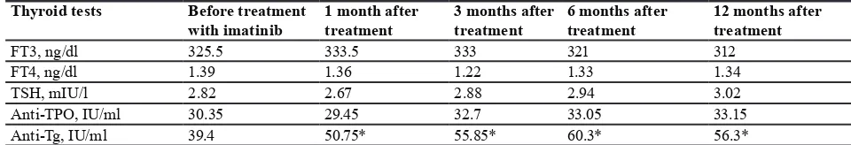
(70%) patients were male and 6 (30%) were female. The mean values for TSH, FT4, FT3, Anti-TPO, and Anti-Tg before treatment were 2.82 mIU/L, 1.39 ng/dl, 325.50 ng/dl, 30.35IU/ml, and 39.40 IU/ml, respectively (table 1). Mean values for TSH, FT4, FT3 and Anti-TPO 1, 3, 6 and 12 months after initiation of Imatinib were not
statistically significant. There was a significant difference
in mean values of Anti-Tg 1, 3, 6 and 12 months after initiation of treatment with imatinib (P=0.014, 0.008, 0.003 and 0.002, respectively).
Discussion
CML is the most common myeloproliferative disorder in adults with a characteristic cytogenetic abnormality
known as t(9:22).10 De Groot et al.6 reported the results of
a study on 11 patients who had received imatinib (1 with gastrointestinal stromal tumor and 10 with medullary thyroid carcinoma (MTC); eight of these patients had previously undergone thyroidectomy and were on thyroid hormone replacement therapy. Increased thyroid hormone requirements were observed while on imatinib therapy. In another study by de Groot et al.8 nine out
of 15 advanced MTC patients who were treated with imatinib, had previously undergone total thyroidectomy and were on thyroid hormone replacement therapy, all them had increased thyroid hormone requirements while on therapy. On the other hand, patients with intact thyroid glands remained euthyroid while on imatinib. Therefore, both studies showed that all patients with intact thyroid glands receiving imatinib had no thyroid dysfunction. In a study from Desai et al.11 on 42 patients treated with
sunitinib for a median of 37 weeks (range, 10 to 167), 4 patients (10%) developed isolated TSH suppression and 7 patients (17%) experienced transient, mild TSH elevations. The risk for hypothyroidism increased with the duration of treatment with sunitinib. Six (40%) of 15 hypothyroid patients had suppressed TSH concentrations before developing hypothyroidism suggesting thyroiditis. Kim et al.9 retrospectively reviewed thyroid function tests
in 10 patients who were treated with dasatinib, 2 patients were on levothyroxine prior to starting therapy, 5 patients developed hypothyroidism (4 subclinical, 1 clinical), and two patients had subclinical hyperthyroidism, none of which required treatment. Dora et al.12 found no
adverse effect of imatinib on thyroid function and lack of correlation between TSH levels with dose, duration, or even cumulative dose of imatinib therapy suggests that this drug has no side effect on thyroid function.
The mechanism of imatinib-induced subclinical or clinical hypothyroidism was stimulation of T3 and T4 clearance owing to elevated activity of liver microsomal enzyme, uridine-diphosphate-glucuronyltransferase (UGTs), which needed to be stabilized.13 Sorafenib has
been associated with hypothyroidism in patients with previously normal thyroid function, with an incidence of 18% in one study14 and 67% in another study.15 Other TKIs
have also been associated with thyroid disease in patients with previously intact thyroid function.9 In a retrospective
study of 64 patients treated for CML, hypothyroidism was seen in 13%, 50%, and 22% of patients treated with imatinib, dasatinib, or nilotinib, respectively.9 The
incidence of preceding transient thyrotoxicosis was also high suggesting a phase of thyroiditis preceding the
Table 1: Thyroid tests in patients with chronic myeloid leukemia before and after Imatinib therapy Thyroid tests Before treatment
with imatinib 1 month after treatment 3 months after treatment 6 months after treatment 12 months after treatment
FT3, ng/dl 325.5 333.5 333 321 312
FT4, ng/dl 1.39 1.36 1.22 1.33 1.34
TSH, mIU/l 2.82 2.67 2.88 2.94 3.02
Anti-TPO, IU/ml 30.35 29.45 32.7 33.05 33.15 Anti-Tg, IU/ml 39.4 50.75* 55.85* 60.3* 56.3*
*P value<0.05 was significant; TSH, thyroid-stimulating hormone; FT4, free thyroxine; FT3, free triiodothyronine; Anti-TPO,
anti-thyroid peroxidase; Anti-Tg, anti-thyroglobulin
Thyroid dysfunction during imatinib therapy
loss of function.9 The main mechanism of TKI-induced
hypothyroidism is unclear. Rare cases of thyrotoxicosis preceding the development of hypothyroidism suggest that there is a preceding thyroiditis. Some suggestions for the mechanism of hypothyroidism associated with TKIs include direct toxic effects on thyrocytes, reduced TPO activity,16 impaired iodine uptake,17 or stimulation of
Hashimoto thyroiditis;18 although Hashimoto thyroiditis
is improbable to be the main mechanism because of the low prevalence of Anti-TPO antibodies in patients with sunitinib-induced hypothyroidism.17,19 The most likely
explanation is that the thyroid dysfunction is related to the effects of these factors on tyrosine kinases involved in vascular function such as VEGFR. This could cause
attenuation of the thyroid blood flow to this extremely vascular gland. If the blood flow decreases rapidly, an
ischemic thyroiditis could result leading to a transient
period of thyrotoxicosis. If the decreased blood flow
develops more slowly, gradual thyroid destruction may occur with resulting hypothyroidism.14 Supporting
evidence for this theory includes the finding that thyroid
cells express VEGF and VEGFR mRNA and studies on mice have shown glandular capillary regression with TKI exposure.20 Two recent case reports demonstrated
reduced thyroid volume and vascularity by doppler ultrasound,17,21,22 with rapid increase in size of the thyroid
following cessation of sunitinib. The reduced thyroid
volume (because of reduced blood flow) may also explain
the impaired radioactive iodine uptake in vivo17 but not
in vitro.23
Conclusion
Based on results of this study, there was no significant
change on thyroid function tests during imatinib therapy and all variables were within normal ranges. However, larger studies with larger sample size are recommended to prove imatinib-induced hypothyroidism.
Conflict of Interest: None declared.
References
1. Payandeh M, Sadeghi M, Sadeghi E. Treatment and Survival in Patients with Chronic Myeloid Leukemia in a Chronic Phase in West Iran. Asian Pac J Cancer Prev. 2015;16(17):7555-9.
2. Le Tourneau C, Faivre S, Raymond E. New developments in multitargeted therapy for patients with solid tumours. Cancer Treat Rev. 2008;34(1):37-48.
3. Daub H. Kinase inhibitors: narrowing down the real targets. Nat Chem Biol. 2010 Apr;6(4):249-50. 4. Illouz F, Laboureau-Soares S, Dubois S, Rohmer V,
Rodien P. Tyrosine kinase inhibitors and modifications
of thyroid function tests: a review. Eur J Endocrinol. 2009;160(3):331-6.
5. Rios MB, Ault P. Identification of side effects
associated with intolerance to BCR-ABL inhibitors in patients with chronic myeloid leukemia. Clin J Oncol Nurs. 2011;15(6):660-7.
6. de Groot JW, Zonnenberg BA, Plukker JT, van Der
Graaf WT, Links TP. Imatinib induces hypothyroidism in patients receiving levothyroxine. Clin Pharmacol Ther. 2005;78(4):433-8.
7. Riesenbeck LM, Bierer S, Hoffmeister I, Köpke
T, Papavassilis P, Hertle L, et al. Hypothyroidism correlates with a better prognosis in metastatic renal cancer patients treated with sorafenib or sunitinib. World J Urol. 2011;29(6):807-13.
8. de Groot JW, Zonnenberg BA, van Ufford-Mannesse PQ, de Vries MM, Links TP, Lips CJ, et al. A phase II trial of imatinib therapy for metastatic medullary thyroid carcinoma. J Clin Endocrinol Metab. 2007;92(9):3466-9.
9. Kim TD, Schwarz M, Nogai H, Grille P, Westermann J, Plöckinger U, et al. Thyroid dysfunction caused by second-generation tyrosine kinase inhibitors in Philadelphia chromosome-positive chronic myeloid leukemia. Thyroid. 2010;20(11):1209-14.
10. Payandeh M, Sadeghi E, Khodarahmi R, Sadeghi M. Appearance and Disappearance of Chronic Myeloid Leukemia (CML) in Patient with Chronic Lymphocytic Leukemia (CLL). Int J Hematol Oncol Stem Cell Res. 2014;8(4):49-53.
11. Desai J, Yassa L, Marqusee E, George S, Frates MC, Chen MH, et al. Hypothyroidism after sunitinib treatment for patients with gastrointestinal stromal tumors. Ann Intern Med. 2006;145(9):660-4. 12. Dora JM, Leie MA, Netto B, Fogliatto LM, Silla
L, Torres F, et al. Lack of imatinib-induced thyroid dysfunction in a cohort of non-thyroidectomized patients. Eur J Endocrinol. 2008;158(5):771-2. 13. de Groot JW, Links TP, van der Graaf WT. Tyrosine
kinase inhibitors causing hypothyroidism in a patient on levothyroxine. Ann Oncol. 2006;17(11):1719-20. 14. Tamaskar I, Bukowski R, Elson P, Ioachimescu
AG, Wood L, Dreicer R, et al. Thyroid function test abnormalities in patients with metastatic renal cell carcinoma treated with sorafenib. Ann Oncol. 2008;19(2):265-8.
15. Miyake H, Kurahashi T, Yamanaka K, Kondo Y, Muramaki M, Takenaka A, et al. Abnormalities of thyroid function in Japanese patients with metastatic renal cell carcinoma treated with sorafenib: a prospective evaluation. Urol Oncol. 2010;28(5):515-9. 16. Wong E, Rosen LS, Mulay M, Vanvugt A, Dinolfo M, Tomoda C, et al. Sunitinib induces hypothyroidism in advanced cancer patients and may inhibit thyroid peroxidase activity. Thyroid. 2007;17(4):351-5. 17. Mannavola D, Coco P, Vannucchi G, Bertuelli R,
Carletto M, Casali PG, et al. A novel tyrosine-kinase selective inhibitor, sunitinib, induces transient hypothyroidism by blocking iodine uptake. J Clin Endocrinol Metab. 2007;92(9):3531-4.
18. Alexandrescu DT, Popoveniuc G, Farzanmehr H, Dasanu CA, Dawson N, Wartofsky L. Sunitinib-associated lymphocytic thyroiditis without circulating antithyroid antibodies. Thyroid. 2008;18(7):809-12. 19. Rini BI, Tamaskar I, Shaheen P, Salas R, Garcia
J, Wood L, et al. Hypothyroidism in patients with metastatic renal cell carcinoma treated with sunitinib.
Allahyari A et al.
J Natl Cancer Inst. 2007;99(1):81-3.
20. Kamba T, Tam BY, Hashizume H, Haskell A, Sennino B, Mancuso MR, et al. VEGF-dependent plasticity of fenestrated capillaries in the normal adult microvasculature. Am J Physiol Heart Circ Physiol. 2006;290(2):H560-76.
21. Makita N, Miyakawa M, Fujita T, Iiri T. Sunitinib induces hypothyroidism with a markedly reduced vascularity. Thyroid. 2010;20(3):323-6.
22. Rogiers A, Wolter P, Op de Beeck K, Thijs M, Decallonne B, Schöffski P. Shrinkage of thyroid volume in sunitinib-treated patients with renal-cell carcinoma: a potential marker of irreversible thyroid dysfunction? Thyroid. 2010;20(3):317-22.
23. Salem AK, Fenton MS, Marion KM, Hershman JM. Effect of sunitinib on growth and function of FRTL-5 thyroid cells. Thyroid. 2008;18(6):631-5.