Fang He, PhD Julie M. Jones, PhD Claudia
Figueroa-Romero, PhD Dapeng Zhang, PhD Eva L. Feldman, MD,
PhD
Stephen A. Goutman, MD
Miriam H. Meisler, PhD Brian C. Callaghan, MD Peter K. Todd, MD, PhD
Correspondence to Dr. Todd: [email protected]
Supplemental data at Neurology.org/ng
Screening for novel hexanucleotide repeat
expansions at ALS- and FTD-associated
loci
ABSTRACT
Objective:To determine whether GGGGCC (G4C2) repeat expansions at loci other thanC9orf72
serve as common causes of amyotrophic lateral sclerosis (ALS).
Methods:We assessed G4C2repeat number in 28 genes near known ALS and frontotemporal
dementia (FTD) loci by repeat-primed PCR coupled with fluorescent fragment analysis in 199 patients with ALS (17 familial, 182 sporadic) and 136 healthy controls. We also obtained blood from patients with ALS4 for evaluation of repeats surrounding theSETXgene locus.C9orf72 expansions were evaluated in parallel.
Results:Expansions of G4C2repeats inC9orf72explained 8.8% of sporadic and 47% of familial
ALS cases analyzed. Repeat variance was observed at one other locus,RGS14, but no large expansions were observed, and repeat sizes were not different between cases and controls. No G4C2repeat expansions were identified at other ALS or FTD risk loci or in ALS4 cases.
Conclusions:G4C2expansions near known ALS and FTD loci other thanC9orf72are not a
com-mon cause of ALS.Neurol Genet2016;2:e71; doi: 10.1212/NXG.0000000000000071
GLOSSARY
ALS5amyotrophic lateral sclerosis;FTD5frontotemporal dementia;G4C25GGGGCC;GWAS5genome-wide association studies;SNP5single-nucleotide polymorphism.
A GGGGCC (G
4C
2) hexanucleotide repeat expansion in the first intron of
C9orf72
is the most
common monogenic cause of amyotrophic lateral sclerosis (ALS) and frontotemporal dementia
(FTD).
1,2There are normally between 2 and 23 G
4
C
2repeats at this locus. The repeat expands
to hundreds in affected individuals,
2although 30 repeats may be sufficient to elicit G
4
C
2-specific pathology.
3A growing body of evidence suggests that the
C9orf72
repeat elicits toxicity primarily through
gain-of-function mechanisms that are independent of the genetic locus where the repeat resides.
Specifically,
Drosophila
and mouse models of ALS with expression of the repeat outside its
normal genomic context suggest that instability and expansion of G
4C
2repeats elsewhere in
the genome could also cause ALS or FTD. A precedent for this phenomenon is found in
spinocerebellar ataxia, in which CAG repeat expansions in a diverse set of genes elicit
over-lapping clinical phenotypes.
4We therefore hypothesized that cryptic repeat expansions at loci other than
C9orf72
could
also contribute to ALS and FTD pathogenesis. Using repeat-primed PCR assays, we evaluated
whether G
4C
2repeats near known ALS and FTD loci identified by linkage analysis or
genome-wide association studies (GWAS) exhibited expansions in a cohort of patients with
ALS and controls in the University of Michigan ALS Patient Biorepository. Our results
confirm that repeat instability and large expansions at
C9orf72
are common in sporadic
From the Department of Neurology (F.H., C.F.-R., B.C.C., E.L.F., S.A.G., P.K.T.) and Department of Human Genetics (J.M.J., M.H.M.), University of Michigan, Ann Arbor; Veteran Association Health System (B.C.C., P.K.T.), Ann Arbor; and National Center for Biotechnology Information (D.Z.), National Institutes of Health, Bethesda, MD.
Funding information and disclosures are provided at the end of the article. Go to Neurology.org/ng for full disclosure forms. The Article Processing Charge was paid by the authors.
ALS in the United States, but expansions at
other disease-associated loci are rare in this
population and are unlikely to be a common
cause of ALS.
METHODS Standard protocol approvals, registrations, and patient consents.This study was approved by the Institu-tional Review Board of the University of Michigan. Individual pa-tients and controls who contributed these DNA samples provided written informed consent via representatives from the Coriell Institute, University of Michigan ALS Patient Biorepository, or individually to a member of the research group.
ALS and control patient cohorts and DNA isolation. DNA samples for repeat-primed PCR and genomic PCR were from the following sources: 1mg of genomic DNA from 199 patients with ALS and 136 healthy controls from the University of Michigan ALS Patient Biorepository. Patients with ALS met the revised El Escorial criteria5and were recruited from the University of Michigan ALS Clinic; controls were recruited via the University of Michigan clinical trials Web site (https:// umclinicalstudies.org/). Demographic data were analyzed using SAS9.5 software (SAS Institute Inc., Cary, NC) and summarized in table e-1 at Neurology.org/ng. Two hundred fifty nanograms of genomic DNA from 86 patients with sporadic ALS from Coriell Cell Repository panel #NDPT026 (Coriell Institute) was used for determination ofC9orf72repeat status only. One hundred micrograms of genomic DNA from a patient with ALS4 and 1 non-ALS sibling from a previously reported large pedigree6 was extracted from 4 mL of whole blood using a commercial DNA isolation kit (DNeasy Blood & Tissue kit; Qiagen, Netherlands). Genomic DNA from a second published ALS4 case with a family history7was extracted from patient fibroblast cells obtained from a collaborator’s laboratory using the same kit.
Candidate gene selection. We performed a BLAST search (National Center for Biotechnology Information) against the human genome for G4C2repeat sequences using a sequence of 5 G4C2pure
repeats (GGGGCCGGGGCCGGGGCCGGGGCCGGGGCC) as a start query. The identified repeat loci were overlaid with pub-lished genetic loci associated with ALS or FTD (see a recent review8 and table 1). We constrained our analysis to repeat loci within 2 mega base pairs (Mb) of either the mapped critical region for an ALS or FTD candidate gene/locus or with single-nucleotide polymorphisms (SNPs) that achieved statistical significance on GWAS in sporadic ALS cohorts. Additional candidate repeats located more than 2 Mb outside of disease-associated loci were identified by requiring at least 3 pure repeats in a gene with abundant neuronal expression in brain based on BioGPS and Proteomic DB database analysis.9,10 Three additional candidate genes with G4C2repeats within the previously
identified critical region of ALS4 but missed by our initial in silico analysis were added after we obtained access to case samples.
G4C2 repeat determination. G4C2 repeat numbers in the
longer allele were determined by repeat-primed PCR as previously reported,11followed by capillary electrophoresis and fragment analysis. The primer sequences are included in table e-2. The individual reverse primers for each candidate gene were designed using Primer 3 tools (http://biotools.umassmed.edu/ bioapps/primer3_www.cgi) and were labeled with either 6-FAM or 5-HEX fluorescent dye. The PCR products were diluted in highly deionized formamide (HiDi formamide; ThermoFisher Scientific, Waltham, WA) containing size standard ROX-1000 (BioVenture, TN). Fragment analysis was performed at the University of Michigan Sequencing Core facility
on an ABI 3730 Sequencer. The data were analyzed using GeneMarker (SoftGenetics, PA) to determine the maximal repeat number.
Sanger sequencing. For samples with fewer than 35 G4C2
repeats inC9orf72, DNA flanking the repeat regions was PCR-amplified with the following primers:C9orf72forward: 59-CCG CAG CCT GTA GCA AGC-39andC9orf72reverse: 59-AGT CGC TAG AGG CGA AAG C-39 using the same thermal cycling program as the repeat-primed PCR. The PCR products were gel purified and subjected to Sanger sequencing at the University of Michigan Sequencing Core facility to determine the exact repeat number.
For samples with a 31-nucleotide insertion in theVAV2gene, DNA flanking the repeat region was PCR-amplified with the following primers: VAV2 forward: 59-GCC CAG GAC AGG AGG CCT CAG CA-39and VAV2 reverse: 59- CTC AGG GCC GGG AGG AAG CAC CT-39using the same thermal cycling program as the repeat-primed PCR. The PCR products were gel-purified and subjected to Sanger sequencing as described above. For repeats atHuwe1andRGS14, PCR primers flanking the repeat regions were used to determine specific repeat sizes (see supplemental data).
Southern blot confirmation of repeat expansions at
C9orf72.For Southern blot analysis, 25-mg aliquots of lym-phoblast genomic DNA were obtained for 5 selected Coriell samples. Two additional lymphoblast cell lines were obtained from the Coriell Institute and were grown up to 15 million cells, and approximately 100mg of lymphoblast genomic DNA was extracted using a DNA isolation kit. Ten fibroblast cell lines with G4C2repeat expansion and 2 control fibroblast lines (1
ALS sample without expansion and 1 control) were obtained from the University of Michigan ALS Patient Biorepository and 100 mg of fibroblast genomic DNA was extracted from 15 million cells.
Statistical analysis.Two-tailed Fisher exact tests orx2tests were performed to test for association of repeat length with ALS and to verify genotype frequencies in theVAV2gene. For RGS14 repeat length, a 2-tailed nonparametric t test was performed to compare the median repeat size differences. For the correlation ofC9orf72repeat size and patient age at onset of ALS, the Pearson correlation test was performed.
RESULTS We first conducted an in silico experi-ment to identify all human genes containing at least 2 G4C2 repeats. Our rationale was that transcribed
repeats at such loci could become unstable and lead to disease-causing expansions, unless the chromosomal context of theC9orf72repeat was critical to disease pathogenesis. Our initial analysis revealed that short G4C2 repeats are quite common in the human
to a percentage of sporadic cases) would identify most repeat expansions that confer a substantial contribution to the genetic burden of ALS in the United States. Consistent with this idea, the C9orf72 locus was identified on multiple GWAS surveys in ALS and FTD in sporadic populations prior to identification
of the causative repeat.12–14 Using this approach, we
identified 21 candidate genes, including C9orf72, containing at least 2 G4C2 repeats that are located
within 2 Mb of known ALS-FTD loci or loci identified in GWAS by at least 2 associated SNPs (figure 1A).
We developed a modified fluorescent fragment analysis after repeat-primed PCR to evaluate instabil-ity at each of these loci, as previously described for C9orf72repeat expansions.11We also assessed repeat
size atC9orf72. These assays were tested on control samples, and the actual repeat sizes were confirmed by standard PCR and Sanger sequencing. All 21 assays demonstrated accurate detection of repeat size, based on comparison with Sanger sequencing (data not shown). This assay readily detected G4C2expansions
atC9orf72in samples from ALS cases known to har-bor the repeat.
We applied these assays to a sample collection from the University of Michigan ALS Patient Repos-itory containing 199 ALS cases, of which 17 had a family history of ALS, and 136 healthy controls (University of Michigan). The demographic charac-teristics of this collection are shown in table e-1. Of note, there were no differences in the age at sample collection, sex composition, or racial/ethnic makeup between ALS cases and controls, and both cases and controls had similar frequencies of a family history of a non-ALS neurodegenerative condition (table e-1). In addition, we used samples from Coriell Cell Repository panel NDPT026, containing 86 sporadic ALS cases, for additional screening for repeat expan-sions inC9orf72(Coriell Institute). Consistent with published findings, we observed a wide variation of G4C2 repeat length at C9orf72 in both cases and
controls (table 1, table e-3, figure 1B). Pathogenic repeat expansions were observed in 47% of the famil-ial cases and 8.8% (17/182) of sporadic ALS cases in the University of Michigan ALS Patient Repository (figure 1B and table e-3).C9orf72repeat expansions were observed at a similar frequency (6/86; 7.0%) in ALS samples from the Coriell Institute. To validate these findings and determine the actual repeat expan-sion size in cases with low repeat numbers, genomic PCR and selective Southern blotting analysis were performed. One sample contained exactly 32 repeats in the longer allele, which was confirmed by Sanger sequencing. Other samples exhibited a single band by genomic PCR reflecting the shorter, PCR-amplifiable allele. Southern blotting on genomic DNA extracted from patient-derived fibroblasts or lymphocytes in a subset of cases demonstrated a wide range of path-ologic repeat expansion sizes from 200 to more than 1,500 repeats (figure e-1). Repeat expansion size from peripheral blood samples or cultured cells did not correlate with clinical age at onset (R 5 0.173 in
Table 1 G4C2repeat loci evaluated in this study
Gene Location Repeat no. ALS/FTD gene loci or SNPa Differentin ALS?
ARMC2 Intron 2 FIG4/ALS11 No
ATXN2 Coding 2 ATXN2 No
C8orf76 Intron 2 rs4581057, rs10106208 No
C9orf72 Intron 2–35 C9orf72 Yes
C16orf72 Intron 2 rs1551960, rs7185240 No
CACNA1G Coding 2 rs8068533, rs1061947 No
DOCK1 Intron 2 rs7082776, rs4363506 No
GPR123 Intron 2–4 OPTN/ALS12 No
HUWE1b Intron 12–14 UBQLN2/ALS15 No
ITPR3 Intron 3–4 rs9380343, rs963733 No
KMT2C Intron 2 rs4725431, rs10260404 No
MBD2c Coding 3 ALS3 No
NEURL1B Intron 2 rs871503, rs4868146 No
PAPD5 Coding 3 rs12929266, rs1075875 No
PRRC2B Coding 2 SETX/ALS4 No
SUPT5H Coding 2 rs12327672, rs11669124, rs2287735
No
SYNM Intron 3 rs931892, rs3803478 No
TAF4d Coding 6 VAPB/ALS8 No
TPP1 Coding 2 rs2063082, rs16917433 No
TTC28 Intron 2 rs6005863, rs5762919 No
ZNF423e Intron 4 rs1075875, rs1505112 No
BSNf Coding 5 NA No
GAS7 Intron 3 NA No
RGS14 Intron 4–15 NA No
STK39 Coding 3–5 rs13015447 No
PRRC2B Coding 2 SETX/ALS4 No
PRRX2 Intron 3–5 SETX/ALS, rs395119 No
SURF4 Intron 2 SETX/ALS4 No
VAV2 Intron 2 SETX/ALS4 No
Abbreviations: ALS 5 amyotrophic lateral sclerosis; FTD 5 frontotemporal dementia; G4C25GGGGCC; GWAS5genome-wide association study; NA5not applicable; SNP5
single-nucleotide polymorphism.
aG
4C2repeats found within 2 Mb of GWAS SNPs from references 12–14,28–36. bMBD2has 2 separate G
4C2repeats that are 2 and 3 repeats long. Data shown for the
3-repeat region. The 2-3-repeat region was invariant.
cTAF4has 6 imperfect G
4C2repeats ((C2G4)3CCGGGC(C2G4)2). dZNF423has 2 separate G
4C2repeats that are 2 and 4 repeats long. Data are shown for
the 4-repeat region. The 2-repeat region was invariant.
eBSNhas 2 separate G
4C2repeats. One is 3 repeats long and the other has 5 imperfect
repeats (CCGGGGCCGGGGCCGGGGCCCGGGCCGGGG). Data are shown for the 5-repeat region. The 3-repeat region was invariant.
f
HUWE1 has 12 imperfect G4C2 repeats ((C2G4)3CCAGGG(C2G4)4CCAGGGCCGGTG
Pearson correlation test,p50.572, figure e-2), con-sistent with previous reports.15
In contrast to C9orf72, G4C2 repeats assayed at
other loci exhibited little or no variance in repeat number in the University of Michigan ALS Patient
Biorepository samples. Of the 20 novel repeats ana-lyzed, 16 showed no variance at all across the cohort (80%). Four genes displayed small variations in repeat number (table 1), but none of these cases contained expansions of more than 2 repeats greater than the
Figure 1 G4C2repeat numbers from controls and patients with ALS
prevalent alleles. The degree of repeat length variation was not significantly different between control and ALS groups, demonstrating a lack of association between repeat number variation and ALS/FTD in our cohort (figure 1, B–E, p,0.001 forC9orf72, p50.76 forGPR123,p50.69 forHUWE1,p5 0.374 for ITPR3;x2test forC9orf72andITPR3, and
Fisher exact test forGPR123 andHUWE1used for the rest for different allele frequencies).
To expand the initial screening we selected 4 addi-tional candidate genes that are highly expressed in brain and contain at least 3 G4C2repeats in controls. In these
candidate genes, no large repeat expansions were found in ALS or control groups (table 1, figure 1, F and G). However, an intronic repeat in RGS14 displayed a much larger variation than those in other loci studied, with between 4 and 15 repeats present in both controls and ALS cases. The distribution of repeat lengths in RGS14was independent of clinical ALS (figure 1F) or C9orf72expansion status (control 8.4961.74 vs ALS 8.161.93,p50.082, 2-tailed studentttest).
As a third approach, we examined additional loci harboring G4C2 repeats located near the ALS4 locus
in patients with this disorder. ALS4 is a form of juvenile ALS with very slow progression that is thought to result from dominant mutations inSETX.6The mechanism of
these dominant mutations is unclear, because recessive mutations inSETXcause a distinct neurodegenerative disorder, ataxia with oculomotor apraxia type 2, which does not include motor neuron degeneration.16Within
the critical region defined by linkage analysis,6there are
4 genes containing short G4C2 repeats: PRRX2,
PCCR2B,SURF4, andVAV2(figure 2A). To investi-gate whether a cryptic repeat expansion might provide an alternative explanation for the ALS4 phenotype, we analyzed repeat numbers in 2 patients with ALS4 and an unaffected control from 2 previously described ALS4 families.6,7No repeat expansions were observed (figure
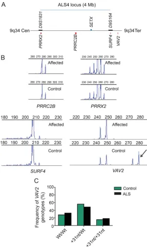
2B). Of these 4 loci, only 1 (PRRC2B) was included in our initial repeat screen in the University of Michigan ALS Patient Repository. Assessment of the additional 3 loci in this larger cohort identified no repeat expansions, only a minor repeat length variance in gene PRRX2 (table 1, figure 1H). We did observe an amplicon length variant of 31 bp inVAV2by fragment analysis in the ALS4family (figure 2B). Sanger sequencing identified it as a 31-bp intronic insertion in the longer allele that contains an imperfect duplication of intronic sequence (GCCGGGGCCGTGTGGCCCTCACGCAGT-GACC). This insertion was common in controls and ALS cases in the University of Michigan ALS Patient Biorepository panel (figure 2C), and ap-pears to be nonpathogenic (p50.398,x2test).
DISCUSSION G4C2 repeat expansions in C9orf72
are the most common known cause of ALS and
FTD. Here we tested whether G4C2repeat instability
at other loci could be a common cause of ALS. Our results confirmed that G4C2 repeat expansions and
instability are common at theC9orf72locus in both familial and sporadic ALS cases in 2 patient cohorts (University of Michigan ALS Patient Repository and the NDPT026 ALS collection from Coriell). How-ever, for 27 other genomic loci that harbor G4C2
repeats, no pathogenic G4C2expansions were
identi-fied. Thus, repeat instability at these alternative loci are unlikely to be major contributors to the genetic burden in ALS in the United States.
This study is not exhaustive in the number of repeat loci assayed or the number of patient and familial cohorts examined. Therefore, we cannot rule out G4C2repeat expansions as rare causes of ALS. For
practical reasons, we assayed fewer than 10% of the G4C2repeats located near protein-coding genes and
a much smaller fraction of the GC-rich repeats in the human genome. Specifically, we focused our analysis on repeats located adjacent to loci already implicated in ALS or FTD. Our rationale for this choice was that utilization of GWAS conducted in similar patient populations as well as known genetic loci in rare fam-ilies (which have subsequently been shown to con-tribute to a percentage of sporadic cases) would colocalize with repeat expansions that confer a sub-stantial contribution to the genetic burden of ALS in the United States. Consistent with this idea, the C9orf72locus was identified on multiple GWAS sur-veys in ALS and FTD in sporadic populations prior to identification of the causative repeat. Moreover, we reasoned that cryptic repeat expansions might have escaped identification during the initial analyses of these loci, several of which lack an identified causative gene or a clear pathogenic mechanism for the cur-rently implicated gene. As such, our findings that none of these alternative loci harbor repeat expansions in 199 ALS cases suggest that G4C2repeat expansions
at non-C9orf72 loci are unlikely to be a common cause of ALS in our patient population, given that most common highly penetrant mutations would be detected by this approach. However, it is important to acknowledge that for highly penetrant loci identi-fied in familial ALS cases, it might be necessary to examine the original published cohorts, which might carry rare or private mutations. We took this step for ALS4 and found no G4C2 repeat instability at 4
neighboring loci. Application of this approach to other isolated pedigrees could more definitively rule out expansions at these genetically implicated loci.
The lack of G4C2repeat expansions at alternative
335 individuals, and variance in repeat size was largely absent from the 1,000-genome database for the 321 intragenic G4C2 repeat loci not assayed (data not
shown). With one exception, the repeat size variants observed were limited to 1 or 2 repeats. Studies in vitro suggest that stretches of G4C2as short as 4
repeats are capable of forming intramolecular
G quadruplexes, which might contribute to repeat instability through activation of DNA mismatch repair pathways and mistemplating.17 In general,
our data are consistent with this threshold, since only genes with at least 4 repeats had variation of more than 1 repeat. However, some of the G4C2repeats we
studied were larger than this threshold but were invariant, suggesting that the length of the repeat is insufficient to explain instability. While future work will be needed to determine which factors drive selec-tive instability, it may reflect specific aspects of the GC-rich sequence surrounding the repeat at the C9orf72loci. This concept is supported by the find-ing that all pathologic repeat expansions atC9orf72 observed to date have occurred on a single haplotype background.18
Our approach here focused solely on the role of G4C2 repeat expansions in ALS. However, G4C2
repeat expansions in genes other thanC9orf72may be enriched in neurologic disorders other than ALS. Phenotypes of polyglutamine disorders associated with CAG repeat expansions, for example, are not all the same.19In this context, it is interesting to note
that theC9orf72repeat expansion exhibits neurologic symptoms in addition to motor neuron disease and frontotemporal cognitive dysfunction, which are still being explored.11,20
We observed noteworthy repeat instability in both cases and controls in one other gene: a G4C2intronic
repeat inRGS14with a mean repeat length of 9 and a range of 4–14 repeats.RGS14encodes a member of the regulator of G-protein signaling family of pro-teins. It is highly expressed in the developing brain and has known functions in hippocampal learning and synaptic plasticity.21 Although we observed no
association between the size of this repeat and clinical ALS, our study cannot rule out a role for expansion in rare ALS families or other racial/ethnic populations. As such, reevaluation of this locus may be worthy of further study in familial ALS, FTD, and other neu-rodegenerative disorders.
Other repeat expansions are implicated in causing or modifying motor neuron disease phenotypes. A GGCCTG repeat expansion inNOP56causes spino-cerebellar ataxia type 3622and has motor neuron
dis-ease as a clinical feature. Moderate CAG repeat expansions (encoding polyglutamine) in ATXN223
and GCG repeat expansions (encoding polyalanine) inNIP1A24 are associated with an increased risk of
ALS. However, other known pathogenic CAG and CGG repeat expansions do not influence the risk of ALS or FTD, suggesting that the observed effects may be gene specific rather than repeat sequence spe-cific.25–27
We screened 28 loci harboring G4C2repeats near
known ALS/FTD loci in 199 patients with ALS and
Figure 2 Analysis of G4C2repeats near theALS4locus
(A) Schematic of theALS4locus defined by chromosome markersD9S1831andD9S164
(black boxes)6drawn to scale showing the location of 4 G
4C2repeat-containing genes (red
boxes) and the proposed disease-causing geneSETX(blue box). (B) Chromatograms showing the G4C2repeat peaks in the 4 G4C2repeat-containing genes from an affected ALS4 case
did not find pathogenic repeat expansions other than that previously reported in C9orf72. While future studies in larger cohorts and select families are still needed, our results suggest that G4C2repeat
expan-sions outside the C9orf72 gene are not a common cause of ALS, supporting the importance of identify-ingciselements that explain the genetic instability at theC9orf72locus.
AUTHOR CONTRIBUTIONS
F.H., M.H.M., D.Z., B.C.C., and P.K.T. conceptualized and designed the study. F.H., J.M.J., and C.F.-R. performed the experiments. F.H., J.M.J., M.H.M., B.C.C., and P.K.T. analyzed the data. M.H.M., E.L.F., S.A.G., and B.C.C. provided patient samples. F.H. and P.K.T. wrote the manuscript. All authors revised the manuscript.
ACKNOWLEDGMENT
The authors thank all patients who provided the samples for this study and Blake Swihart and Jayna Duell, RN, for assistance in study coordination. The authors thank Suzzane Genik for technical assistance in fragment anal-ysis and Zixing Wang for assistance on data processing. They thank the University of Michigan ALS Patient Biorepository for access to patient fi-broblasts. The authors thank Michio Hirano for providing patient DNA and Phillip Chance and David Cornblath for helpful discussions. This repository is funded by the Center for Disease Control (CDC) Agency for Toxic Substances and Disease Registry (contract 200-2013-56856), the Program for Neurology Research & Discovery, and the A. Alfred Taubman Medical Research Institute. The authors also thank colleagues in P.K.T.’s laboratory for help in experiments and data interpretation and proofreading of the manuscript.
STUDY FUNDING
This project was funded by NIH R01GM24872 to M.H.M.; grants from the Centers for Disease Control and Prevention (200-2013-56856) and the A. Alfred Taubman Medical Research Institute to E.L.F.; and grants from the Michigan Alzheimer’s Disease Center at the University of Mich-igan, NIH R01NS08681001, and the Department of Veterans Affairs BLRD 1I21BX001841 to P.K.T. The funders played no role in study design, data collection and analysis, decision to publish, or preparation of the manuscript.
DISCLOSURE
Drs. He and Jones report no disclosures. Dr. Figueroa-Romero has received research support from the Katherine Rayner Fund and the A. Alfred Taubman Medical Research Institute. Dr. Zhang receives intramu-ral funding support from the NIH. Dr. Feldman receives funding support from Novo Nordisk, the NIH, the Juvenile Diabetes Research Founda-tion, the US-Israel Binational Science FoundaFounda-tion, the American Diabe-tes Association, and the A. Alfred Taubman Medical Research Institute. Dr. Goutman receives research funding support from Neuralstem, Inc; the Centers for Disease Control and Prevention/Agency for Toxic Sub-stances and Disease Registry; and the Agency for Toxic SubSub-stances and Disease Registry (contract #200-2013-56856). Dr. Meisler receives fund-ing support from the NIH. Dr. Callaghan receives fundfund-ing support from Impeto Medical Inc and the NIH, has received travel/speaker honoraria from the American Academy of Neurology and the World Federation of Neurology, performs medical consultations for Advance Medical, con-sults for a PCORI grant, has received honoraria from the British Medical Journal, has worked with the ALS Association, and has acted as a consul-tant for medical legal cases. Dr. Todd receives funding support from the NIH, the Veterans Administration, the National Ataxia Foundation, the National Fragile X Foundation, and the Muscular Dystrophy Asso-ciation; serves as a paid consultant for Denali Therapeutics and for an NIH grant; serves as an unpaid consultant for the National Fragile X Foundation; has received travel/speaker honoraria from the American Academy of Neurology, the National Fragile X Foundation, and the Japanese Society of Neurochemistry; has received publishing royalties from UpToDate; and has received license fee payments for antibodies
developed by his group from Millipore for distribution to scientists and commercial groups, and for vectors and antibodies developed by his group from Denali Therapeutics. Go to Neurology.org/ng for full dis-closure forms.
Received December 10, 2015. Accepted in final form March 1, 2016.
REFERENCES
1. Renton AE, Majounie E, Waite A, et al. A hexanucleotide repeat expansion in C9ORF72 is the cause of chromosome 9p21-linked ALS-FTD. Neuron 2011;72:257–268. 2. DeJesus-Hernandez M, Mackenzie IR, Boeve BF, et al.
Expanded GGGGCC hexanucleotide repeat in noncoding region of C9ORF72 causes chromosome 9p-linked FTD and ALS. Neuron 2011;72:245–256.
3. Gami P, Murray C, Schottlaender L, et al. A 30-unit hexanucleotide repeat expansion in C9orf72 induces path-ological lesions with dipeptide-repeat proteins and RNA foci, but not TDP-43 inclusions and clinical disease. Acta Neuropathol 2015;130:599–601.
4. Orr HT. Cell biology of spinocerebellar ataxia. J Cell Biol 2012;197:167–177.
5. Brooks BR, Miller RG, Swash M, Munsat TL; World Federation of Neurology Research Group on Motor Neu-ron Disease. El Escorial revisited: revised criteria for the diagnosis of amyotrophic lateral sclerosis. Amyotroph Lat-eral Scler Other Motor Neuron Disord 2000;1:293–299. 6. Chen YZ, Bennett CL, Huynh HM, et al. DNA/RNA helicase gene mutations in a form of juvenile amyotrophic lateral sclerosis (ALS4). Am J Hum Genet 2004;74:1128– 1135.
7. Hirano M, Quinzii CM, Mitsumoto H, et al. Senataxin mutations and amyotrophic lateral sclerosis. Amyotroph Lateral Scler 2011;12:223–227.
8. Marangi G, Traynor BJ. Genetic causes of amyotrophic lateral sclerosis: new genetic analysis methodologies entail-ing new opportunities and challenges. Brain Res 2015; 1607:75–93.
9. Wu C, Macleod I, Su AI. BioGPS and MyGene.info: organizing online, gene-centric information. Nucleic Acids Res 2013;41:D561–D565.
10. Wilhelm M, Schlegl J, Hahne H, et al. Mass-spectrome-try-based draft of the human proteome. Nature 2014;509: 582–587.
11. Meisler MH, Grant AE, Jones JM, et al. C9ORF72 expansion in a family with bipolar disorder. Bipolar Disord 2013;15:326–332.
12. Laaksovirta H, Peuralinna T, Schymick JC, et al. Chro-mosome 9p21 in amyotrophic lateral sclerosis in Finland: a genome-wide association study. Lancet Neurol 2010;9: 978–985.
13. Shatunov A, Mok K, Newhouse S, et al. Chromosome 9p21 in sporadic amyotrophic lateral sclerosis in the UK and seven other countries: a genome-wide association study. Lancet Neurol 2010;9:986–994.
14. van Es MA, Veldink JH, Saris CG, et al. Genome-wide association study identifies 19p13.3 (UNC13A) and 9p21.2 as susceptibility loci for sporadic amyotrophic lateral sclerosis. Nat Genet 2009;41:1083–1087. 15. van Blitterswijk M, DeJesus-Hernandez M,
16. Moreira MC, Klur S, Watanabe M, et al. Senataxin, the ortholog of a yeast RNA helicase, is mutant in ataxia-ocular apraxia 2. Nat Genet 2004;36:225–227. 17. Reddy K, Schmidt MH, Geist JM, et al. Processing of
double-R-loops in (CAG)$(CTG) and C9orf72 (GGGGCC)$ (GGCCCC) repeats causes instability. Nucleic Acids Res 2014;42:10473–10487.
18. Majounie E, Renton AE, Mok K, et al. Frequency of the C9orf72 hexanucleotide repeat expansion in patients with amyotrophic lateral sclerosis and frontotemporal dementia: a cross-sectional study. Lancet Neurol 2012; 11:323–330.
19. Orr HT, Zoghbi HY. Trinucleotide repeat disorders. An-nu Rev Neurosci 2007;30:575–621.
20. Cooper-Knock J, Shaw PJ, Kirby J. The widening spec-trum of C9ORF72-related disease; genotype/phenotype correlations and potential modifiers of clinical phenotype. Acta Neuropathol 2014;127:333–345.
21. Vellano CP, Lee SE, Dudek SM, Hepler JR. RGS14 at the interface of hippocampal signaling and synaptic plasticity. Trends Pharmacol Sci 2011;32:666–674.
22. Kobayashi H, Abe K, Matsuura T, et al. Expansion of intronic GGCCTG hexanucleotide repeat in NOP56 causes SCA36, a type of spinocerebellar ataxia accompa-nied by motor neuron involvement. Am J Hum Genet 2011;89:121–130.
23. Elden AC, Kim HJ, Hart MP, et al. Ataxin-2 intermediate-length polyglutamine expansions are associ-ated with increased risk for ALS. Nature 2010;466: 1069–1075.
24. Blauw HM, van Rheenen W, Koppers M, et al. NIPA1 polyalanine repeat expansions are associated with amyotro-phic lateral sclerosis. Hum Mol Genet 2012;21: 2497–2502.
25. Figley MD, Thomas A, Gitler AD. Evaluating noncoding nucleotide repeat expansions in amyotrophic lateral sclero-sis. Neurobiol Aging 2014;35:936.e1–936.e4.
26. Lee T, Li YR, Chesi A, et al. Evaluating the prevalence of polyglutamine repeat expansions in amyotrophic lateral sclerosis. Neurology 2011;76:2062–2065.
27. Groen EJ, van Rheenen W, Koppers M, et al. CGG-repeat expansion in FMR1 is not associated with amyotrophic lat-eral sclerosis. Neurobiol Aging 2012;33:1852.e1–1852.e3. 28. Dunckley T, Huentelman MJ, Craig DW, et al.
Whole-genome analysis of sporadic amyotrophic lateral sclerosis. N Engl J Med 2007;357:775–788.
29. Schymick JC, Scholz SW, Fung HC, et al. Genome-wide genotyping in amyotrophic lateral sclerosis and neurolog-ically normal controls: first stage analysis and public release of data. Lancet Neurol 2007;6:322–328.
30. Cronin S, Berger S, Ding J, et al. A genome-wide associ-ation study of sporadic ALS in a homogenous Irish pop-ulation. Hum Mol Genet 2008;17:768–774.
31. van Es MA, van Vught PW, Blauw HM, et al. Genetic variation in DPP6 is associated with susceptibility to amyotrophic lateral sclerosis. Nat Genet 2008;40:29–31. 32. Chio A, Schymick JC, Restagno G, et al. A two-stage
genome-wide association study of sporadic amyotrophic lateral sclerosis. Hum Mol Genet 2009;18:1524–1532. 33. Landers JE, Melki J, Meininger V, et al. Reduced
expres-sion of the kinesin-associated protein 3 (KIFAP3) gene increases survival in sporadic amyotrophic lateral sclerosis. Proc Natl Acad Sci USA 2009;106:9004–9009. 34. Iida A, Takahashi A, Kubo M, et al. A functional variant
in ZNF512B is associated with susceptibility to amyotro-phic lateral sclerosis in Japanese. Hum Mol Genet 2011; 20:3684–3692.
35. Kwee LC, Liu Y, Haynes C, et al. A high-density genome-wide association screen of sporadic ALS in US veterans. PLoS One 2012;7:e32768.
DOI 10.1212/NXG.0000000000000071
2016;2;
Neurol Genet
Fang He, Julie M. Jones, Claudia Figueroa-Romero, et al.
Screening for novel hexanucleotide repeat expansions at ALS- and FTD-associated loci
This information is current as of May 11, 2016
Services
Updated Information &
http://ng.neurology.org/content/2/3/e71.full.html
including high resolution figures, can be found at:
Supplementary Material
http://ng.neurology.org/content/suppl/2016/05/11/2.3.e71.DC1
Supplementary material can be found at:
References
http://ng.neurology.org/content/2/3/e71.full.html##ref-list-1
This article cites 36 articles, 2 of which you can access for free at:
Citations
http://ng.neurology.org/content/2/3/e71.full.html##otherarticles
This article has been cited by 3 HighWire-hosted articles:
Permissions & Licensing
http://ng.neurology.org/misc/about.xhtml#permissions
its entirety can be found online at:
Information about reproducing this article in parts (figures,tables) or in
Reprints
http://ng.neurology.org/misc/addir.xhtml#reprintsus
Information about ordering reprints can be found online:
Neurology. All rights reserved. Online ISSN: 2376-7839.