R E S E A R C H A R T I C L E
Open Access
Uncoupling of complex regulatory patterning
during evolution of larval development in
echinoderms
Kristen A Yankura, Megan L Martik, Charlotte K Jennings, Veronica F Hinman
*Abstract
Background:Conservation of orthologous regulatory gene expression domains, especially along the neuroectodermal anterior-posterior axis, in animals as disparate as flies and vertebrates suggests that common patterning mechanisms have been conserved since the base of Bilateria. The homology of axial patterning is far less clear for the many marine animals that undergo a radical transformation in body plan during metamorphosis. The embryos of these animals are microscopic, feeding within the plankton until they metamorphose into their adult forms.
Results:We describe here the localization of 14 transcription factors within the ectoderm during early
embryogenesis inPatiria miniata, a sea star with an indirectly developing planktonic bipinnaria larva. We find that the animal-vegetal axis of this very simple embryo is surprisingly well patterned. Furthermore, the patterning that we observe throughout the ectoderm generally corresponds to that of“head/anterior brain”patterning known for hemichordates and vertebrates, which share a common ancestor with the sea star. While we suggest here that aspects of head/anterior brain patterning are generally conserved, we show that another suite of genes involved in retinal determination is absent from the ectoderm of these echinoderms and instead operates within the
mesoderm.
Conclusions:Our findings therefore extend, for the first time, evidence of a conserved axial pattering to
echinoderm embryos exhibiting maximal indirect development. The dissociation of head/anterior brain patterning from“retinal specification”in echinoderm blastulae might reflect modular changes to a developmental gene regulatory network within the ectoderm that facilitates the evolution of these microscopic larvae.
Background
The astonishing diversity of animal forms, coupled with the complex life histories typical of many marine inverte-brates, presents numerous challenges in inferring the ancestral character of members of the closely related phyla collectively known as the deuterostomes. Modern molecular phylogenies place four phyla within the mono-phyletic deuterostomes: Echinodermata and Hemichor-data comprise a distinct clade called the Ambulacraria [1-3] that is a sister group to Chordata [4]. Xenoturbella is a recent out-group addition to the Ambulacraria [5].
Within the Ambulacraria, the free-swimming, bilater-ally symmetric larvae of echinoderms, especibilater-ally the bipinnaria larva of sea stars and the auricularia larva of
sea cucumbers, share many similarities with the tornaria larva of indirectly developing hemichordates. These microscopic larvae have an apical concentration of sero-tonergic neurons [6] and one or two concentrations, or bands, of cilia used to feed and swim in the plankton [7,8]. Neurons lie beneath this ciliated epithelium and innervate the bands [9]. Similarities in larval form initi-ally provided the basis for many of the hypotheses sur-rounding the evolutionary origins of the chordates and, in particular, the centralized nervous system. These hypotheses, in which a microscopic larval stage is assumed ancestral to the entire deuterostome clade, pro-pose that a centralized nervous system evolved from an infolding of the larval ciliary bands [10-12].
Not all Ambulacrarians develop through a larval stage, however, and recent comparisons of regulatory gene expression have revealed that orthologs of many genes * Correspondence: [email protected]
Department of Biological Sciences, Carnegie Mellon University, Pittsburgh, PA 15213, USA
and signaling molecules involved in vertebrate neural patterning are expressed in spatially restricted domains along the anterior-posterior (AP) axis of the direct developing vermiform hemichordate juvenile Saccoglos-sus kowalevskii[13,14], a species that does not develop via a tornaria larva. This general correspondence in AP position of orthologs between the vertebrate and hemi-chordate nervous systems implies some homology in axial patterning. In addition, the broad ectodermal expression of these genes in hemichordates suggests that a diffuse panectodermal neural domain was the ancestral state of the deuterostome nervous system and that the centralization event occurred later within the lineage leading to the chordates. These findings and those of other researchers (reviewed in [15,16]) therefore negate the need to invoke a ciliated ancestor for deuter-ostomes as suggested by Garstang [10]. In addition, gen-eral homologies in axial expression patterns between vertebrates and direct developing protostomes have been observed as well [17], suggesting that common pat-terning mechanisms have been employed since the radiation of Bilaterians. Indirect developing larval forms have less obviously distinguished body axes and no strong homology of axial patterning and, as a result, appear derived and possibly secondarily simplified in comparison.
Here we examine the expression of regulatory gene orthologs that have known or suspected roles in pat-terning the axial neuroectoderm of many protostome and deuterostome embryos within the indirectly devel-oping sea star,Patiria miniata(previouslyAsterina min-iata), which forms a typical bipinnaria larva. We show that these genes are expressed in diffuse concentric ectodermal domains that pattern the early embryonic axis. Furthermore, we observe in the sea star a general correspondence of domains of orthologous gene expres-sion to those found along the AP and dorsal-ventral (DV) axis of direct developing deuterostomes. In addi-tion, we detect expression of retinal determining gene orthologs in the mesoderm of echinoderm larvae, but not within the ectoderm of gastrulating embryos. We discuss the role of this implied modularity of regulatory patterning during evolution.
Results and discussion
Isolation of sea star transcription factors
P. miniata orthologs of regulatory genes that have known or suspected roles in patterning the axial neu-roectoderm of many protostome and deuterostome embryos were isolated using a candidate gene approach. Recombinants for the following seven genes that encode homeodomain proteins were obtained:retinal homeobox
(rx), optix-like homeobox 3 (six3), gastrulation and brain-specific homeobox(gbx), lim domain homeobox 2
(lhx2) and paired box homeobox 6 (pax6), as well as members of the Nkx gene family, nk2.1 and nk1. We also identified partial sequences ofeyes absent(eya), the ets family genepea3, and two C2H2 zinc-finger genes,
zicandkrupple-like factor 13(klf13). The following four winged-helix forkhead box genes were isolated:foxq2,
foxj1, foxd and foxg. A complete list of orthologs, sequence lengths, and orthology of gene sequences is provided in Additional file 1.
Animal-vegetal patterning of the sea star blastula ectoderm
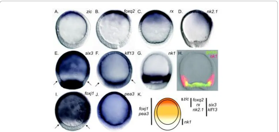
Sea star late blastulae have a morphologically distinct animal-vegetal (AV) axis that is first readily observed when elongation of cells at the vegetal pole results in a noticeable thickening of epithelium termed the vegetal plate[18]. During gastrulation, the vegetal plate invagi-nates to produce the mesoderm and endoderm of the larva, leaving the remaining animal epithelium as ciliated ectoderm [18]. At this stage, no obvious mor-phological differences in ectodermal cell type have been observed, but we nonetheless reveal here a remarkable complexity of regulatory states within the ectoderm (summarized in Figure 1).
Transcripts of sea star regulatory genes are localized throughout the animal ectoderm in overlapping con-centric domains along the AV axis. Some of these tran-scripts, such as those ofzic,foxq2, rx andnk2.1, are found only in the animal-most ectoderm (Figures 2A-2D). Of these genes,zicappears to be most closely loca-lized to the animal pole, while expression offoxq2andrx
overlaps withzic, yet extends further. Transcripts ofsix3
andklf13(Figures 2E and 2F) also are detected in the ani-mal-most ectoderm of blastulae; however, they show a still broader distribution. Although there is no clear cell morphology that demarcates the boundary between the animal ectoderm and the vegetal plate endomesoderm, we observe a ring ofnk1expression above the vegetal plate (Figure 2G) that partially overlaps with endoder-mally localizedgataetranscripts (Figure 2H). Thenk1
expression domain therefore likely marks the vegetal-most ectoderm of the blastula.foxj1andpea3(Figures 2I and 2J) are expressed throughout the ectoderm.
Taken together, the spatial expression of these regula-tory genes demonstrates that the ectoderm of the sea star blastula is patterned along the AV axis in at least five nested concentric domains (summarized in Figure 2K).
Regulatory gene expression within the ectoderm of the ciliary bands and animal pole domain
ciliary band that loops below it and around the aboral surface at the“back”of the embryo (Figure 3A) [7,18]. Transcripts of several genes that were distributed broadly throughout the ectoderm prior to gastrulation are later expressed within the ectoderm of the ciliary bands of the
larva following gastrulation (for example,foxj1andklf13
in Figures 3B-3E,pea3as summarized in Figure 1 and as previously reported forotxandhnf-6/onecutexpression [18,19]). At present, it is unclear if these patterns of expression reflect a migration of ectodermal cells to the
otx pax6
gbx hox1 hox3,4 hox7/8 hox10/13
six3 “foxg”
rx “nk2.1” foxj1
otx hnf-6
foxg
nk1, gbx zic, foxq2, rx, six3, pea3,
nk2.1, klf13, foxj1, foxd, lhx2 six3
klf13 foxq2
rx nk2.1
nk1 foxj1
pea3 otx* hnf-6*
zic
(a)
zic rx foxj1
pea3 otx* hnf-6*
klf13
pax6 foxg
nk1 gbx foxg foxd nk2.1 foxq2
six3 lhx2
(c)
(b)
(f)
(d)
(g)
Mouth
zic, foxq2, rx, six3, pea3, pax6
foxj1 pea3 otx* hnf-6*
klf13 foxg lhx2
nk1 gbx foxd nk2.1 lhx2
zic, foxq2, rx, nk2.1, klf13, foxj1
nk1 hnf-6 six3
foxg lhx2
lhx2
otx
zic, foxq2,
“foxg”, lhx2, “tbr” rx, six3, pax6, “nk2.1”,
“nk1” hox1 hox3 hox4 hox8 hox10-13 “pea3”
“hnf-6”
M R1 R2 R3 R4 R5 R6 R7 R8 F
gbx foxj1
(e)
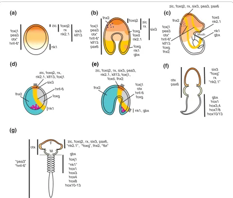
Figure 1Comparison of orthologous neuroectodermal gene expression domains among the deuterostomes.(A-E)Indirectly developing echinoderms.(F)Directly developing hemichordate.(G)Generalized vertebrate. Sea stars (Figures 1A-1C) and sea urchins (Figures 1D and 1E) are viewed laterally; animal pole is up and oral side is right. Figures 1F and 1G are dorsal views; anterior is up. Genes are listed beside their cognate expression domains. Vertical bars in Figures 1A, 1B, 1F and 1G approximate domain boundaries. The orange to yellow gradient in Figures 1A, 1F and 1G reflects a general conservation of anterior (animal)-most axial patterning among the three phyla.(A)Nested, concentric expression domains pattern the animal-vegetal (AV) axis of blastulae; asterisks denote previously reported expression [18,19].(B)Concentric domains ofzic,
sites of the future ciliary bands or if there is another pat-terning mechanism that restricts the earlier broad expres-sion. Other transcripts are first detected at this stage within the ectoderm on the oral side of the gastrula and then later within the ciliary bands (for example, tran-scripts offoxg,foxdandgbx; Figures 3F and 3G, 3H and 3I, and 3J and 3K, respectively). A two-probe whole mountin situhybridization (WMISH) offoxgandlhx2, a gene localized to the aboral ectoderm, further highlights the oral side restriction offoxgtranscripts in the gastrula (Figure 3).
The expression patterns at this later stage also show that the regulatory state of the early larval ciliary bands is heterogeneous, for example, nk2.1 and foxd are expressed in part of the preoral ciliary band directly above the mouth (Figures 3G and 3I, respectively), while
gbxandnk1 are localized to part of the postoral ciliary band below the mouth (Figures 3K and 3L, respectively). Therefore, while the regulatory state of the ciliary band ectoderm can be defined by a suite of transcription fac-tors (that is,klf13,foxj1, pea3,foxg, otxand hnf-6/one-cut), they are further subdivided into pre- and postoral regions on the basis of the localization of foxd, nk2.1,
nk1andgbx.
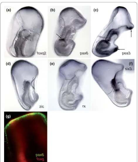
Other transcripts that we detected within the derm of the blastula remained within the animal ecto-derm as gastrulation proceeded in what we define here as the animal pole domain. Unlike sea urchins, the sea star,P. miniata, does not appear to have a morphologi-cally distinct animal pole domain at this stage. Tran-scripts of foxq2, pax6andpea3 (Figures 4A-4C) tightly localize to the animal pole ectoderm, although their vegetal boundaries do not exactly coincide. Transcripts ofzic, rxand six3are expressed within the animal pole domain as well, but even more vegetally throughout the animal ectoderm (Figures 4D-4F). The vegetal boundary of the animal pole domain therefore is not clearly defined by regulatory gene expression. The preoral and postoral ciliary bands run through the sea star animal pole domain as demonstrated by a two-probe fluores-cencein situhybridization (FISH) using the ciliary band marker, foxg, and the animal pole domain gene, pax6
(Figure 4G). Thus, despite its lack of morphological regionalization, the animal pole has a distinct regulatory state, as defined by foxq2, pax6,pea3, zic, rxand six3
expression, suggesting that it is a unique territory within the sea star. It is not yet clear whether these genes remain expressed in all cells of the sea star animal pole
Figure 2Nested concentric expression domains pattern the axial ectoderm of sea star,P. miniata, blastulae. Embryos are oriented with the animal pole up.(A-G)Whole mountin situhybridization (WMISH).(A)zic,(B)foxq2,(C)rx, and(D)nk2.1expression is restricted to the animal-most ectoderm. Transcripts of(E)six3and(F)klf13are detected in the ectoderm and in the vegetal plate endomesoderm. Arrows in(E)
Pre-oral CB
Post-oral CB M
A
M
A
(a)
foxj1
*
foxj1klf13
(b)
(d)
Entire CB
(c)
nk2.1 foxg
Transverse Pre-oral CB
Transverse Post-oral CB
gbx
(j)
nk1
(g)
foxj1
(h)
foxd
(f)
foxg
(l) (m)
lhx2 foxg
klf13
(e)
foxd
(i)
gbx
(k)
Figure 3Heterogeneous regulatory patterning of the larval ciliary bands as visualized byWMISH.(A)Schematic describes the position of the two larval ciliary bands (red) from oral (left) and lateral (right) views. A, anus; CB, ciliary band; M, mouth.(B-F)WMISH. Expression of(Band
C)foxj1and(DandE)klf13is initially broad throughout(BandD)the ectoderm of gastrulae, then later is restricted to(CandE)the larval ciliary bands. Arrows in Figure 3B show the vegetal limits offoxj1expression. Arrows in Figure 3D point to a clearing above the vegetal pole where transcripts ofklf13were detected.klf13transcripts are additionally detected in an ectodermal territory near the mouth (arrows in Figure 3E).(F)foxgis first expressed within two ectodermal domains on the oral side of gastrulae.(G)FISHofnk2.1(green) and ciliary band markerfoxg
domain during later stages of larval development or if expression becomes refined to only subsets of cells within this domain.
Comparisons of ectodermal patterning between sea urchin and sea star embryos
At first inspection, the expression patterns of many genes appear markedly different in the earlier blastula stages of sea urchin and sea star embryos. The later restrictions within the animal pole or ciliary bands are, with some exceptions, more similar (Figure 1). We sug-gest that the sea urchin embryo may simply undergo a relatively more rapid specification of these territories, with an associated loss of intermediate domains that we observe in the sea star. Indeed, a careful examination of expression patterns in sea urchin has recently shown that the apical plate in sea urchin consists of at least two regulatory domains: an inner animal pole domain
flanked by a ring of six3 expression [20]. These two domains in the sea urchin hatched blastula may there-fore represent a more apically compressed version of the nested, concentric regulatory domains found in the sea star blastula.
Some of the patterning differences between sea urchin and sea star ectoderm also seem to account for the dif-ferences observed in the localization of the pan-neuronal marker, synaptotagmin-B [21]. For example, similar to the patterns of gene expression that we describe here, synaptotagmin-B is detected broadly throughout the ectoderm of the sea star gastrula, but in the larva it is found primarily in neurons associated with the ciliary bands and animal pole [9]. In the sea urchin, however, synaptotagmin-B is already localized to the animal pole domain and the presumptive ciliary band by the gastrula stage [9].
Although expression of many of the genes within the ciliary bands of the sea star appears conserved in the sea urchin,nk2.1andfoxdshow clear differences in expres-sion that may be associated with the evolutionary transi-tion from a double looping of the ciliary band around the body of the sea star bipinnaria and hemichordate tornaria to a single looping of the ciliary band observed in sea urchins. This single ciliary band in the sea urchin develops at the junction between oral and aboral ecto-derm. nk2.1and foxd are expressed in part of the pre-oral ciliary band of the pre-oral hood of the sea star, while sea urchin orthologs of these are found in the animal plate ectoderm (compare Figures 1C-1E). Interestingly, both of these genes in sea urchin appear enriched on the oral side of the embryos [22,23]. Thus, we speculate that the preoral ciliary band may have been compressed into the oral-side animal plate territory in sea urchins and that this region within the sea urchin may therefore constitute a different territory than the remaining ani-mal plate.
Conservation of anterior (animal)-most regulatory patterning with other deuterostomes
Comparisons of the regulatory gene expression patterns that we observed in these indirectly developing sea star embryos with those known in directly developing bilater-ians illuminate additional surprising patterns of conserva-tion. We observe a general mapping of gene expression patterns along body axes (compare Figures 1A-1C with Figures 1F-1G). For instance, in the sea star,foxq2,rx,
pax6andsix3orthologs are apically expressed within the ectoderm.foxq2 expression in the amphioxus, a basal chordate, is restricted to the anterior-most end of the embryo [24]. Orthologs ofrxandpax6are expressed in the anterior-most neuroectoderm in the hemichordate
Saccoglossus[13], and they also pattern the anteriorly localized eye primordium in vertebrates [25,26]. The foxg
pax6
zic rx
six3 pax6
foxq2
(d) (e)
(g)
(b) (a)
pea3
(f) (c)
Figure 4Gene expression molecularly defines the animal pole domain in the sea star. Embryos are shown laterally, with the animal pole up and oral side to the right.(A-F)WMISH. Expression of(A)foxq2,(B)pax6,(C)pea3,(D)zic,(E)rxand(F)six3within the apical-most ectoderm defines the animal pole domain within late gastrulae (Figures 4A, 4D and 4F) and early larvae (Figures 4B, 4C and 4E). The vegetal limits of this domain are variable (see dotted lines in Figures 4E and 4F). Transcripts ofpea3additionally localize within the ectoderm of the larval ciliary bands (arrows in Figure 4C).
pax6expression in mesodermally derived coelom (arrow in Figure 4B).(G)FISHdemonstrates that the ciliary bands, as marked byfoxg
Drosophila rxortholog is required for brain development [27]. Orthologs ofsix3andotxare expressed in anterior neuroectoderm in members of all three deuterostome phyla [13,28,29]. The most vegetal ectoderm in sea stars is characterized by the presence ofnk1 andgbx tran-scripts. In vertebrates, agbxortholog establishes the mid-brain-hindbrain boundary [30]. The zebrafish ortholog of
nk1, sax2, is expressed within the midbrain-hindbrain boundary as well, although its expression is not exclusive to this territory [31]. Expression ofnk1andgbx in sea stars, and possibly sea urchins, marks the vegetal (poster-ior)-most ectoderm.
There is some evidence of additional conservation between the DV and oral-aboral axes as well. The mouse ortholog ofnk2.1(nkx2.1) is involved in the formation of motor neurons in the ventral telencephalon [32]. Sacco-glossus nk2.1orthologs also show a ventral bias in expres-sion [13]. Furthermore, foxg plays a role in ventral forebrain development, whilelhx2specifies dorsal telence-phalic fates [32]. We similarly show that expression of sea star orthologs offoxgandnk2.1is restricted to the oral (ventral) ectoderm, whilelhx2orthologs are expressed within the aboral (dorsal) ectoderm (Figure 1B).
While in these comparisons we do not intend to con-vey a tight homology in gene expression patterns across deuterostome phyla, we predict that similarities in the overall patterning are an ancestral innovation and per-haps evidence of maintenance of some elements of a developmental gene regulatory network (GRN) inherited from a common ancestor. Conservation, however, is not maintained for orthologs of genes expressed within regions posterior to the midbrain-hindbrain boundary in chordates and hemichordates asnk1 marks the vegetal-most ectoderm. Also, the overlapping expression ofhox
gene orthologs needed to pattern the posterior of many embryos are found only later in echinoderm develop-ment within the mesoderm of the rudidevelop-ment [33].
Separation of“retinal”from“anterior neural”regulatory patterning
Vertebrate orthologs of transcription factors such as
pax6,six3andrxplay known roles in pattering and spe-cifying anterior vertebrate sensory systems, most notably the eyes [25,26,34]. Furthermore, orthologs ofpax6, the
six gene family members andeya operate in a similar gene network for retinal determination in both verte-brates andDrosophila(as reviewed in [35]).
Having established that orthologs of many regulatory genes involved in anterior neural specification are also expressed within the anterior ectoderm of sea star embryos, we sought to determine if orthologs of tran-scription factors involved within the retinal determina-tion network are also expressed within echinoderm embryos.
We have already shown that the sea starpax6ortholog is expressed within the animal pole domain (Figure 4B), although it is not expressed within the ectoderm of the sea urchin embryo (Figure 5). Transcripts of bothpax6
andeyain both sea urchins and sea stars, however, are detected in the mesoderm of midgastrulae and then more prominently in one mesodermally derived coelom in late gastrulae (Figures 5A-5D and Figures 5G-5L). While we were unable to obtain asix1.2ortholog from the sea star, this gene is expressed also within the meso-dermal coelom in the sea urchin (Figures 5E and 5F).
In the sea urchin, expression of two members of the light-sensing rhodopsin family of G-coupled protein receptors,opsin1andopsin4, has been shown as early as 1 week [36]. We were unable to obtain sea star opsin sequences; however, we confirmed the expression of
opsin1andopsin41-week-old sea urchin larvae (Figures 5M and 5N). The morphology of the late larval sea urchin embryos makes it difficult to decipher the precise location of these transcripts within the embryo. We therefore sought to determine ifopsins collocalize with
eya, which we show is expressed likely within one or both coeloms (depending on developmental timing) in 1-week-old larvae (Figure 5O). Using a two-probeFISH, we observe that transcripts ofopsin1colocalized with those ofeyain 1-week-old sea urchin larvae (Figures 5P-5R). Expression of retinal determination orthologs within the mesoderm of gastrulae and larvae allow for the possi-bility that these genes operate within a common GRN.
The tightly coupled GRNs for anterior neural and visual sensory structures that are found in vertebrates and also in invertebrates, such asDrosophila, therefore are spatially separated in echinoderms. The presence of gene transcripts of pax6,rxand six3, but not, for exam-ple, eya, within the animal ectoderm of sea star bipin-naria larva may indicate a partial retention of an ancestral retinal determination network that once oper-ated within this embryonic territory. This might also explain the absence of apically localized rhabdomeric eyespots, which are characteristic of the indirectly devel-oping tornaria larvae of some hemichordates but were likely lost in the echinoderm lineage [16].
Conclusions
Inferring ancestral states
time. Thus, echinoderm embryos may have conserved sets of genetic regulatory relationships for“head/anterior brain” within the ectoderm of the early blastula and others for“retinal determination” within the mesoder-mal coelom of the gastrula.
Much of the difficulty in inferring the ancestral state of the deuterostomes and the mysteries of the origin of the phylum to which we belong arises from the com-plex life histories found within extant lineages [37]. Given the conservation of complex sensory and AP patterning between protostomes such as Drosophila
and vertebrates, the parsimonious explanation is that ancestral developmental regulatory interactions, per-haps even entire GRN subcircuits, have been uncoupled along the lineage of echinoderms, possibly coincident with a simplification in early development. However, until a greater breath of taxa have resolved GRNs, we cannot know the flexibility with which mod-ular subcircuits can be deployed during evolution of alternative body plans or if intercalation of GRN sub-circuits occurs independently or coincident with an increase in complexity.
PmEya
SpSix1.2
SpPax6
SpEya
(a)
(b)
(c)
(d)
(e)
(f)
(g)
(h)
(i)
(j)
(k)
(l)
(q)
SpEya
SpOpsin1 SpOpsin1SpEya
(p)
(r)
SpOpsin1
(m)
(n)
SpOpsin4
PmPax6
(o)
SpSm50
SpEya
Figure 5Retinal determination orthologs are expressed within sea urchin, (Strongylocentrotus purpuratus, Sp), and sea star mesoderm.
Methods
Sea star and sea urchin embryo culture and characterization of gene expression
P. miniataembryos, previously namedAsterina miniata, and Strongylocentrotus purpuratus embryos were cul-tured as described previously [18,38]. Partial gene sequences were obtained via screening a 3-day-old (late-gastrula stage)P. miniata arrayed cDNA library using
S. purpuratussequence-specific probes and low strin-gency conditions as previously described [39]. Whole mountin situ hybridization (WMISH) was performed as described previously [18].
Two-colorFISH
WMISH was performed essentially as described by Hin-manet al. [18], with modifications to detect riboprobes using fluorescence as described by Denkers et al. [40]. In brief, both digoxygenin (DIG) and 2,4-dinitrophenol (DNP) labeled riboprobes were used. Hybridized probes were detected using anti-DIG antibody (1:2,000; Roche: Indianapolis, IN, USA) and anti-DNP antibody (1:1,000; PerkinElmer: Chicago, IL, USA), both conjugated to horseradish peroxidase, and the Tyramide Signal Ampli-fication (TSA) Plus Fluorescence Systems Kit (PerkinEl-mer). A CyIII- or fluorescein-labeled tyramide was deposited near the in situriboprobe in a reaction cata-lyzed by horseradish peroxidase, allowing for fluores-cence detection of DIG- and DNP-labeled riboprobes.
Additional material
Additional file 1: Table 1. List of sea star,P. miniata, orthologs and orthology of gene sequences.
Acknowledgements
The authors thank Dr Daniel Brown, Brenna McCauley, Alys Cheatle and three anonymous reviewers for helpful comments on the manuscript. This work was partially supported by National Science Foundation grant 0844948 to VFH, and CKJ and MLM were funded by a Howard Hughes Medical Institute Undergraduate Education Research Grant.
Authors’contributions
VFH and KAY conceived of the study, participated in its design and drafted the manuscript. KAY cloned sea star orthologs and performedWMISHand
FISH. CKJ cloned sea urchin orthologs and performed WMISH. MLM cloned sea star orthologs and performedWMISHandFISH. All authors read and approved the final manuscript.
Competing interests
The authors declare that they have no competing interests.
Received: 5 August 2010 Accepted: 30 November 2010 Published: 30 November 2010
References
1. Bromham LD, Degnan BM:Hemichordates and deuterostome evolution: robust molecular phylogenetic support for a hemichordate + echinoderm clade.Evol Dev1999,1:166-171.
2. Castresana J, Feldmaier-Fuchs G, Yokobori S, Satoh N, Paabo S:The mitochondrial genome of the hemichordateBalanoglossus carnosusand the evolution of deuterostome mitochondria.Genetics1998,
150:1115-1123.
3. Cameron CB, Garey JR, Swalla BJ:Evolution of the chordate body plan: new insights from phylogenetic analyses of deuterostome phyla.Proc Natl Acad Sci USA2000,97:4469-4474.
4. Turbeville JM, Schulz JR, Raff RA:Deuterostome phylogeny and the sister group of the chordates: evidence from molecules and morphology.Mol Biol Evol1994,11:648-655.
5. Bourlat SJ, Juliusdottir T, Lowe CJ, Freeman R, Aronowicz J, Kirschner M, Lander ES, Thorndyke M, Nakano H, Kohn AB, Heyland A, Moroz LL, Copley RR, Telford MJ:Deuterostome phylogeny reveals monophyletic chordates and the new phylum Xenoturbellida.Nature2006,444:85-88. 6. Byrne M, Nakajima Y, Chee FC, Burke RD:Apical organs in echinoderm
larvae: insights into larval evolution in the Ambulacraria.Evol Dev2007, 9:432-445.
7. Hyman LH:The Invertebrates: Echinodermata.New York: McGraw-Hill; 1955.
8. Hyman LH:The Invertebrates: Smaller Coelomate Groups.New York: McGraw-Hill; 1959.
9. Nakajima Y, Kaneko H, Murray G, Burke RD:Divergent patterns of neural development in larval echinoids and asteroids.Evol Dev2004,6:95-104. 10. Garstang W:Preliminary note on a new theory of the phylogeny of the
Chordata.Zool Anz1894,17:122-125.
11. Lacalli TC:Apical organs, epithelial domains, and the origin of the chordate central nervous system.Am Zool1994,34:533-541.
12. Nielsen C:Origin of the chordate central nervous system - and the origin of chordates.Dev Genes Evol1999,209:198-205.
13. Lowe CJ, Wu M, Salic A, Evans L, Lander E, Stange-Thomann N, Gruber CE, Gerhart J, Kirschner M:Anteroposterior patterning in hemichordates and the origins of the chordate nervous system.Cell2003,113:853-865. 14. Lowe CJ, Terasaki M, Wu M, Freeman RM Jr, Runft L, Kwan K, Haigo S,
Aronowicz J, Lander E, Gruber C, Smith M, Kirschner M, Gerhart J: Dorsoventral patterning in hemichordates: insights into early chordate evolution.PLoS Biol2006,4:e291.
15. Lacalli TC:Protochordate body plan and the evolutionary role of larvae: old controversies resolved?Can J Zool2005,83:216-224.
16. Brown FD, Prendergast A, Swalla BJ:Man is but a worm: chordate origins.
Genesis2008,46:605-613.
17. Arendt D, Denes AS, Jekely G, Tessmar-Raible K:The evolution of nervous system centralization.Philos Trans R Soc Lond B Biol Sci2008,
363:1523-1528.
18. Hinman VF, Nguyen AT, Davidson EH:Expression and function of a starfish Otx ortholog, AmOtx: a conserved role for Otx proteins in endoderm development that predates divergence of the eleutherozoa.
Mech Dev2003,120:1165-1176.
19. Otim O, Hinman VF, Davidson EH:Expression of AmHNF6, a sea star orthologue of a transcription factor with multiple distinct roles in sea urchin development.Gene Expr Patterns2005,5:381-386.
20. Wei Z, Yaguchi J, Yaguchi S, Angerer RC, Angerer LM:The sea urchin animal pole domain is a Six3-dependent neurogenic patterning center.
Development2009,136:1179-1189.
21. Burke RD, Osborne L, Wang D, Murabe N, Yaguchi S, Nakajima Y: Neuron-specific expression of a synaptatogmin gene in the sea urchin Strongylocentrotus purpuratus.J Comp Neurol2006,496:244-251. 22. Tu Q, Brown CT, Davidson EH, Oliveri P:Sea urchin Forkhead gene family:
phylogeny and embryonic expression.Dev Biol2006,300:49-62. 23. Takacs CM, Amore G, Oliveri P, Poustka AJ, Wang D, Burke RD, Peterson KJ:
Expression of an NK2 homeodomain gene in the apical ectoderm defines a new territory in the early sea urchin embryo.Dev Biol2004, 269:152-164.
24. Yu JK, Holland ND, Holland LZ:AmphiFoxQ2, a novel winged helix/ forkhead gene, exclusively marks the anterior end of the amphioxus embryo.Dev Genes Evol2003,213:102-105.
25. Bailey TJ, El-Hodiri H, Zhang L, Shah R, Mathers PH, Jamrich M:Regulation of vertebrate eye development by Rx genes.Int J Dev Biol2004, 48:761-770.
27. Davis RJ, Tavsanli BC, Dittrich C, Walldorf U, Mardon G:Drosophilaretinal homeobox (drx) is not required for establishment of the visual system, but is required for brain and clypeus development.Dev Biol2003, 259:272-287.
28. Simeone A, Acampora D, Mallamaci A, Stornaiuolo A, D’Apice MR, Nigro V, Boncinelli E:A vertebrate gene related to orthodenticle contains a homeodomain of the bicoid class and demarcates anterior neuroectoderm in the gastrulating mouse embryo.EMBO J1993, 12:2735-2747.
29. Oliver G, Mailhos A, Wehr R, Copeland NG, Jenkins NA, Gruss P:Six3, a murine homologue of the sine oculis gene, demarcates the most anterior border of the developing neural plate and is expressed during eye development.Development1995,121:4045-4055.
30. Rhinn M, Brand M:The midbrain-hindbrain boundary organizer.Curr Opin Neurobiol2001,11:34-42.
31. Bae YK, Shimizu T, Muraoka O, Yabe T, Hirata T, Nojima H, Hirano T, Hibi M: Expression ofsax1/nkx1.2andsax2/nkx1.1in zebrafish.Gene Expr Patterns
2004,4:481-486.
32. Hebert JM, Fishell G:The genetics of early telencephalon patterning: some assembly required.Nat Rev Neurosci2008,9:678-685. 33. Arenas-Mena C, Cameron AR, Davidson EH:Spatial expression of Hox
cluster genes in the ontogeny of a sea urchin.Development2000, 127:4631-4643.
34. Carl M, Loosli F, Wittbrodt J:Six3 inactivation reveals its essential role for the formation and patterning of the vertebrate eye.Development2002, 129:4057-4063.
35. Wawersik S, Maas RL:Vertebrate eye development as modeled in Drosophila.Hum Mol Genet2000,9:917-925.
36. Raible F, Tessmar-Raible K, Arboleda E, Kaller T, Bork P, Arendt D, Arnone MI: Opsins and clusters of sensory G-protein-coupled receptors in the sea urchin genome.Dev Biol2006,300:461-475.
37. Swalla BJ:Building divergent body plans with similar genetic pathways.
Heredity2006,97:235-243.
38. Ettensohn CA, Wessel GM, Wray GA:Development of Sea Urchins, Ascidians, and Other Invertebrate Deuterostomes: Experimental Approaches.San Diego: Elsevier Academic Press; 2004.
39. Hinman VF, Nguyen AT, Cameron RA, Davidson EH:Developmental gene regulatory network architecture across 500 million years of echinoderm evolution.Proc Natl Acad Sci USA2003,100:13356-13361.
40. Denkers N, Garcia-Villalba P, Rodesch CK, Nielson KR, Mauch TJ:FISHing for chick genes: triple-label whole-mount fluorescence in situ hybridization detects simultaneous and overlapping gene expression in avian embryos.Dev Dyn2004,229:651-657.
41. Materna SC, Howard-Ashby M, Gray RF, Davidson EH:The C2H2zinc finger
genes ofStrongylocentrotus purpuratusand their expression in embryonic development.Dev Biol2006,300:108-120.
42. Burke RD, Angerer LM, Elphick MR, Humphrey GW, Yaguchi S, Kiyama T, Liang S, Mu X, Agca C, Klein WH, Brandhorst BP, Rowe M, Wilson K, Churcher AM, Taylor JS, Chen N, Murray G, Wang D, Mellott D, Olinski R, Hallbook F, Thorndyke MC:A genomic view of the sea urchin nervous system.Dev Biol2006,300:434-460.
43. Rizzo F, Fernandez-Serra M, Squarzoni P, Archimandritis A, Arnone MI: Identification and developmental expression of theetsgene family in the sea urchin (Strongylocentrotus purpuratus).Dev Biol2006,300:35-48. 44. Howard-Ashby M, Materna SC, Brown CT, Chen L, Cameron RA,
Davidson EH:Identification and characterization of homeobox transcription factors genes inStrongylocentrotus purpuratus, and their expression in embryonic development.Dev Biol2006,300:74-89. 45. Su YH, Li E, Geiss GK, Longabaugh WJ, Kramer A, Davidson EH:A
perturbation model of the gene regulatory network for oral and aboral ectoderm specification in the sea urchin embryo.Dev Biol2010, 329:410-21.
46. Otim O, Amore G, Minokawa T, McClay DR, Davidson EH:SpHnf6, a transcription factor that executes multiple functions in sea urchin embryogenesis.Dev Biol2004,273:226-243.
47. Poustka AJ, Kuhn A, Radosavljevic V, Wellenreuther R, Lehrach H, Panopoulou G:On the origin of the chordate central nervous system: expression ofonecutin the sea urchin embryo.Evol Dev2004,6:227-236. 48. Minokawa T, Rast JP, Arenas-Mena C, Franco CB, Davidson EH:Expression
patterns of four different regulatory genes that function during sea urchin development.Gene Expr Patterns2004,4:449-456.
49. Yuh CH, Brown CT, Livi CB, Rowen L, Clarke PJC, Davidson EH:Patchy interspecific sequence similarities efficiently identify positive cis-regulatory elements in the sea urchin.Dev Biol2002,246:148-161. 50. Aruga J:The role ofZicgenes in neural development.Mol Cell Neurosci
2004,26:205-221.
51. Munchberg SR, Ober EA, Steinbeisser H:Expression of the Ets transcription factors erm and pea3 in early zebrafish development.Mech Dev1999, 88:233-236.
52. Hong SK, Kim CH, Yoo KW, Kim HS, Kudoh T, Dawid IB, Huh TL:Isolation and expression of a novel neuron-specific onecut homeobox gene in zebrafish.Mech Dev2002,112:199-202.
53. Bulfone A, Smiga SM, Shimamura K, Peterson A, Puelles L, Rubenstein JL: T-brain-1: a homolog of Brachyury whose expression defines molecularly distinct domains within the cerebral cortex.Neuron1995,15:63-78. 54. Aamar E, Dawid IB:Isolation and expression analysis offoxj1andfoxj1.2
in zebrafish embryos.Int J Dev Biol2008,52:985-991.
55. Tümpel S, Wiedemann LM, Krumlauf R:Hoxgenes and segmentation of the vertebrate hindbrain.Curr Top Dev Biol2009,88:103-137.
doi:10.1186/1741-7007-8-143
Cite this article as:Yankuraet al.:Uncoupling of complex regulatory patterning during evolution of larval development in echinoderms.BMC Biology20108:143.
Submit your next manuscript to BioMed Central and take full advantage of:
• Convenient online submission
• Thorough peer review
• No space constraints or color figure charges
• Immediate publication on acceptance
• Inclusion in PubMed, CAS, Scopus and Google Scholar
• Research which is freely available for redistribution