Medical Journal of the Islamic Republic ofIran
THE CORRELATION BETWEEN SPERM
MORPHOLOGY AND MOTILITY IN FERTILE AND
INFERTILE MEN
ZAHRA SADIGlll GILANI, Ph.D.
AND
MOHAMMAD ALI SADIGHI
GILANI,* M.D.
From the Department of Anatomy, Iran University of Medical Sciences, Tehran, and the * Department of Urology, Shariati Hospital, Tehran University of Medical Sciences, Tehran, Islamic Republic of Iran.
ABSTRACT
The swim-up procedure was used to collect a sperm fraction with improved motility and normal morphology from fertile and infertile semen samples. The untreated and swim-up fractions were analyzed with the Hamilton-Thorn Motility Analyser. Comparisons were made to see the effect of this sperm selection technique on sperm motility and morphology. They were then examined by transmission electron microscopy and the various sperm morphologies were recorded. Fertile and infertile samples were compared for differences in these parameters.
There were no significant differences between fertile and infertile samples in the frequencies of various sperm morphologies in the untreated and swim-up fractions. There was a trend towards fertile samples having a higher motility of the untreated ejaculate, as well as more normal heads and tails and fewer morphological abnormalities than infertile samples.
Sperm motility was found to be affected by sperm morphology, with an increase in normal sperm morphology resulting in an increase in motility. Normal sperm tail morphology appears to be the most important factor in motility, since this was the only morphology to show a significant increase in the swim-up fractions in all semen samples. Head morphology is less important to motility, but it is thought to play a major role in fertilization of the egg.
Keywords: Morphology, motility, sperm.
MIIRI, Vol. II, No.4, 319-324,1998.
Volumell Number 4 Winter 1376 February 1998
INTRODUCTION
The World Health Organization
(1978)
analyzeddata
from more than
7000
infertile couples and proposed the following criteria for classifying a normal semen sample. The sperm concentration must be greater than or equal to20
million per mL, there should be no agglutination, at least30%
of the sperm must be morphologically normal and75%
of the sperm must be alive. The seminal fluid must be normal in appearance and viscosity, and at least
50%
of the sperm must be fully motile (WHO1992).
Most authors accept that a correlation exists between fertilizing ability and seminal parameters.9.11.13.26 There is good evidence that morphologically normal spermatozoa offer the best fertilizing potential,4.20 and morphologically abnormal spermatozoa are also functionally abnormal.4,7.16.17Correlation Between Sperm Morphology and Motility and Fertility
A* B
Donor
UT SUP UT
total cell no. 842 164 347
concen tration 175 34 60 (106/mL)
motile cell no. 471 109 36
concentration 98 23 6.2 (J06/mL)
% cells recovered 19.5
% motility 56 66 10
% difference in 10 motility between
UT and SUP
V AP (Il-m/s) 37 39 28
VSL (Il-mls) 33 32 27
mean STR (%) 83 75 88
mean ALH (Il-m) 3 4.9 1.8
* = fertile
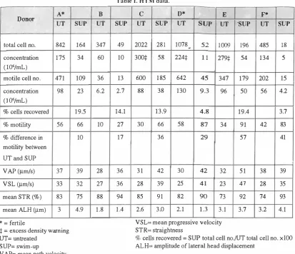
Table I. HTM data.
SUP 49 10 13 2.7 14.1 27 17 36 36 94 1.4
C D* E
UT SUP UT SUP UT
2022 281 1078 � 52 1009
300:j: 58 224:j: 11 279:j:
600 185 642 45 347
88 38 130 9.3 96
13.9 4.8
30 66 58 87 34
36 29
31 42 30 42 32
28 39 25 41 23
85 91 82 90 73
2.6 3.0 2.1 1.3 3.1
VSL= mean progressive velocity STR= straightness
F*
'SUP UT
196 485
54 134
179 202
50 56
19.4
91 42
57
51 38
47 28
92 74
3.7 3.2 SUP 18 5 15 4.2 3.7 83 41 I 39 35 93 4.1
:j: = excess density warning UT= untreated
SUP= swim-up
% cells recovered = SUP total cell no.jUT total cell no. xl00 ALH= amplitude of lateral head displacement
V AP= mean path velocity
The work of many in vitro fertilization (IVF) laboratories is therefore aimed at making use of procedures which isolate higher percentages of motile spermatozoa with normal morphology from fresh ejaculate. Of the many methods for semen preparation used in IVF, the swim-up procedure is the most commonly used for samples showing either normal or subnormal seminal parameters.2.12.23
There is much controversy over the importance of using motility or morphology in defining a sample as fertile or infertile. 19 The swim-up technique appeared to be successful in selecting a fraction of sperm with improved motility and normal morphology.
MATERIALS AND METHODS
Donor
Nonnal head
Double head
Abnormal/multiple vacuoles
Nuclear inclusion
Macrocephalic
Microcephalic
Granulated nucleus
Abnormal acrosome
Cytoplasmic droplet
Tahle n. Head morphologv.
A· D C
UT SUP UT SUP UT
25.6 44.9 7.2 24.6 11.5
0.9 0.0 0.0 0.5 1.0
15.2 13.4 2.9 11.8 15.0
1.4 3.7 6.2 4.4 9.5
2.8 2.3 1.0 0.5 0.1
0.0 4.2 0.5 0.0 0.1
16.6 4.2 17.7 16.3 15.0
33.6 19.9 42.6 32.0 32.5
3.7 7.2 22.0 9.8 15.5
D· SUP UT
14.6 3.2
0.0 0.0
19.0 26.9
4.4 3.7
0.0 0.5
0.0 0.0
6.8 7.8
41.5 54.3
13.8 3.8
SUP 3.9 0.5 20.0 8.8 0.0 0.0 17.6 46.3 2.9
The semen samples (A, B, C, D) were obtained from donors attending the infertility clinic, and semen samples (E,
F)
were obtained from donors not attending the clinic. The semen was collected by masturbation after a period of at least forty-eight hours of abstinence. Each sample was divided into two: half was left untreated and half was used(%) FrequenCies
ofthe various sperm
morphologies before and after swim-up in the four semen samples, when examined by transmission electron microscopy.*=fertile UT= untreated SUP=swim-up
z.
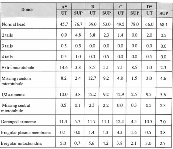
Sadighi Gilani and M.A. Sadighi GilaniTable III. Tail morphology.
Donor A* B C D*
UT SUP UT SUP UT SUP UT SUP
Nonnal head 45.7 76.7 39.0 53.0 49.5 78.0 66.0 68.1
2 tails 0.9 4.8 3.8 2.3 1.4 0.0 2.0 0.5
3 tails 0.5 0.5 0.0 0.0 0.0 0.0 0.0 0.0
4 tails 0.5 1.0 0.0 0.5 0.0 0.0 0.5 0.0
Extra microtubule 14.6 3.8 8.5 5.1 7.1 8.5 1.0 2.3
Missing random 8.2 2.4 12.7 9.2 4.8 1.5 3.0 4.6
microtubule
1/2 axoneme 10.0 3.8 12.2 9.2 12.9 2.5 9.5 5.6
Missing central 0.5 0.1 2.3 2.2 0.0 0.3 0.5 2.3
microtubule
Deranged axoneme 11.3 5.7 11.7 11.1 12.4 4.5 10.5 7.0
Irregular plasma membrane 0.1 0.0 1.4 1.3 4.3 1.6 0.5 0.8
Irregular mitochondria 5.0 0.7 5.6 4.2 3.8 2.1 3.0 2.7
(%) Frequencies of the various sperm morphologies before and after swim-up in the four semen samples, when examined by transmission electron microscopy.
*=fertile UT= untreated SUP=swim-up
in the swim-up procedure. Both untreated and swim-up
fractions were fIrst analyzed using the Hamilton-Thorn
Motility Analyser
(HTM)
and then prepared for transmission
electron microscopy (TEM). The analyzer measured the
various seminal parameters, and when the sperm
concentration was greater than 150 million sperm per mL
or when the machine showed an excess density warning,
1: 1 (v/v) dilution of the sample with human tubular fluid
(RTF)
was necessary. For swim-up, 1
mL
of liquefIed
semen from the fresh ejaculate was placed in a test tube and
1 mL of
RTFwas layered on top and it was left in an
incubator for 1 hour. A drop of this swim-up fraction was
analyzed in the HTM and then prepared for TEM in the
same way as the untreated ejaculate. MAR test was carried
out on the samples to detect the presence of antisperm
antibodies. Each sample was centrifuged and resuspended
in 2.5% glutaraIdehyde in 0.1 M cacodylate buffer (PH 7.2)
for 2.5 hours. After fIxation the samples were washed in
cacodylate buffer (PH 7.2) twice for 5 minutes and postftxed
in 1 % osmium tetroxide for 1 hour. Dehydration using
increasing concentrations of methanol was followed by
inftItration in Spurr resin. The blocks were thin sectioned
using a Reichert OMU3 ultramicrotome. Each grid was
stained by 2% uranyl acetate and 3% lead citrate. The
sections were examined by TEM (Philips
4(0).
200 head
and 200 tail morphologies were scored and examined
separately from the swim-up and untreated preparations
from donors. The microscopic field of view was moved up
and down in an ordered way
tocover the grid consistently
and avoid counting any sperm heads or tails twice.
RESULTS
HTMData
There was a dramatic decrease in the total cell number
between the untreated and swim-up fractions in all samples
analyzed, although no apparent difference was seen between
fertile and infertile samples (Table I). In all samples there
was an increase in percentage of motility following swim
up, but the amount varied. Normospermic Donor A showed
the smallest increase (10%). Donor B who was
asthenozoospermic showed an increase in motility of 17%
after swim-up, but he was still below WHO criteria for
normal motility even after sperm selection (27%). Donor
Cwas also asthenozoospermic, but showed a larger increase
of36% motility following swim-up. Normospermic Donor
Correlation Between Sperm Morphology and Motility and Fertility
Fig. 1. Longitudinal section through sperm (xlO,SOO). The acrosome (A) surrounds the nucleus (N). The principle segment (PS) is thicker than the equatorial segment (ES). The redundant nuclear membrane (RNM) can be seen in the neck region. The annulus
(AN)
marks the boundary between the mid piece (MP) and the principle piece (PP). The axoneme(AX)
is visible, as well as the ribs of the fibrous sheath (FS). Mitochondria (M) form a sheath around the mid piece of the flagella.D had the highest motility prior to swim-up
(58%)
but his
motility increase was less than in Donor C, E and
F.Asthenozoospermic Donor E showed the'largest increase
in percentage motility following swim-up
(�7%),
followed
by normospermic Donor F who showed the second largest
increase in motility of
41 %.
Overall there was no significant
difference in motility increase after swim-up between
fertile and infertile donors. Donors A, C, E and F showed
an increase in mean amplitude of lateral head displacement
(ALH) after swim-up, the largest increase of
1.9%
being
for normospermic Donor A. Donors B and D showed a
decrease in mean ALH after swim-up. Allexcept A showed
an increase in mean straightness (STR) after swim-up, and
the highest increase of
19%
was recorded for Donors E and
F. There was an increase in mean progressive velocity
(VSL) in all donors except A following sperm selection,
together with a universal increase in mean path velocity
(V
AP).
The largest increase in both of these was in Donor
E. There were no significant differences between the motion
characteristics after swim-up between clinically fertile and
infertile donors.
Electron micrographs
The acrosome (A) surrounds the anterior end of the
nucleus (N). The principle segment (PS) of the acrosome is
thicker than the equatorial segment (ES). The redundant
nuclear membrane (RNM) can be seen in the neck region.
The annulus (AN) marks the boundary between the mid
piece
(MP)
and the principle piece (PP), the axoneme (AX)
is visible, as well as the ribs of the fibrous sheath (FS). The
mitochondria (M) form a sheath around the mid piece of the
Fig. 2. Longitudinal section through head and neck region of sperm (x22,SOO). The plasma membrane (PM) surrounds the entire sperm cell. The acrosome is bounded by the inner acrosomal membrane (lAM) and the outer acrosomal membrane (OAM). The nuclear membrane lines the implantation fossa (IF) and the proximal centriole (PC) is embedded in the capitulum. The segmented columns (SC) which make up the capitulum attach to the anterior end o f the nine nuter cnarse fibers (CF).
A
H
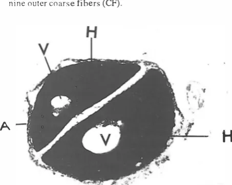
Fig. 3. Double
heaueu
spl!rmlxIS,UUU). rhl! l!lllargl:Li aUUSUl11c(A) and large vacuoles (V) deform the heads (H). Vacuoles can contain cytoplasmic organelles, lipid droplets or arrays of membranes.
flagella (Fig. O. The acrosome is bounded by the inner
acrosomal membrane (lAM) and the outer acrosomal
membrane (OAM). Between the nuclear membrane and the
inner acrosomal membrane is the subacrosomal space. The
plasma membrane (PM) surrounds the entire sperm. In the
neck region, the nuclear membrane lines the implantation
fossa
(IF)
and the proximal centriole (PC) is embedded in
the capitulum. The segmented columns (SC) which make
up the capitulum attach to the anterior end of the nine outer
coarse fibers (CF) (Fig.
2).
In double heads, the acrosome
(A) is partially enlarged and both heads (H) share the
plasma membrane. Normal sperm can contain small
Z.
Sadighi Gilani and M.A. Sadighi GilaniFig. 4. Longitudinal section through double-tailed sperm (x45 ,000). The double tail with ribs of fibrous sheath, microtubule doublets (D) and central microtubules (CM) are bounded by the same plasma membrane (PM).
eM
D-�
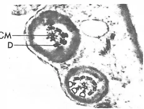
FIg. S. Transverse section through principle piece of sperm tail with half an axoneme and sperm tail with missing microtubule
doublets (x67,500). Deletion involves microtubule doublets (D), but the central microtubule (CM) is not included.
vacuoles, but in some cases these vacuoles
(V)
may be so large that they deform the head or reduce the amount of nuclear chromatin (Fig.3).
Sperm were scored with2, 3
and4
tails, and axonemal abnormalities were the most commontail
abnormality. The double tail with ribs of fibrous sheath, microtubule doublets (D) and central microtubules (CM) are bounded by the same plasma membrane (PM) (Fig.4).
Deletions involved the absence of one or more microtubule doublets from random position in the axoneme. Deletion of the central microtubules involved either the absence of both or one. In some flagella, exactly half of the axoneme is missing, although the central pair of microtubules (CM) is not included in this description (Fig.5).
ComparativeTEM
data about head and tail morphology are shown in Tables II and III. AlthoughHTM
data for donorsE
andF
are included, there is no comparativeTEM
data for them since too few cells were recovered in swim-up fractions forTEM
study.DISCUSSION
Donor A has shown 'the smallest percentage increase in motility after swim-up because his spenn were initially of a good motility and exceeded the WHO criteria for a fertile sample. Donor C's motility increase of
36%
transformedthis semen sample into a fertile one, with an improved motility of
66%.
The motility increase from58
to87%
in Donor D was surprising because he had a high level of antisperm antibodies detected in the fresh ejaculate. Antisperm antibodies are thought to cause a decrease in spenn numbers and motility.3,22.25 However DonorD'shigh percentage of motility after swim-up could be because antisperm antibodies were free in the semen.14,2' Donor D was known to be only IgG positive which may explain why his sperm remained highly motile. Donor A showed the largest increase in normal heads after swim-up which was expected since this semen sample had the most normal parameters. However, the very small increase ill nonnal head morphology seen in Donor C, together with the large increase in motility after swim-up, throws some doubt on the importance of normal head morphology to motility.8 The high frequency of abn6rmal acrosomes in both the untreated and swim-up fractions of Donor D was perhaps related to the presence of antisperm antibodies. Also, since Donor C was the only sample to show an increase in abnormal acrosomes after swim-up despite a high increase in percentage of motility, these results for C and D suggest either that abnonnal acrosomes do not affect motility or that they are artefacts of fixing."Surprisingly, Donor D had the highest percentage of nuclear inclusions and granulated nuclei after swim-up as well as a high percentage of motility. Therefore it appears that these head abnormalities do not affect motility. The fact that the majority of samples showed a decrease in deletions of microtubules reflects the deleterious effect of this abnormality on sperm motility. Derangement of the normal axonemal topography is thought to have an adverse effect on sperm motility as the tail cannot perfonn the sliding mechanism needed for propagation of flagellar waves. '5,27
According to WHO criteria, Donor A and Donor D can be classified as fertile, and Donor
B
and Donor C are both asthenozoospermic. Normal head morphology does not correlate with motility because DonorB
had a higher frequency of normal heads after swim-up than C and D, yet his motility after swim-up was less than half that ofC's and a third of thatofD's. The swim-up technique appeared to be successful in selecting a fraction of sperm with improved motility and normal morphology, although swim-up does not remove all the abnormal morphologies in an ejaculate, neither does in vivo selection in the female genital tract.' Thus swim-up is a good emulation of this sperm selection process. The main differences between aCorrelation Between Sperm Morphology and Motility and Fertility
clinically fertile and infertile sample using WHO criteria are that a fertile sample has both a higher initial motility of untreated ejaculate and a lower percentage of ultrastructurally abnormal morphology, especially axonemal abnormalities, than an infertile sample. As expected, motility was seen to be affected by sperm morphology with an increase in normal morphology resulting in an increase in motility.6 All samples show an increase in motility following swim-up which correlates with an increase in normal tail morphology and a decrease in the number of tail abnormalities. However tail morphology appears to be most important for sperm motility and the only significant difference was an increase in the number of normal tails after swim-up, while head morphology does not correlate with motility but plays a major role in fertilization of the egg.18 The increase in normal tail, decrease in missing microtubules and decrease in deranged axonemes in all samples after swim-up is most likely to be responsible for increased motility after swim-up. This indicates a positive correlation between motility and normal tail morphology.
REFERENCES
1. Ahlgren M: Sperm transport and survival in the human fallopian tube. Gynec Invest 6: 206-214, 1975.
2. Aitken RJ: Assessment of sperm function for IVF. Hum Reprod 3: 89-95, 1988.
3. Barret CLR, Havelock LM, Harrison PE: Antisperm antibodies are more prevalent in men with low sperm motility. Int J Androl 12: 110-116, 1989.
4. Bartoov B, Eltes F, Weissenberg R: Morphological characterization of abnormal human spermatozoa using transmission electron microscopy. Arch Androl 5: 305-322, 1980.
5. Bartoov B, Eltes F, Langsam J: Ultrastructural studies in morphological assessment of human spermatozoa. Int J Androl 5: 81-96, 1982.
6. Chan S, Wang C, Chan S: Predictive value of sperm
morphology and movement characteristics in the outcome
of in vitro fertilization of human oocyts. J In Vitro Fertil
Embryo Transfer 6: 142-148, 1989.
7. Fukuda M, Morales P, Overstreet J: Acrosomal function of human spermatozoa with normal and abnormal head morphology. Gamete Res 24: 59-65,1989.
8. Katz D F, Diel L, Overstreet JW: Differences in the movement of morphologically normal and abnormal human seminal spermatozoa. BioI Reprod 26: 566-570, 1982.
9. Kruger TF, Menkue1d R, Stander FS: Sperm morphology features as a prognostic factor in in vitro fertilization. Fertil Steril 46: 1118-1123, 1986.
10. Liu DY, Duplessis YP, Nayudu PL: The use of in vitro
fertilization to evaluate putative tests of human sperm function. Fertil Steril 49: 272-277, 1988.
11. Liu DY, Baker HW: Relationship between human sperm
acrosine, acr o some, morphology and fertilization in vitro.
Human Reprod 5: 298-303, 1990.
12. Lopata A, PatuHo MJ, Chang A: A method for collection of motile spermatozoa from human semen. Fetil Steri127: 677-684, 1976.
13. Mahadevan MM, Trounson N: The influence of seminal characteristic o n the success rate of human in vitro
fertilization. Fertil Steril 42: 400-405, 1984.
14. Matson PL, Junk SM, Spittle JW: Effect of antispermatozoal antibodies in seminal plasma upon spermatozoal function. Int J Androl 11: 101-106,1988.
15. McClure RD, Brawer J, Robaire B: Ultrastructure ofirnmotile spermatozoa in infertile males: a spectrum of structural defects. Fertil Steril 40: 395-399, 1983.
16. McGowam MP, Baker HWG, Kovacs G: Selection of h igh fertility donors for artifical insemination programmes. Clin Reprod Fertil 2: 269-274, 1983.
17. Mortimer D, Leslie E, Kelly R: Morphological selection of human spermatozoa "in vivo" and "in vitro". J Reprod Fertil 64: 391-399, 1982.
18. Oehninger S, Acosta R, Morshedi M: Relationship between morphology a n d motion characteristics of human spermatozoa in semen and in the swim-up sperm fractions. Int J Androl ll: 446-452, 1990.
19. Parinaud J, Vietez G, Montaffian H: Relevance of acrosome function in the evaluation of semen fertilizing ability. Fertil Steril 63: 598-603, 1995.
20. Pousette A, AkerIofE, Rosenborg L: Increase in progressive motility and improved morphology of human spermatozoa following their migration through percoll gradients. Int J Androl 9: 1-13,1986.
21. Rajah SV, P arslow JM, Howell RJ: The effect of autoantibodies to sperm in infertile men on in vitro fertilization treatment. J Reprod Fertil 10: 123-127, 1992. 22. Steen Y, Forssman L, Lonnerstedt E: Anti-sperm IgA
autoantibodies against the equatorial segment of the human spermatozoon are associated with i mpaired sperm penetration. lnt J Ferti! Menopausal Stud 2: 109-119,1994. 23. Van Der Yen HH, Jeyendran RS, A-Hasani S: Glass wool column filtration of human semen: relation to swim-up procedure and outcome ofIVF. Hum Reprod 3: 85-88, 1988. 24. WHO: Lab manual for the exhibition of human sperm and sperm -cervical mucus interactions. 3rd Edition, Public Press Syndicate for the University of Cambidge, 1992.
25. WolfJ, De Almeida, Ducot B: High level of sperm-associated antibodies impair human sperm oolemma interaction after sub-zonal insemination. Fertil Steril 63: 584-590, 1995.
26. Y ovich JL, Stander JD: The limitation of in vitro fertilization
for males with severe oligospermia and abnormal sperm
morphology. J In vitro Fertil Embryo Transfer 1: 172-179,
1984.
27. ZamboniL: The ultrastructural pathology of the spermatozoon as a cause of infertility: the role of electron microscopy in evaluation of semen quality . Ferti! Steril 48: 711-733, 1987.