R E S E A R C H
Open Access
Development of polyclonal antiserum
against movement protein from
Potato
leafroll virus
and its application for the virus
detection
Fang Yang
†, M. Rashid
†, Xiao-Yan Zhang, Zong-Ying Zhang, Ying Wang, Da-Wei Li, Jia-Lin Yu and Cheng-Gui Han
*Abstract
The serological method is one of the most important techniques extensively used in crop production to detect different pathogens, especially plant viruses. An antiserum is essential for serological tests. The 17 kDa movement protein (MP) ofPotato leafroll virus(PLRV) is related to the membranous structures and localized to the
plasmodesmata, but there is no report on preparation of PLRV-MP antiserum for detection of PLRV. To prepare PLRV-MP antiserum, reverse transcription polymerase chain reaction (RT-PCR) was carried out to amplify the PLRV-MP gene, which was constructed into a prokaryotic vector to express the protein inEscherichia colifor immunization of rabbits, after purification. Western blotting revealed that this developed antiserum could effectively detect PLRV, but with better results from the perspective of color development and economics by using antiserum at the ratio range of 1:10000 to 1:40000, presenting high sensitivity and specificity to PLRV. The serological
detection results for PLRV of the field samples were identical to the RT-PCR detection. This is the first report on the development of PLRV-MP antiserum that has been successfully used for both laboratory and field detection of PLRV. The results provide a fundamental tool for further research on the function of PLRV-MP.
Keywords:Potato leafroll virus, MP, Prokaryotic expression and purification, Antiserum development, Serological detection
Background
Potato (Solanum tuberosum L.) is one of the most important crops and is cultivated in more than 100 countries all over the world (He et al. 2012). It is in-fected by many pests, including at least 40 viruses and two viroids (Jeffries et al. 2006). One of the most severe viral diseases is caused by PLRV, which is distributed widely in the potato-growing areas of the world (Gillen and Novy2007). PLRV is a representative species of the genus Polerovirus belonging to the family Luteoviridae (King et al.2012) and was first described by Quanjer et al. in 1916 (Loebenstein et al.2001). It is transmitted by aphids in a circulative non-propagative manner and is
mainly restricted to the phloem tissues of infected plants (Mayo and Ziegler-Graff1996; Lee et al.2005). This viral disease is reportedly responsible for individual plant yield losses of over 50% and causes an annual global yield loss of 20 million tonnes (Wales et al. 2008). Primary symptoms of PLRV infection are yellowing of leaves, which may roll inward (Batool et al. 2011). Sec-ondary symptoms in plants grown from infected potato tubers are stunting of shoot and leaflets rolling upwards, starting with the oldest leaves (Warren et al.2005).
PLRV is a positive-sense single-stranded RNA virus comprising approximately 5.9 kb with six ORFs (open reading frame) encoding for proteins (King et al. 2012). The fourth ORF encodes a 17 kDa phloem-limited protein (P4, or MP) that is associated with virus cell-to-cell move-ment (Sokolova et al.1997) and also has a role in regula-tion of PLRV replicaregula-tion (Tacke et al. 1991). The PLRV-MP can form homodimers and this dimerization
* Correspondence:[email protected]
†Fang Yang and M. Rashid contributed equally to this work.
State Key Laboratory for Agrobiotechnology and Key Laboratory of Pest Monitoring and Green Management, MOA, China Agricultural University, Beijing 100193, People’s Republic of China
occurs in the N-terminus (Tacke et al. 1993). The PLRV-MP is phosphorylated near its C-terminus by a membrane-associated protein kinase, occurring in mem-branous structures possibly at the deltoid plasmodesmata (Sokolova et al.1997).
Serology testing is one of the most important tech-niques widely used in plant virology for its specificity in disease diagnosis and relative ease of accomplishment (Derrick 1973; Lin et al. 1990). The effectiveness of a successful serological detection mostly depends on the availability and specificity of the antiserum. Most plant viruses contain MP genes in their genome that are asso-ciated with cell-to-cell movement of the virus through the plasmodesmata (Wolf et al. 1989; Haupt et al.2005; Akamatsu et al. 2007). Anti-MP antisera used to detect these MPs in different plant viruses have been reported (Xie et al.2007; Calegario et al.2012; Li et al.2015; Koo-livand et al.2016). As far as we know, there is no report on the preparation of PLRV-MP antiserum used for the detection of PLRV and its MP. Therefore, our study was conducted to purify bacterially-expressed recombinant PLRV-MP fusion protein and prepare its specific anti-serum, which was successfully used to detect the MP in PLRV-infected plants.
Results
Prokaryotic expression and purification of PLRV-MP recombinant protein, and preparation of polyclonal antiserum
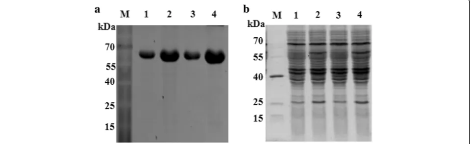
The PLRV-MP gene was amplified by PCR, and ligated with a prokaryotic expression vector pDB.His.MBP to produce pDB.His.MBP–PLRV-MP. Then the MP fusion protein was expressed in Escherichia coli (strain: BL21) and purified. During the prokaryotic expression and purification of PLRV-MP protein, the osmotic fluid was detected using Sodium Dodecyl Sulfate Polyacrylamide Gel Electrophoresis (SDS-PAGE). The results showed a
specific protein band near the 67 kDa protein marker (Fig. 1a), consistent with the size of the His.MBP– PLRV-MP fusion protein (Fig. 1b), indicating that the fusion protein was successfully purified and the His.MBP tag was banded at 42 kDa (Fig. 1b). Perhaps due to the rupture of the PLRV-MP and His.MBP tags, the fusion protein degraded during the purification process. The concentration of the fusion protein was approximately 2 mg/mL. The purified fusion protein (4 mL) was used to prepare the polyclonal antiserum in the Beijing Genom-ics Institute (Beijing, China), and a total of more than 50 mL of antiserum was obtained.
Titer analysis
The titer of PLRV-MP antiserum was analyzed by West-ern blotting with PLRV-infectedNicotiana benthamiana leaves. A weak protein band of 17 kDa (Sokolova et al.
1997; Lee et al.2002) was detectable down to an
anti-serum dilution of 1:1280000. From the viewpoint of color development, the antiserum functioned best at the ratio range of 1:10000 to 1:40000 (Fig.2).
Sensitivity analysis
In order to analyze the sensitivity of the PLRV-MP anti-serum (anti-MPPLRV), proteins extracted from the PLRV-infected N. benthamiana leaves were serially di-luted and subjected to Western blot analysis. When the total protein was diluted by 40-fold, a positive band was clearly detected for the antiserum dilution factors 1:10000 and 1:20000 (Fig. 3a, b), while purified MP fu-sion protein of approximately 0.025 ng could be detected with the antiserum diluted to 1:20000 (Fig.3c).
Specificity analysis
Total protein was extracted fromN. benthamianaleaves infected by PLRV,Turnip yellows virus(TuYV) and Bras-sica yellow virus (BrYV), respectively, and healthy leaf
was used as a negative control. Western blotting with anti-MPPLRVat ratios of 1:10000, 1:20000, and 1:30000 revealed positive results only in reactions with PLRV-infected N. benthamiana leaf (Fig. 4), indicating that the antiserum was specific to PLRV.
Detection of PLRV from field samples using PLRV-MP antiserum and comparison with RT-PCR detection
Potato leaf samples from individual plants collected in Qianqin, Inner Mongolia were tested by Western blotting with anti-MPPLRV at a ratio of 1: 20000. The positive bands exhibited in three leaf samples were consistent with that of the positive control (PLRV-infectedN. benthami-analeaf ) (Fig.5a) and identical to that of RT-PCR detec-tion (Fig. 5b), indicating that this anti-MPPLRV was applicable for detection of PLRV in field crops.
Discussion
Protein expression is the way in which proteins are syn-thesized, modified and regulated in living organisms such as bacteria, yeast, plant or animal cells. The E. coli pro-karyotic expression becomes a representative expression
system as it has a clear genetic background, rapid reproduction, high expression levels, ease of purifica-tion of expression products, good stability, strong anti-pollution ability, low cost, and a wide range of ap-plications (Hockney 1994). Many recombinant poly-clonal antibodies specific to plant viruses have been previously generated for serological detection of viruses such as Tomato spotted wilt virus (Vaira et al. 1996), Potato virus Y (Folwarczna et al. 2008), Alfalfa mosaic virus (Khatabi et al. 2012), and Wheat streak mosaic virus (Tatineni et al. 2014). Recombinant antisera for Egyptian isolates of both Potato virus X(Soliman et al.
2006) and PLRV (Aseel and Hafez 2017; El-Attar et al.
2010) have also been produced. In our experiment, E.
coli was used to express His.MBP–PLRV-MP from which PLRV-MP fusion protein was purified and anti-MPPLRVsuccessfully produced.
Viral MP plays an important role in the cell-to-cell movement of the infectious material. Recent research showed that the interaction of PLRV-MP with the pro-tein 3a affected their localization to the mitochondria and plastids (DeBlasio et al.2018). Serology is one of the Fig. 2Titer determination of PLRV-MP antiserum by Western blotting. Antiserum used at 10 different dilutions (1:4000, 1:8000, 1:10000, 1:16000, 1:20000, 1:32000, 1:40000, 1:64000, 1:80000, and 1:128000) against PLRV, each of which consists of a left lane of negative control and a right lane containing proteins extracted from PLRV-infectedNicotiana benthamianaleaves
most important techniques for virus detection (Aseel and Hafez 2017) and the antiserum (either monoclonal or polyclonal) is the basis for the serology test. In most cases, the anti-coat protein (CP) antisera are used for virus detection; however, virus detection by the anti-CP antisera may not show whether the virus is in active (replication) or inactive stage. The positive detection of the MP with anti-MP antisera may indicate that the virus is in the multiplication stage. Polyclonal and monoclonal MP antibodies specific to Barley yellow dwarf virus (Xie et al. 2007; Li et al. 2015), polyclonal MP antibody specific to Citrus leprosis virus C (Cale-gario et al. 2012), and polyclonal MP antibody specific toGrapevine fanleaf virus (Koolivand et al.2016) have been reported to detect the corresponding viruses, but most were effective only in laboratory use. Western blotting with anti-MPPLRVin our study showed that the antiserum could detect PLRV efficiently, although ra-tios of 1:10000 to 1:40000 were better for color devel-opment. It was highly sensitive down to below 0.025 ng of protein at the antiserum ratio of 1:30000 and strictly specific to PLRV. The antiserum could also detect PLRV from field samples and the detection result was identical to that of RT-PCR.
Conclusions
Because viral MP plays an important role in the process of virus infection, efforts to express PLRV-MP in E. coli and to produce a specific polyclonal antiserum against this protein will provide an important tool for identifica-tion of PLRV infecidentifica-tion and further research on the localization, expression, interaction with host and vari-ous biochemical modifications of PLRV-MP are needed.
Methods
Plant material and growth conditions
Wild-typeN. benthamianaplants were grown at 24 ± 1 °C with a photoperiod of 16/8 h of light/dark cycle. Po-tato leaf samples were collected from Qianqi, Inner Mongolia.
Construction of PLRV-MP prokaryotic expression vector
The pT-PLRV-MP was constructed by amplification of pCa-PLRV containing the infectious cDNA clone of PLRV derived from the plasmid pBNUP110 (Franco-Lara et al.
1999) with the primer pair PLMPNdeF (5′-CATATGTCA
ATGGTGGTGTACAA-3′) and PLMPXhoR (5′-CTCG
AGTCATCCGCGCTTGATAAG-3′). The plasmids were then digested withNdeI andXhoI followed by ligation into Fig. 4Specificity analysis of PLRV-MP antiserum (anti-MPPLRV) by Western blotting. Marker: PageRuler Prestained Protein Ladder, Mock: protein extracted from the healthy leaf was used as a negative control, and the rest of the lanes are for the protein extracted fromNicotiana benthamianaleaves infected byBrassica yellow virus,Turnip yellows virusandPotato leafroll virus, respectively
Fig. 5Comparison of detections of PLRV from field crops using anti-MPPLRVand RT-PCR.aLane M: PageRuler Prestained Protein Ladder.
the expression vector pDB.His.MBP (DNASU Plasmid Re-pository, Arizona, USA), and predigested with NdeI and XhoI to obtain the prokaryotic expression clone pDB.His.MBP–PLRV-MP containing the target gene.
Prokaryotic expression and purification of PLRV-MP protein, and preparation of polyclonal antiserum
Oscillation culture of pDB.His.MBP–PLRV-MP was ad-justed to an OD600 of 0.6–0.8. Isopropyl-β
-D-thiogalacto-side (IPTG; Sigma-Aldrich, St. Louis, MO, USA) was added to make a final concentration of 0.1 mM. After in-cubation at 18 °C for 6 h, it was centrifuged at 4000×g for 10 min. The cells were resuspended with high-salt buffer (20 mM Tris-HCl, 500 mM NaCl, pH 8.0) and centrifuged at 12,000×g for 1 h. The supernatants were collected on a Ni-affinity column (Qiagen, Hilden, Germany) and the proteins washed with elution buffer (20 mM Tris-HCl, 150 mM NaCl, pH 8. 0). Each purification process was subjected to SDS-PAGE and the appropriate eluent was concentrated to obtain the pDB.His.MBP–PLRV-MP fu-sion protein.
The purified fusion protein was sent to the Beijing Genomics Institute to prepare the polyclonal antiserum (anti-MPPLRV) by immunizing rabbits. Sensitivity and specificity of the antiserum developed against recombin-ant PLRV-MP were evaluated by Western blotting.
Western blot detection
Proteins were separated by SDS-PAGE using the proto-col of Zhuo et al.2014. Briefly, proteins were transferred to a nitrocellulose membrane (GE Healthcare, Bucking-hamshire, UK) by electrotransfer (200 mA, 90 min) with a mini trans-blot electrophoretic transfer cell (Bio-Rad, Hercules, CA, USA), and the nitrocellulose membranes were incubated in 1 × TBST buffer (20 mM Tris-HCl pH 7.5, 150 mM NaCl, 0.05% Tween-20) containing 5% skim milk powder at 37 °C. After blocking for 1 h, anti-MPPLRV was added at a certain dilution and incu-bated at 37 °C for 1 h, and washed three times with 1 × TBST for 10 min each time. The nitrocellulose membrane was then incubated with anti-rabbit in goat (IgG; Sigma-Aldrich) diluted at 1:3000 for 1 h at 37 °C as a sec-ondary antibody followed by washing with 1 × TBST. The band was visualized by using nitro-blue tetrazolium and 5-bromo-4-chloro-3-indolyphosphate (Sigma-Aldrich).
Determination of anti-MPPLRVtiter
Total protein was extracted from PLRV-infected N. benthamiana leaves and healthy N. benthamiana leaf (as a negative control) as described by Zhuo et al.2014. Anti-MPPLRV with a dilution factor of 4000, 8000, 10000, 16000, 20000, 32000, 40000, 64000, 80000, and 128000 was used in Western blotting to determine the titer value.
Sensitivity analysis of the anti-MPPLRV
The experiment was performed by extracting the total protein from PLRV-infected N. benthamiana leaves and multiple dilution samples were prepared by diluting the stock sample multiples of 10, 20, 40, 80, 160, 320, 640, 1280, and 2560 with protein extraction buffer. Western blotting was performed using anti-MPPLRVat the ratio of 1:10000 and 1:20000 respectively.
Another experiment was performed by loading 20, 10, 5, 2, 1, 0.5, 0.25, 0.1, 0.05, 0.025, 0.0125, and 0.005 ng of purified protein respectively and anti-MPPLRV was di-luted at the ratio of 1:20000.
Anti-MPPLRVspecificity analysis
Total protein was extracted fromN. benthamianaleaves infected by BrYV, TuYV and PLRV respectively with healthy leaf as a negative control. Specificity of the anti-serum was detected by Western blotting.
Field application of the antiserum
Leaf samples collected from individual standing field po-tato plants in Qianqi, Inner Mongolia, were tested for PLRV detection using our newly developed anti-MPPLRV, with healthy leaf sample as a negative control and PLRV-infectedN. benthamianaleaf as a positive control. The result was compared with RT-PCR detection results.
Abbreviations
BrYV:Brassica yellow virus; CP: Coat protein; IPTG: Isopropyl-β -D-thiogalactoside; MP: Movement protein; ORF: Open reading frame; PLRV:Potato leafroll virus; RT-PCR: Reverse transcription polymerase chain reaction; SDS-PAGE: Sodium Dodecyl Sulphate Polyacrylamide Gel Electrophoresis; TBST: Tris-buffered saline; TuYV:Turnip yellow virus
Acknowledgments
We thank Professor Peter Palukaitis (Seoul Women’s University) for providing the plasmid pBNUP110 containing the infectious cDNA clone of PLRV, Dr. David Baulcombe (John Innes Centre, UK) for providingN. benthamiana plants and Dr. Salah Bouzoubaa (University of Strasbourg, France) for providingE. colistrain MC1022.
Funding
This work was supported by the National Natural Science Foundation of China (31671995 and 31371909) and the 111 project (B13006).
Availability of data and materials
The datasets used and/or analyzed during the current study are available from the corresponding author on reasonable request.
Authors’contributions
C-GH conceived the study and revised the manuscript. FY and MR performed the experiments. FY drafted the manuscript and MR wrote the manuscript. X-YZ constructed a plasmid. ZYZ, YW, DWL and JLY contributed reagents/materials/ analysis tools. All authors read and approved the final manuscript.
Ethics approval and consent to participate
Not applicable
Consent for publication
Competing interests
The authors declare that they have no competing interests.
Received: 17 September 2018 Accepted: 20 January 2019
References
Akamatsu N, Takeda A, Kishimoto M, Kaido M, Okuno T, Mise K. Phosphorylation and interaction of movement protein and coat proteins ofBrome mosaic virusin infected barley protoplasts. Arch Virol. 2007;152:2087–93. Aseel DG, Hafez EE. The comparison of antibodies raised against PLRV with two
different approaches- viral particles purification and recombinant production of CP. J Plant Pathol Microbiol. 2017;8:407.https://doi.org/10.4172/2157-7471. 1000407.
Batool A, Khan MA, Farooq J, Mughal SM, Iftikhar Y. Elisa-based screening of potato germplasm againstPotato leafroll virus. J Agric Res. 2011;49:57–63. Calegario RF, Labate MTV, Peroni LA, Stach-Machado DR, Andrade MO,
Freitas-Astúa J, et al.In vitroexpression and antiserum production against the movement protein ofCitrus leprosis virusC (CiLV-C). Trop Plant Pathol. 2012; 37:136–41.
DeBlasio SL, Xu Y, Johnson RS, Rebelo AR, MacCoss MJ, Gray SM, et al. The interaction dynamics of twoPotato leafroll virusmovement proteins affects their localization to the outer membranes of mitochondria and plastids. Viruses. 2018;10:585.https://doi.org/10.3390/v10110585.
Derrick KS. Quantitative assay for plant viruses using serologically specific electron microscopy. Virology. 1973;56:652–3.
El-Attar AK, Riad BY, Saad A, Soliman AM, Mazyad HM. Expression of the coat protein gene ofPotato leafroll virusinEscherichia coliand development of polyclonal antibodies against recombinant coat protein. Arab J Biotech. 2010; 13:85–98.
Folwarczna J, Plchová H, Moravec T, Hoffmeisterová H, Dedic P, Cerovská N. Production of polyclonal antibodies to a recombinant coat protein ofPotato virus Y. Folia Microbiol (Praha). 2008;53:438–42.
Franco-Lara LF, McGeachy KD, Commandeur U, Martin RR, Mayo MA, Barker H. Transformation of tobacco and potato with cDNA encoding the full-length genome ofPotato leafroll virus: evidence for a novel virus distribution and host effects on virus multiplication. J Gen Virol. 1999;80:2813–22. Gillen AM, Novy RG. Molecular characterization of the progeny ofSolanum
tuberosumidentifies a genomic region associated with resistance toPotato leafroll virus. Euphytica. 2007;155:403–15.
Haupt S, Cowan GH, Ziegler A, Roberts AG, Oparka KJ, Torrance L. Two plant-viral movement proteins traffic in the endocytic recycling pathway. Plant Cell. 2005;17:164–81.
He Z, Larkin R, Honeycutt W, editors. Sustainable potato production: global case studies. 1st ed. Dordrecht: Springer; 2012.
Hockney RC. Recent developments in heterologous protein production in Escherichia coli. Trends Biotechnol. 1994;12:456–63.
Jeffries C, Barker H, Khurana SMP. Potato viruses (and viroids) and their management. In: Gopal J, Khurana SMP, editors. Handbook of potato production, improvement and post-harvest management. New York: The Haworth’s food products press; 2006. p. 387–448.
Khatabi B, He B, Hajimorad MR. Diagnostic potential of polyclonal antibodies against bacterially expressed recombinant coat protein ofAlfalfa mosaic virus. Plant Dis. 2012;96:1352–7.
King AMQ, Adams MJ, Carstens EB, Lefkowitz EJ. Virus taxonomy: classification and nomenclature of viruses: ninth report of the international committee on taxonomy of viruses. San Diego: Academic Press, Elsevier; 2012.
Koolivand D, Bashir NS, Behjatnia SA, Joozani RJ. Production of polyclonal antibody againstGrapevine fanleaf virusmovement protein expressed in Escherichia coli. Plant Pathol J. 2016;32:452–9.
Lee L, Kaplan IB, Ripoll DR, Liang D, Palukaitis P, Gray SM. A surface loop of the potato leafroll virus coat protein is involved in virion assembly, systemic movement, and aphid transmission. J Virol. 2005:1207–14.
Lee L, Palukaitis P, Gray SM. Host-dependent requirement for thePotato leafroll virus 17 kDa protein in virus movement. Mol Plant Microbe Inter. 2002;15:1086–94. Li N, Chen Z, Liu Y, Liu Y, Zhou X, Wu J. Development of monoclonal antibodies
and serological assays specific forBarley yellow dwarf virusGAV strain. Virol J. 2015;12:136–44.
Lin NS, Hsu YH, Hsu HT. Immunological detection of plant viruses and a mycoplasma-like organism by direct tissue blotting on nitrocellulose membranes. Phytopathology. 1990;80:824–8.
Loebenstein G, Berger PH, Brunt AA, Lawson RH. Virus and virus-like diseases of potatoes and production of seed potatoes. Netherlands: Kluwer Academic Publishers; 2001. p. 69–72.
Mayo MA, Ziegler-Graff V. Molecular biology of luteoviruses. Adv Virus Res. 1996; 46:413–60.
Sokolova M, Prufer D, Tacke E, Rohde W. The potato leafroll virus 17 kDa movement protein is phosphorylated by a membrane-associated protein kinase from potato with biochemical features of protein kinase C. FEBS Lett. 1997;400:201–5.
Soliman AM, Barsoum BN, Mohamed GG, El-Attar AK, Mazyad HM. Expression of the coat protein gene of the Egyptian isolate ofPotato virus XinEscherichia coliand production of polyclonal antibodies against it. Arab J Biotech. 2006; 9:115–28.
Tacke E, Prufer D, Schmitz J, Rohde W. The potato leafroll luteovirus 17 K protein is a single-stranded nucleic acid-binding protein. J Gen Virol. 1991;72:2035–8. Tacke E, Schmitz J, Prufer D, Rohde W. Mutational analysis of the nucleic
acid-binding 17 kDa phosphoprotein ofPotato leafroll luteovirusidentifies an amphipathic a-helix as the domain for protein-protein interactions. Virology. 1993;197:274–82.
Tatineni S, Kovacs F, French R.Wheat streak mosaic virusinfects systemically despite extensive coat protein deletions: identification of virion assembly and cell-to-cell movement determinants. J Virol. 2014;88:1366–80.
Vaira AM, Vecchiati M, Masenga V, Accotto GP. A polyclonal antiserum against a recombinant viral protein combines specificity with versatility. J Virol Methods. 1996;56:209–19.
Wales S, Platt HW, Cattlin N. Diseases, pests and disorders of potatoes. London: Manson Publishing Ltd; 2008. p. 75–6.
Warren M, Kruger K, Schoeman AS.Potato virus Y(PVY)and Potato leafroll virus (PLRV): literature review for potatoes South Africa. Pretoria: Department of Zoology and Entomology, Faculty of Natural and Agricultural Sciences, University of Pretoria; 2005.
Wolf S, Deom CM, Beachy RN, Lucas WJ. Movement protein ofTobacco mosaic virusmodifies plasmodesmatal size exclusion limit. Science. 1989;246:377–9. Xie JJ, Wang XF, Liu Y, Peng YF, Zhou GH. Expression, purification and antiserum
preparation of BYDV GAV movement protein, and its application. In: Peng YL, Kang ZS, editors. Proceeding of the annual meeting of Chinese society for plant pathology. Yangling: Northwest A&F University Press; 2007. p. 215–7. in Chinese.