and fluorescence-assisted cell sorting
Fei Gao
1,2, Zhenzhen Hao
1, Xianhua Sun
1, Lina Qin
3, Tong Zhao
3, Weiquan Liu
2, Huiying Luo
1, Bin Yao
1*and Xiaoyun Su
1*Abstract
Background: In the biofuel industry, cellulase plays an indispensable role in hydrolyzing cellulose into fermentable glucose. Trichoderma reesei is a popular filamentous fungus with prominent ability to produce cellulase. While classical mutagenesis and modern multiplex genome engineering are both effective ways to improve cellulase production, successful obtaining of strains with improved cellulase-producing ability requires screening a large number of strains, which is time-consuming and labor intensive.
Results: Herein, we developed a versatile method coupling expression of the red fluorescence protein (DsRed) in T. reesei and fluorescence-assisted cell sorting (FACS) of germinated spores. This method was first established by expressing DsRed intracellularly under the control of the major cellulase cbh1 promoter in T. reesei, which allowed us to rapidly isolate cellulase hyperproducers from T. reesei progenies transformed with a dedicated transcriptional activator ace3 and from an atmospheric and room temperature plasma-created mutant T. reesei library. Since intracel-lularly expressed DsRed was expected to isolate mutations mainly affecting cellulase transcription, this method was further improved by displaying DsRed on the T. reesei cell surface, enabling isolation of strains with beneficial genetic alterations (overexpressing hac1 and bip1) affecting regulatory stages beyond transcription. Using this method, T. reesei cellulase hyperproducers were also successfully isolated from an Agrobacterium-mediated random insertional mutant library.
Conclusions: The coupled DsRed-FACS high-throughput screening method proved to be an effective strategy for fast isolation of T. reesei cellulase hyperproducers and could also be applied in other industrially important filamentous fungi.
Keywords: Trichoderma reesei, DsRed, FACS, Cellulase, High-throughput screening
© The Author(s) 2018. This article is distributed under the terms of the Creative Commons Attribution 4.0 International License (http://creativecommons.org/licenses/by/4.0/), which permits unrestricted use, distribution, and reproduction in any medium, provided you give appropriate credit to the original author(s) and the source, provide a link to the Creative Commons license, and indicate if changes were made. The Creative Commons Public Domain Dedication waiver (http://creativecommons.org/ publicdomain/zero/1.0/) applies to the data made available in this article, unless otherwise stated.
*Correspondence: [email protected]; [email protected]
1 Key Laboratory for Feed Biotechnology of the Ministry of Agriculture,
oxidase [7], endo-mannanase [8], antibodies, and inter-ferons [9, 10].
On the route towards lignocellulosic biofuels, cellu-lase plays an indispensable role by hydrolyzing cellulose into fermentable glucose. T. reesei has long been used to produce cellulase and currently is still one popular microbe [11]. Varying strategies have been utilized to improve cellulase production in T. reesei, mainly by engi-neering the T. reesei strains and modifying the fermen-tation processes. Cellulase (and other kinds of enzymes or proteins) is synthesized from amino acid precursors. Therefore, the concept of metabolic engineering can be employed to improve cellulase expression and secretion in T. reesei basically by means of random mutagenesis and genetic engineering [12]. To improve production of cellulase widely useful in the biofuel industries, chemi-cal and physichemi-cal mutagenesis were initially carried out, which successfully generated hyperproducer mutants such as RUT-C30 [13] and CL-847 [14]. Additionally, Agrobacterium-mediated random insertional disruption of chromosomal genes was also successful in generat-ing T. reesei hyperproducers [15]. Genetic engineering by overexpressing selected genes stimulating cellulase expression [7, 16] and removing the ones repressing cel-lulase expression [17, 18] controlling transcription, trans-lation, and secretion as well as intracellular redox balance and cell metabolism [19–22] have become two common strategies to improve cellulase production in T. reesei. It is noticed that multiplex genome engineering, i.e., manipulating more than one gene at a time, creates rich biological diversity from which a cellulase hyperproducer can be isolated [16]. By including more genes to manipu-late, this biodiversity may even ascend, increasing the possibility to obtain hyperproducers. However, the con-curring higher complexity demands a larger amount of transformants to be analyzed.
Both random mutagenesis and genetic engineering methods require considerable time and labor in screen-ing. For T. reesei, this challenge is further complicated by the facts that T. reesei is multicellular and filamentous, cellulase is extracellularly expressed, and integration
ditional shake flask to microtiter plate culture; however, the quantity of strains to be screened is still large [28]. Herein, we sought to overcome these bottlenecks and establish a new high-throughput screening method Spe-cifically, we used the red fluorescence protein DsRed as a reporter molecule and demonstrated that the intracellu-larly expressed, and more importantly, surface-displayed DsRed coupled with FACS can be used for high-through-put screening of cellulase hyperproducers from T. reesei progeny libraries generated by random mutagenesis or genetic engineering. It is expected that our results may provide a robust engineering framework for future efforts to engineer T. reesei, and other industrially important filamentous fungi as well, for enhanced secretion of cel-lulase and other valuable proteins.
Results
Constructing the plasmids for intracellular expression and surface‑display of DsRed
Intracellular expression of DsRed dictates isolation of genetically engineered T. reesei cellulase hyperproducers
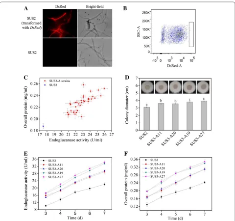
The plasmid encoding the codon-optimized DsRed under the control of cbh1 promoter was transformed into T. reesei SUS2. DsRed was successfully expressed in T. reesei and red fluorescence appeared to be evenly distributed in the hyphae cytosome, which could be clearly visual-ized under fluorescence microscope (Fig. 2A). High level expression of DsRed resulted into change of the colony color from pale white to red on a MM-lactose plate, which could even be observed by naked eyes (Additional file 1). Next, we tested whether higher level of DsRed expression incurred by genetic modification of T. reesei would be positively correlated with higher cellulase pro-duction. For this purpose, the transcriptional activator ace3 was constructed downstream of the strong consti-tutive pdc1 promoter to obtain pPdc1-ace3 (Additional file 2). This plasmid was transformed into a uridine-aux-otrophic derivative of the DsRed-expressing SUS2 trans-formant, namely SUS3. Note that SUS2 and SUS3 have nearly identical cellulase-producing abilities (data not shown). Overexpression of ace3 is known to stimulate cellulase expression in T. reesei by two- to fourfold [21]. Thus, 40 spores with highest red fluorescence signal (top 0.1%) were collected for further analyses (Fig. 2B). Since SUS3 (and its parent strain SUS2) is a dedicated strain for egl2- overexpression, we measured the endoglucanase activity and used it as a cellulase indicator. All these selected ace3-transformants behaved better in prelimi-nary flask fermentation, producing more endoglucanase as well as extracellular proteins (Fig. 2C).
SUS2 and four representative ace3-transformants with moderately to highly enhanced cellulase-producing abil-ity were grown on cellulose plates for comparison of halos (an indicator of cellulose hydrolysis rate, roughly repre-senting the cellulase activity). All ace3-transformants
formed a halo (diameter: 3.50–3.95 cm) larger than that of SUS2 (diameter: 3.20 cm), indicative of improved cel-lulase producing ability (Fig. 2D). In flask fermentation, these four strains exhibited higher endoglucanase activi-ties and protein concentrations from day 3 post-Avicel induction (Fig. 2E, F). These results indicated that the intracellular expressed DsRed under the control of cbh1 promoter could be used to dictate selection of the T. ree-sei high cellulase producers. Using quantitative PCR, the gene copy numbers of ace3 in the four transformants were determined to be two for SUS3-A11, SUS3-A19, and SUS3-A20 and three for SUS3-A27, higher than one in the parental strain (data not shown).
Rapid isolation of T. reesei cellulase hyperproducers generated from ARTP random mutagenesis
Through successful isolation of ace3-transformants with improved cellulase producing ability, the validity of the high-throughput screening method was demonstrated. However, we realized that random mutagenesis has been used as an effective way for T. reesei strain improvement [14] and is still being used widely. Indeed, most indus-trial strains are mutagenized derivatives of the QM6a strain [13]. Therefore, we tested if this DsRed-based FACS method could also be used for isolation of cellulase hyperproducers from a randomly mutagenized T. reesei library.
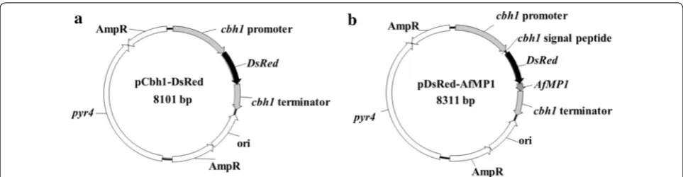
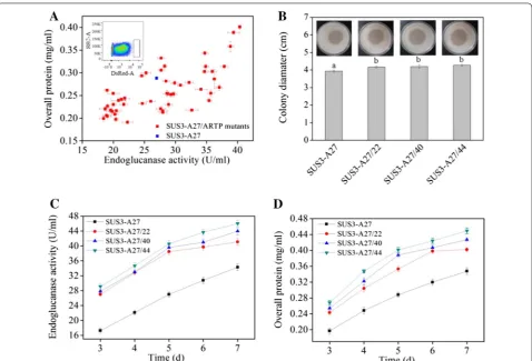
The atmospheric and room temperature plasma muta-tion system, or ARTP, uses radio-frequency atmospheric-pressure glow discharge plasma jets to create mutations in the DNA sequence and has been used to mutate more than 40 kinds of microorganisms including bacteria, fungi, and microalgae [33]. SUS3-A27, the best strain of the ace3-transformants, was used for mutagenesis. The ARTP-treated spores were grown for 14 h in MM-lactose/sophorose liquid medium for germination. This culture period allows expression of cellulase genes (and Fig. 1 Schematic diagrams of the plasmids for expressing DsRed. a The pCbh1-DsRed plasmid for expressing DsRed intracellularly. b The
here DsRed also) [34] but precludes formation of long intertwining hyphae clog, which would be troublesome for flow cytometry analysis. Fifty-one germinated spores with highest red fluorescence signals were picked out from FACS (Fig. 3A). Unlike the ace3-transformation, some of the sorted mutants behaved poorer than the par-ent strain in the preliminary screening in flask cultivation
(Fig. 3A). This indicated that, stronger red fluorescence signal at the early stage of germination for these mutants from random mutagenesis do not necessarily parallel with higher cellulase producing ability. In spite of this inconsistence, certain mutants exhibited higher cellu-lase activity as well as overall protein concentrations than those of the parent strain (Fig. 3A). Three representative Fig. 2 Isolation of cellulase hyperproducers from ace3-transformants by FACS using the intracellularly expressing DsRed as an indicator. A
mutant strains (A27/22, A27/40, and SUS3-A27/44) were used for further detailed analyses. These mutants formed cellulose-hydrolyzing halos with a diam-eter of 4.15–4.30 cm, respectively, larger than that of SUS3-A27 (3.95 cm) on MM-Avicel plate (Fig. 3B). In addition, on the 7th day post-induction, SUS3-A27/22, SUS3-A27/40, and SUS3-A27/44 produced 41.1, 43.9, and 46.0 U/ml endoglucanase and 0.41, 0.43, and 0.45 mg/ml extracellular proteins, respectively, which were all higher than the values of SUS3-A27 (34.2 U/ml for endoglucanase activity, Fig. 3C) and 0.35 mg/ml (for extracellular protein concentration, Fig. 3D).
Surface‑display of DsRed enabled identifying genetic alterations beneficial for cellulase secretion
The intracellularly expressed DsRed hardly allows iden-tification of beneficial mutations or genetic modifica-tions affecting the stages beyond transcription. However,
this obstacle could be overcome if the expressed DsRed reporter protein also undergoes the secretory pathway while being attached to the cell. This can be achieved by displaying DsRed on the T. reesei cell surface. For this purpose, we fused the DsRed gene in frame between the sequences coding for the CBH1 signal peptide and AfMP1 GPI anchor. The CBH1 signal peptide will lead the DsRed protein through the secretory pathway and the covalently linked GPI anchor can keep DsRed attached to the cell surface. These traits are expected to facilitate high-throughput isolation of cellulase hyperproducers generated by genetic alterations at any regulatory stages.
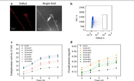
incubating the germinated transformant spores with a monoclonal antibody against DsRed and FITC-labeled goat anti-mouse secondary antibody followed by flow cytometry analyses of both DsRed and FITC signals (Additional file 3). One should note that the red fluo-rescence of the DsRed-AfMP1 transformant (SUS4) was weaker than that expressing intracellular DsRed. This was also reflected by the color change of the DsRed-AfMP1– transformant (Additional file 1). To analyze if surface-dis-played DsRed could be used to screen beneficial genetic alterations at stages beyond transcription, six genes (bip1, hac1, ftt1, sso2, sar1, and ypt1) regulating cellulase folding and secretion were constructed into plasmids individually under strong constitutive promoters. Bip1 is an ER-resident molecular chaperone assisting folding of nascent proteins, while Hac1 is a global transcription regulator controlling the unfolded protein response [35]. Ftt1, Sso2, Sar1, and Ypt1 are proteins involved in diverse stages of the cellulase secretion pathway in T. reesei [22]. Equal amounts of the six plasmids were combined and one-time transformed into a uridine auxotroph of SUS4. The transformant spores were pooled for FACS. Sixty
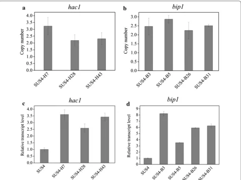
spores with the highest red fluorescence signal were collected (Fig. 4b). Seven representative strains were determined to produce more cellulase (Fig. 4c) and extra-cellular proteins (Fig. 4d). Interestingly, we discovered by PCR that these strains were transformants overexpress-ing hac1 and bip1 only but not the other four genes (data not shown). Quantitative PCR analysis indicated that 2 to 3 copies of hac1 (Fig. 5a) and bip1 (Fig. 5b) genes were existent in these transformants while there was only one copy in the parent strain (data not shown). The RT-qPCR analysis further proved that both the bip1 and hac1 genes were transcribed to levels as high as 2.5- to 3.6-fold (hac1, Fig. 5c) and 3.5- to 8.2-fold (bip1, Fig. 5d) in these transformants.
Isolation of cellulase hyperproducers from an insertional mutagenesis library of T. reesei aided by surface‑displayed DsRed and FACS
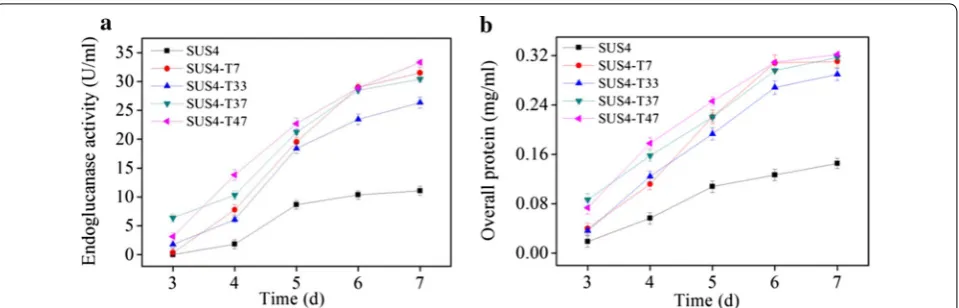
Agrobacterium-mediated transformation was used to randomly insert the pyr4 gene into the chromosome of the uridine auxotroph of SUS4. It was expected that, when the function of a gene negatively affecting cellulase Fig. 4 Isolation of cellulase hyperproducers from T. reesei transformed with a mixture of six plasmids by FACS directed by surface-displayed DsRed. a
Microscopic observation of the T. reesei hyphae displaying DsRed on cell surface. T. reesei was cultivated at 28 °C for 24 h in MM-lactose/sophorose. b
expression was disrupted, the recombinant strain would display a cellulase hyperproducer phenotype. The spores from the transformants were pooled, germinated in MM-lactose/sophorose, and sorted by FACS to obtain sixty spores with highest red fluorescence. In shake flask fer-mentation, four representative strains displayed higher endoglucanase activities (26.3–33.3 U/ml, Fig. 6a) and extracellular protein concentration (0.29–0.32 mg/ml, Fig. 6b) than those of the parent strain (11.1 U/ml and 0.15 mg/ml, respectively) at the end of fermentation.
Discussion
As a response to the lack of easy-to-ferment sugars such as glucose, T. reesei secrets cellulase which enables it to acquire carbon and energy from its natural habitat. Bio-synthesis of cellulase is complex: induction of cellulase
genes can all be potentially utilized in T. reesei for speeded strain improvement. However, all these endeav-ors require a high-throughput method as a prerequisite to isolate the cellulase hyperproducers.
The high-throughput method described herein for screening T. reesei cellulase hyperproducers was based on coupling the usage of DsRed and FACS. One advantage of DsRed is that the positive colonies turn to red, mak-ing them easily identified by naked eyes from the trans-formants (Additional file 1). Apparently, the depth of red color is positively associated with the inducing extent of cbh1 promoter. Using this system, we demonstrated as proof-of-concept that cellulase hyperproducers could be isolated from ace3-transformants and from a mutagen-ized library. Cellulose plate assay and preliminary shake flask fermentation confirmed the effectiveness of this method with the number of colonies largely reduced to as few as ~ 50, saving tremendous time and labor than the previous screening method [14]. Despite these successes, expressing DsRed intracellularly is limited to discovery of genetic modifications majorly favoring transcription, in sharp contrast with the complex nature of cellulase expression regulation in T. reesei [37, 38]. Therefore, we overcome this hurdle by further developing a surface-display technology of DsRed and demonstrated that the updated method managed to isolate beneficial genetic modifications (hac1- and bip1-overexpression) favoring nascent protein folding and secretion. The overexpress-ing cassettes for sso2, ftt1, sar1, and ypt1 could not be amplified from the isolated cellulase hyperproducers. It is possible that the expression of these genes may not be a bottleneck for cellulase expression in the investigated strain and under the specific culture condition. However, it could also be that the cellulase hyperproducers bear-ing one of these genes occasionally escaped the FACS
isolation in the current study. These hypotheses undoubt-edly need further investigations.
Different from small metabolites whose production can be either auto-regulated [39] or monitored by bio-sensor-based screening [40], cellulase is a secreted, spe-cial metabolite which cannot be readily quantified using any known, existent biosensors. Coupling DsRed-AfMP1 expression with FACS passes this barrier, enabling fast isolation of T. reesei cellulase hyperproducers from both engineered and mutagenized libraries. We noticed that, however, expression of DsRed-AfMP1 chimera reduced cellulase secretion in the host strain, suggesting that DsRed-AfMP1 might compete with the endogenous cel-lulase for the same secretory pathway. This trait is unde-sirable from the perspective of strain improvement. However, with this surface-display system, the impaired T. reesei can be quickly improved back to normal or even to a higher level of production after one or a few rounds of engineering. Moreover, once the strain is modified to reach a satisfying level of cellulase production, the expression of DsRed can be easily eliminated using the RNAi-mediated gene silencing [6] or gene knockout using the CRISPR/Cas9 system [41], further releasing the occupied carbon, energy, and secretion pathway compo-nents for cellulase.
thermophila [46].
Conclusions
In this study, we established a high-throughput method for fast isolation of T. reesei cellulase hyperproducers. Specifically, coupling expression of DsRed with FACS allowed us to rapidly isolate cellulase hyperproducers from ace3-transformants and from a random mutagen-ized T. reesei library. Furthermore, displaying DsRed on T. reesei cell surface enabled isolation of cellulase hyper-producers with genetic variations for enhanced expres-sion of proteins involved in nascent protein folding and secretion (bip1 and hac1) beyond transcription. This ver-satile system saves tremendous time and labor than the previous screening methods and can, therefore, be used as a robust engineering framework for future metabolic engineering of T. reesei, as well other industrially impor-tant filamentous fungi for enhanced secretion of cellulase and other valuable proteins.
Methods
Strains, plasmids, and culture conditions
The Escherichia coli Trans I-T1 strain from Transgen (Beijing, China) was used as a host for plasmid con-struction and propagation. The Saccharomyces cerevi-siae AH109 (Clontech, San Francisco, CA) auxotrophic strain was used as the host for constructing the plasmids containing the expressing cassettes via DNA assembler [47]. The T. reesei SUS2 strain is uridine-auxotrophic and derived from SUS1 [8] by transforming extra cop-ies of the endogenous cel5A (egl2) gene under control of the cbh1 promoter. The Agrobacterium tumefaciens AGL-1 strain was used as the T-DNA donor for A. tume-faciens-mediated transformation (AMT) of T. reesei. The plasmids containing the codon-optimized DsRed gene (pSKLR) [25] and the AfMP1 gene (pGFP-Mp1) [32] are kindly gifts from Prof. Zhiyang Dong and Haomiao Ouy-ang, respectively, from the Institute of Microbiology Chi-nese Academy of Sciences. The plasmid pTi was provided by Dr. XinXin Xu from Biotechnology Research Institute, Chinese Academy of Agricultural Sciences.
The E. coli and A. tumefaciens were cultured in the Luria–Bertani (LB) medium supplemented with an
the MM-2% Avicel medium. The basal medium (BM), induction medium (IM), and co-cultivation medium (CM) used for AMT were prepared as described previ-ously [48].
Plasmid construction
with the primer pairs pyr4F/R (Additional file 4) and ligated into the XmaJI and PacI restriction sites of the plasmid pTi using the ClonExpress® II One Step Cloning Kit (Vazyme, Nanjing, China) to obtain the plasmid pTi-pyr4 (Additional file 5).
Transformation of T. reesei
The plasmids were introduced into T. reesei using the polyethylene glycol (PEG)-mediated protoplast trans-formation method [51]. Briefly, T. reesei was grown in MM-glucose (2%) at 30 °C for 24 h. The young mycelia were collected, mixed with 10 mg/ml of Lysing Enzymes from Trichoderma harzianum (L1412, Sigma-Aldrich, St. Louis, MO), and frequently observed under microscope until large amounts of protoplasts were released. The uri-dine autotroph transformants were selected on MM-glu-cose plates and screened for integration of the expressing cassette into the chromosome of T. reesei by PCR.
Agrobacterium-mediated transformation was used for insertional mutagenesis of T. reesei [52]. Briefly, A. tume-faciens containing the binary vector pTi-pyr4 was grown at 28 °C for 2 days in liquid BM supplemented with kana-mycin (50 μg/ml). The bacterial cell suspensions were diluted to an optical density at 600 nm (OD600) of 0.2 in IM with 200 μM acetosyringone (AS) and grown for 6 h to an OD600 of 0.4–0.8. Equal volumes of A. tumefaciens cells and the T. reesei protoplasts (107/ml) were mixed. Next, 200 μl of this blend were plated on a 90-mm diam-eter cellophane paper on top of CM-agar in presence of 200 μM AS. After co-cultivation at 25 °C for 48 h, the cel-lophane paper was transferred to the selection medium (BM-agar supplemented with 1 M sorbitol and 200 μg/ ml cefotaxime but lacking uridine). The transformant colonies were transferred onto PDA for sporulation. The spores were mixed and used for FACS screening.
were washed again with PBS and incubated with 10 μg/ ml of fluorescein isothiocyanate (FITC)-labeled goat anti-mouse IgG secondary antibody (Abcam) for 1 h. The cells were finally washed twice with PBS and passed through flow cytometry for analyses of both DsRed and FITC signals.
ARTP‑mediated mutagenesis of T. reesei
For ARTP-mediated mutagenesis, T. reesei spores har-vested from 5-day-old culture of the strain SUS3-A27 on PDA plate were suspended in distilled water to a concen-tration of 109 per ml. Ten μl of this spore suspension were spread on the surface of a sterilized sample plate and sub-jected to ARTP treatment. The radio-frequency power input was set as 100 W and the helium gas flow rate was 10 SLM (standard liter per minute) with a plasma action distance of 2 mm. The processing time was set at 3 min. After ARTP treatment, the spores were trans-ferred to 1 ml of distilled water. The spores were plated on PDA plates and allowed to grow for re-sporulation for 5–7 days. Finally, spores from these plates were mixed for FACS.
FACS
Fresh spores were inoculated into liquid MM-lactose/ sophorose (2% for lactose and 0.003% for sophorose, w/v) and cultured at 28 °C with vigorous shaking for 14 h. High-speed sorting was performed on a FACS Aria sorter at a rate of 5000 events per second, 30 psi with an 85 μm nozzle. Single germlings with the brightest (top 0.1%) DsRed signal were sorted into individual wells of 6-well plates and incubated at 28 °C for sporulation.
Induction of cellulase expression in T. reesei
For shake flask fermentation, fresh spores (1 × 10⁷) of T.
reesei were individually inoculated into 50 ml of liquid MM-glucose (2%) and cultured at 28 °C with agitation at 200 rpm for 48 h. The mycelia were collected and washed twice with MM to remove residual glucose. One gram of the mycelia was then transferred to 100 ml of MM-Avicel
tion was incubated at 50 °C for 10 min and the released reducing sugars were determined using the 3,5-dinitro-salicylic acid (DNS) method. One unit of endoglucanase activity was defined as the amount of enzyme that liber-ated 1 μmol of reducing sugar per minute. The protein concentration was determined using the BCA-200 Pro-tein Assay Kit (Pierce, Rockford, IL).
Fungal growth and microcrystalline cellulose hydrolysis
The T. reesei spores (3 × 10⁵) were individually spotted on
agar plates containing MM-Avicel (2%) and incubated at 28 °C for 4–7 days until halo around the colony could be clearly visualized. The halo diameters were measured and compared.
Reverse transcription quantitative PCR analysis
For reverse transcription quantitative PCR (RT-qPCR), the mycelia of T. reesei cultured in MM-Avicel (2%) for 24 h were collected and pulverized in liquid nitrogen using a pestle and mortar. The total RNA was extracted using the TRIzol reagent (Thermo Fisher Scientific, Waltham, MA). The cDNA was synthesized using the First Strand cDNA Maxima Synthesis kit (TOYOBO, Shanghai, China). RT-qPCR was performed in an Applied Biosystems™ QuantStudi™ 6 Flex Real-Time PCR System (Applied Biosystems, San Diego, CA) using a TransScript Green One-Step SuperMix (TransGen, Beijing, China). The actin gene was used as an endogenous reference. The primers used for RT-qPCR were listed in Additional file 4. The following amplification conditions were used: 95 °C for 10 min for initial denaturation, 40 cycles of 94 °C for 10 s, 60 °C for 20 s, and 72 °C for 20 s.
Determining copy numbers by qPCR
To determine the copy numbers of the integrated ace3, bip1 or hac1 gene in the transformants, the genomic DNA was extracted from the mycelia by a Fungal DNA kit (Omega bio-tek, USA) and used as the template for quantitative PCR (qPCR). The qPCR method was per-formed as that described by Solomon [53]. The cbh1 gene was used to represent a single copy gene. The qPCR was performed with the SYBR Green Real-time PCR
Additional file 1. Observation of representative T. reesei strains expressing
DsRed by naked eyes. The culture medium is MM-lactose plus agar.
Additional file 2. Schematic diagram of pPdc1-ace3.
Additional file 3. Verification of DsRed-AfMP1 expression on T. reesei cell surface.
Additional file 4. Primers used in this study.
Additional file 5. Schematic diagram of pTi-pyr4. LB left border, RB right border.
Abbreviations
FACS: fluorescence-assisted cell sorting; GFP: green fluorescence protein; AMT: Agrobacterium tumefaciens-mediated transformation; YPDA: yeast extract-peptone dextrose medium with adenine; PDA: potato dextrose agar; MM: minimal medium; BM: basal medium; IM: induction medium; CM: co-cultivation medium; AS: acetosyringone; ARTP: atmospheric and room temperature plasma; DNS: 3,5-dinitrosalicylic acid; CBH1: cellobiohydrolase I; CBH2: cellobiohydrolase II; EG1: endoglucanase I; EG2: endoglucanase II; GPI: glycosylphosphatidylinositol; SLM: standard liter per minute.
Authors’ contributions
FG performed research, analyzed data, and wrote the paper. ZH, XhS, and HL analyzed the data, LQ, TZ, and WL provided technical assistance, BY designed research, XS designed research, analyzed data, and wrote the paper. All authors read and approved the final manuscript.
Author details
1 Key Laboratory for Feed Biotechnology of the Ministry of Agriculture, Feed
Research Institute, Chinese Academy of Agricultural Sciences, No. 12 South Zhongguancun Street, Beijing 100081, People’s Republic of China. 2 College
of Biological Sciences, China Agricultural University, Beijing 100193, China.
3 Institute of Microbiology, Chinese Academy of Sciences, Beijing 100101,
China.
Competing interests
The authors declare that they have no competing interests.
Availability of supporting data
All data supporting the conclusions of this article are included within the manuscript and additional files.
Consent for publication
All authors provide their consent for publication of their manuscript in Bio-technology for Biofuels.
Ethics approval and consent to participate Not applicable.
Funding
reesei with the Hormoconis resinae glucoamylase P (gamP) gene: produc-tion of a heterologous glucoamylase by Trichoderma reesei. Curr Genet. 1993;24:223–8.
4. Saarelainen R, Mantyla A, Nevalainen H, Suominen P. Expression of barley endopeptidase B in Trichoderma reesei. Appl Environ Microbiol. 1997;63:4938–40.
5. Miettinen-Oinonen A, Torkkeli T, Paloheimo M, Nevalainen H. Overexpres-sion of the Aspergillus niger pH 2.5 acid phosphatase gene in a heterolo-gous host Trichoderma reesei. J Biotechnol. 1997;58:13–20.
6. Qin LN, Cai FR, Dong XR, Huang ZB, Tao Y, Huang JZ, Dong ZY. Improved production of heterologous lipase in Trichoderma reesei by RNAi medi-ated gene silencing of an endogenic highly expressed gene. Bioresour Technol. 2012;109:116–22.
7. Wu Y, Sun X, Xue X, Luo H, Yao B, Xie X, Su X. Overexpressing key com-ponent genes of the secretion pathway for enhanced secretion of an
Aspergillus niger glucose oxidase in Trichoderma reesei. Enzyme Microb Technol. 2017;106:83–7.
8. Sun X, Xue X, Li M, Gao F, Hao Z, Huang H, Luo H, Qin L, Yao B, Su X. Efficient coproduction of mannanase and cellulase by the transformation of a codon-optimized endomannanase gene from Aspergillus niger into
Trichoderma reesei. J Agric Food Chem. 2017;65:11046–53.
9. Landowski CP, Huuskonen A, Wahl R, Westerholm-Parvinen A, Kanerva A, Hanninen AL, Salovuori N, Penttila M, Natunen J, Ostermeier C, et al. Enabling low cost biopharmaceuticals: a systematic approach to delete proteases from a well-known protein production host Trichoderma reesei. PLoS ONE. 2015;10:e0134723.
10. Landowski CP, Mustalahti E, Wahl R, Croute L, Sivasiddarthan D, Wester-holm-Parvinen A, Sommer B, Ostermeier C, Helk B, Saarinen J, Saloheimo M. Enabling low cost biopharmaceuticals: high level interferon α-2b production in Trichoderma reesei. Microb Cell Fact. 2016;15:104. 11. Garvey M, Klose H, Fischer R, Lambertz C, Commandeur U. Cellulases
for biomass degradation: comparing recombinant cellulase expression platforms. Trends Biotechnol. 2013;31:581–93.
12. Kubicek CP, Mikus M, Schuster A, Schmoll M, Seiboth B. Metabolic engineering strategies for the improvement of cellulase production by
Hypocrea jecorina. Biotechnol Biofuels. 2009;2:19.
13. Peterson R, Nevalainen H. Trichoderma reesei RUT-C30-thirty years of strain improvement. Microbiology. 2012;158:58–68.
14. Durand H, Clanet M, Tiraby G. Genetic improvement of Trichoderma reesei for large scale cellulase production. Enzyme Microb Technol. 1988;10:341–6.
15. Zhong YH, Wang XL, Yu HN, Liang SR, Wang TH. Application of T-DNA insertional mutagenesis for improving cellulase production in the fila-mentous fungus Trichoderma reesei. Bioresour Technol. 2012;110:572–7. 16. Wang S, Liu G, Wang J, Yu J, Huang B, Xing M. Enhancing cellulase
pro-duction in Trichoderma reesei RUT C30 through combined manipulation of activating and repressing genes. J Ind Microbiol Biot. 2013;40:633–41. 17. Nakari-Setala T, Paloheimo M, Kallio J, Vehmaanpera J, Penttila M,
Salo-heimo M. Genetic modification of carbon catabolite repression in Tricho-derma reesei for improved protein production. Appl Environ Microbiol. 2009;75:4853–60.
18. Chen F, Chen X, Su X, Qin L, Huang Z, Tao Y, Dong Z. An Ime2-like mito-gen-activated protein kinase is involved in cellulase expression in the filamentous fungus Trichoderma reesei. Biotechnol Lett. 2015;37:2055–62.
1991;9:327–67.
24. Harkki A, Uusitalo J, Bailey M, Penttila ME, Knowles JKC. A novel fungal expression system: secretion of active calf chymosin from the filamen-tous fungi Trichoderma reesei. Nat Biotechnol. 1989;7:596–603.
25. Qin L, Jiang X, Dong Z, Huang J, Chen X. Identification of two integration sites in favor of transgene expression in Trichoderma reesei. Biotechnol Biofuels. 2018;11:142.
26. Throndset W, Kim S, Bower B, Lantz S, Kelemen B, Pepsin M, Chow N, Mitchinson C, Ward M. Flow cytometric sorting of the filamentous fungus Trichoderma reesei for improved strains. Enzyme Microb Technol. 2010;47:335–41.
27. Pakula TM, Laxell M, Huuskonen A, Uusitalo J, Saloheimo M, Penttila M. The effects of drugs inhibiting protein secretion in the filamentous fungus Trichoderma reesei. Evidence for down-regulation of genes that encode secreted proteins in the stressed cells. J Biol Chem. 2003;278:45011–20.
28. Giese H, Kruithof P, Meier K, Sieben M, Antonov E, Hommes RWJ, Buchs J. Improvement and scale-down of a Trichoderma reesei shake flask protocol to microtiter plates enables high-throughput screening. J Biosci Bioeng. 2014;118:702–9.
29. Baird GS, Zacharias DA, Tsien RY. Biochemistry, mutagenesis, and oligomerization of DsRed, a red fluorescent protein from coral. Proc Natl Acad Sci USA. 2000;97:11984–9.
30. Kim YS, Jung HC, Pan JG. Bacterial cell surface display of an enzyme library for selective screening of improved cellulase variants. Appl Environ Microbiol. 2000;66:788–93.
31. Wen F, Sun J, Zhao H. Yeast surface display of trifunctional minicellu-losomes for simultaneous saccharification and fermentation of cellulose to ethanol. Appl Environ Microbiol. 2010;76:1251–60.
32. Ouyang H, Chen X, Lu Y, Wilson IB, Tang G, Wang A, Jin C. One single basic amino acid at the omega-1 or omega-2 site is a signal that retains glycosylphosphatidylinositol-anchored protein in the plasma membrane of Aspergillus fumigatus. Eukaryot Cell. 2013;12:889–99.
33. Zhang X, Zhang XF, Li HP, Wang LY, Zhang C, Xing XH, Bao CY. Atmos-pheric and room temperature plasma (ARTP) as a new powerful mutagenesis tool. Appl Microbiol Biotechnol. 2014;98:5387–96. 34. Aro N, Ilmen M, Saloheimo A, Penttila M. ACEI of Trichoderma reesei is a
repressor of cellulase and xylanase expression. Appl Environ Microbiol. 2003;69:56–65.
35. Saloheimo M, Valkonen M, Penttila M. Activation mechanisms of the
HAC1-mediated unfolded protein response in filamentous fungi. Mol Microbiol. 2003;47:1149–61.
36. Klein T, Niklas J, Heinzle E. Engineering the supply chain for protein production/secretion in yeasts and mammalian cells. J Ind Microbiol Biotechnol. 2015;42:453–64.
37. Druzhinina IS, Kubicek CP. Genetic engineering of Trichoderma reesei cel-lulases and their production. Microb Biotechnol. 2017;10:1485–99. 38. Glass NL, Schmoll M, Cate JH, Coradetti S. Plant cell wall deconstruction
by ascomycete fungi. Annu Rev Microbiol. 2013;67:477–98.
39. Zhang F, Carothers JM, Keasling JD. Design of a dynamic sensor-regulator system for production of chemicals and fuels derived from fatty acids. Nature Biotechnol. 2012;30:354–9.
•fast, convenient online submission
•
thorough peer review by experienced researchers in your field
• rapid publication on acceptance
• support for research data, including large and complex data types
•
gold Open Access which fosters wider collaboration and increased citations maximum visibility for your research: over 100M website views per year
•
At BMC, research is always in progress.
Learn more biomedcentral.com/submissions
Ready to submit your research? Choose BMC and benefit from:
45. Woodyer R, Simurdiak M, van der Donk WA, Zhao HM. Heterologous expression, purification, and characterization of a highly active xylose reductase from Neurospora crassa. Appl Environ Microb. 2005;71:1642–7. 46. Visser H, Joosten V, Punt PJ, Gusakov AV, Olson PT, Joosten R, Bartels J,
Visser J, Sinitsyn AP, Emalfarb MA, et al. Development of a mature fungal technology and production platform for industrial enzymes based on a
Myceliophthora thermophila isolate, previously known as Chrysosporium lucknowense C1. Ind Biotechnol. 2011;7:214–23.
47. Shao Z, Zhao H. DNA assembler, an in vivo genetic method for rapid construction of biochemical pathways. Nucleic Acids Res. 2009;37:e16. 48. Xu X, Li J, Shi P, Ji W, Liu B, Zhang Y, Yao B, Fan Y, Zhang W. The use of
T-DNA insertional mutagenesis to improve cellulase production by the thermophilic fungus Humicola insolens Y1. Sci Rep. 2016;6:31108.
2008;55:5–8.
54. Vasara T, Keranen S, Penttila M, Saloheimo M. Characterisation of two 14-3-3 genes from Trichoderma reesei: interactions with yeast secretory pathway components. Biochim Biophys Acta. 2002;1590:27–40. 55. Veldhuisen G, Saloheimo M, Fiers MA, Punt PJ, Contreras R, Penttila M, van
den Hondel CA. Isolation and analysis of functional homologues of the secretion-related SAR1 gene of Saccharomyces cerevisiae from Aspergillus niger and Trichoderma reesei. Mol Gen Genet. 1997;256:446–55. 56. Saloheimo M, Wang H, Valkonen M, Vasara T, Huuskonen A, Riikonen
M, Pakula T, Ward M, Penttila M. Characterization of secretory genes