M E T H O D O L O G Y
Open Access
Use of substructure-specific carbohydrate binding
modules to track changes in cellulose accessibility
and surface morphology during the
amorphogenesis step of enzymatic hydrolysis
Keith Gourlay, Valdeir Arantes and Jack N Saddler
*Abstract
Background:Cellulose amorphogenesis, described as the non-hydrolytic“opening up”or disruption of a cellulosic substrate, is becoming increasingly recognized as one of the key steps in the enzymatic deconstruction of cellulosic biomass when used as a feedstock for fuels and chemicals production. Although this process is thought to play a major role in facilitating hydrolysis, the lack of quantitative techniques capable of accurately describing the molecular-level changes occurring in the substrate during amorphogenesis has hindered our understanding of this process.
Results:In this work, techniques for measuring changes in cellulose accessibility are reviewed and a new
quantitative assay method is described. Carbohydrate binding modules (CBMs) with specific affinities for crystalline (CBM2a) or amorphous (CBM44) cellulose were used to track specific changes in the surface morphology of cotton fibres during amorphogenesis. The extents of phosphoric acid-induced and Swollenin-induced changes to cellulose accessibility were successfully quantified using this technique.
Conclusions:The adsorption of substructure-specific CBMs can be used to accurately quantify the extent of changes to cellulose accessibility induced by non-hydrolytic disruptive proteins. The technique provided a quick, accurate and quantitative measure of the accessibility of cellulosic substrates. Expanding the range of CBMs used for adsorption studies to include those specific for such compounds as xylan or mannan should also allow for the accurate quantitative tracking of the accessibility of these and other polymers within the lignocellulosic biomass matrix.
Keywords:Amorphogenesis, Cellulose Accessibility, Swollenin, Cellulose Disruption, Carbohydrate Binding Modules, Enzymatic Hydrolysis, Biofuels
Background
Over the past 50 years a considerable amount of re-search has been dedicated to determining the roles of the hydrolytic proteins involved in the solubilisation and depolymerisation of the carbohydrates within the ligno-cellulosic biomass matrix [1-3]. In its native state, cel-lulose chains typically exist in tightly packed bundles encased within a complex sheath of hemicelluloses and lignin [4-6]. In order for cellulases to hydrolyze
the glycosidic linkages within these chains, they must first be able to diffuse into this dense, heterogeneous matrix and access the cellulose [7].
It is becoming increasingly apparent that enzymatic deconstruction of cellulose occurs through two distinct steps. First, an initial disruption of the substrate, the so-called “cellulose amorphogenesis” phase, is thought to be mediated at least in part by non-hydrolytic disruptive proteins [8]. This step is required to enhance the acces-sibility of the cellulose to the cellulase enzyme mixture [8] while, in the subsequent step, the cellulase enzymes diffuse into and hydrolyze the cellulose. Although the basic functions and mechanisms of the major hydrolytic * Correspondence:[email protected]
Forest Products Biotechnology/Bioenergy Group, Department of Wood Science, Faculty of Forestry, University of British Columbia, 2424 Main Mall, Vancouver, BC V6T 1Z4, Canada
enzymes have been studied extensively, little is known about the role of the non-hydrolytic proteins that have been suggested to be involved in disrupting the substrate prior to hydrolysis [8]. By developing a better under-standing of the role that non-hydrolytic proteins play in the disruption of lignocellulosic materials, it should be possible to design more efficient enzyme preparations, thereby bringing us one step closer to achieving an ef-fective, enzyme/sugar-based biorefinery [5,9-11].
Protein-induced amorphogenesis
Several cellulolytic organisms have been shown to pro-duce non-hydrolytic proteins capable of disrupting cellu-losic and lignocellucellu-losic substrates (Reviewed in [8]). While the exact mechanisms by which these proteins dis-rupt the substrate have yet to be fully resolved, qualitative and semi-quantitative observations have suggested that this disruption can be manifested as a delamination, fibril-lation, swelling, loosening, roughening, pitting, weakening, or decrystallization of cellulosic and lignocellulosic sub-strates. The term“amorphogenesis”has been suggested as a way of describing any combination of these phenomena induced by non-hydrolytic proteins [8].
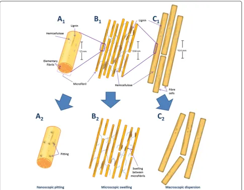
In previous work we [12,13] and other workers [14] have suggested that accessibility challenges at the macroscopic (fibre), microscopic (fibril) and nanoscopic (microfibril) level restrict effective enzymatic hydrolysis. Interestingly, non-hydrolytic disruptive proteins have been shown to dis-rupt the substrate at each of these three organizational levels. Thus it is likely that these proteins play a key role in enhancing the effectiveness of enzymatic hydrolysis. For ex-ample, amorphogenesis induced by non-hydrolytic disrup-tive proteins has been observed at, 1) the macroscopic level through the dispersion of adjacent fibres [15-17], 2) the microscopic level through the loosening/rough-ening/swelling of plant cell walls [16-27], and 3) at the nanoscopic level through the pitting of microfibrils and cellulose decrystallization (Figure 1) [17,23,26,28-30].
Recently, it has been suggested [8] that non-hydrolytic disruptive proteins can be categorized into two distinct groups. Those with an as yet unknown catalytic mechan-ism such as Swollenin [16,17], Loosenin [27], Expansins [21,31] and several Family 1 and Family 2 CBMs [18,19,23,30] and those thought to act through a direct oxidative catalytic mechanism, such as GH61 [32-36] and Family 33 CBMs [37,38] (Table 1). These two groups of proteins are thought to act in distinctly differ-ent ways on the substrate. For example, several of the proteins with uncharacterized catalytic function, such as Swollenin, Expansins and Loosenin, are thought to pro-mote amorphogenesis through disruption of the hydro-gen bonding network of the substrate (i.e. without direct cleavage of the carbohydrate chains) [16,39]. In contrast, the oxidative proteins that act on the substrate do so
through a direct catalytic oxidative mechanism, where radical species are generated in close proximity to the cellulose surface, resulting in the direct oxidative cleav-age of the cellulose chains [33-38].
A common theme between these two groups of pro-teins appears to be the release of soluble oligomers from the substrate. Specifically, Beta-Expansins have been shown to solubilize both hemicellulose and pectin from native maize silk cell walls [31], while the oxidative pro-teins GH61 and CBM33 promote the release of soluble cello-oligosaccharides from model cellulosic substrates [33,37,38]. Further evidence supporting the role of non-hydrolytic proteins in cellulose amorphogenesis comes from reports that disruptive proteins with unknown catalytic mechanisms appear to enhance the enzymatic hydrolysis of native and pretreated substrates [17,20,23-25,27,29,40-43]. In addition to these proteins, hydrolysis enhancing activity has also been observed for the oxida-tive disrupoxida-tive proteins. In particular, GH61 has been shown to significantly reduce the total protein required to achieve 70-80% hydrolysis yields of a pretreated corn stover substrate [32]. These results suggest that non-hydrolytic proteins can act within a similar time scale to that of enzymatic hydrolysis and that they are capable of promoting further amorphogenesis within a substrate that has already been disrupted by a physicochemical pretreatment.
These earlier observations encouraged various re-search groups to try to develop a better understanding of the role that non-hydrolytic proteins might play in promoting the amorphogenesis of lignocellulosic sub-strates, with the expectation that further elucidation of their action might help in the development of more effi-cient commercial enzyme preparations. However, one of the key limitations in better characterizing the non-hydrolytic proteins involved in biomass deconstruction has been the lack of simple quantitative techniques for measuring changes in cellulose accessibility. Various techniques have been used, with mixed success, to try to quantify changes in cellulose accessibility and their rela-tive merits are discussed below. We also describe a novel technique, using the adsorption of substructure-specific CBMs over short time scales (<30 minutes) to quantify cellulose accessibility [44] and to accurately quantify the degree of amorphogenesis induced by non-hydrolytic disruptive proteins.
Techniques for measuring amorphogenesis
enzyme mediated cellulose hydrolysis. This is in part due to the nature of the end product of each step. After enzymatic hydrolysis, the end products (soluble sugars)
are readily quantified by high performance liquid chro-matography or by using colorimetric techniques such as the dinitrosalicylic acid [45] or glucose oxidase assays
Figure 1Simplified schematic representation of amorphogenesis occurring at three levels of biomass organization.Native plant material at the nanoscopic (A1), microscopic (B1) and macroscopic (C1) levels of organization. After amorphogenesis induced by non-hydrolytic disruptive proteins, nanoscopic pitting (A2), microscopic swelling/roughening (B2), and macroscopicfibre dispersion (C2) is observed. These effects occur without significant release of sugars from the substrate.
Table 1 Non-hydrolytic disruptive proteins and their effects on biomass
Proteins with unknown catalytic mechanism Putative function References
Family 1 and 2 CBMs Fibre pitting/roughening, small particle release [18,19,23,30]
Swollenin, Loosenin Fibre swelling, microfibril dispersion, dispersion of cellulose aggregates [16,17,27]
Expansins Loosening of plant cell walls, solubilization of oligomeric sugars [21,31,39]
Expansin-like proteins Loosening of filter paper, dispersion of cellulose aggregates [25,78]
Fibril Forming Protein Fibril release from filter paper [15]
Proteins with putative oxidative catalytic mechanism Putative function References
GH61 Oxidative cleavage of crystalline cellulose [33-36]
CBM33 Oxidative cleavage of crystalline cellulose [37,38]
[46,47]. In contrast, the end products of the amorpho-genesis step are challenging to describe, let alone quan-tify [17].
A major challenge in trying to quantify amorphogen-esis is that the method or technique has to be versatile enough to accurately quantify a range of effects occur-ring at different levels of biomass organization, varying in scale by several orders of magnitude (from the micro-fibril, with typical diameters of 3–5 nm and lengths of 100 s to 1000 s of nm, up to the whole fibre, with dia-meters of 5–50 μm and lengths of 1–4 mm) [5,48,49]. This point was recently highlighted by Jägeret al.(2011), who noted that no single technique has the capacity to simultaneously quantify the effects occurring at multiple levels of cell wall organization [17]. As a result, previous attempts to measure these effects have typically made use of a suite of complementary qualitative and semi-quantitative techniques [15-20,22-30].
The most widely used methods employed to try to confirm disruptive protein mediated amorphogenesis of biomass typically involve the application of qualitative microscopic techniques. Light microscopy has been used to try to assess the macroscopic dispersion of Valonia cell walls and microscopic swelling of cotton fibres induced by the fungal disruptive protein Swollenin [16]. Scanning electron microscopy (SEM) has also been used to show the microscopic roughening of cotton fibres by Swollenin [17] and by the CBMs from the bacteria
Cellulomonas fimi and Clostridium cellulovorans and the fungus Trichoderma reesei [19,22,26]. Additionally, atomic force microscopy (AFM) has been used to show nanoscopic pitting of cotton microfibrils induced by CBM1 from Trichoderma pseudokoningii S-38 [28]. However, while these techniques have provided useful qualitative information on the effects of disruptive pro-teins on model cellulosic substrates, attempts to quantify these effects have so far been limited to either monitor-ing changes in crystallinity [17,23,26,29,30], measurmonitor-ing the release of small particles [19,20,23] or by indirectly quantifying amorphogenesis by measuring changes in the ease of hydrolyzability of the substrate induced by these proteins [17,24,27,42,43]. The various methods previously used to try and quantify amorphogenesis are discussed below.
Crystallinity
There are conflicting opinions on how influential cellulose crystallinity is on limiting enzymatic hydrolysis and the ef-fect that amorphogenesis-inducing proteins might have on enhancing cellulose hydrolysis. Earlier work using Fourier-transform infrared spectroscopy to assess the in-fluence of CBM1 from T. pseudokoningiiS-38 on cotton fibre deconstruction claimed that the addition of CBM1 helped reduce substrate crystallinity [30], while the highly
similar CBM1 from T. reesei when added to Whatman CF11 cellulose fibres did not appear to result in any de-crease in substrate crystallinity when measured using X-ray diffraction [22]. In contrast, the addition of bacterial derived CBM3a fromC. cellulovoransreduced the crystal-linity of cotton fibres when assessed by both Fourier-transform infrared spectroscopy and X-ray diffraction [26] while a recombinant Swollenin, Swo2 from T. pseudoko-ningiiS-38 apparently caused an increase in the crystallin-ity of Avicel PH-101 [50]. Conversely, the application of a recombinant Swollenin from T. reesei resulted in a de-crease in the crystallinity of filter paper, alpha-cellulose and Avicel when measured by powder X-ray diffraction [17].
Although these non-uniform observations might suggest that different combinations of disruptive protein and sub-strate result in different changes in crystallinity, it is more likely that these varied results are due to issues with the methods used to measure crystallinity. These issues include the interpretation of results from the different methods for measuring crystallinity and the applicability of extrapolating crystallinity measurements to suggest the degree of phogenesis. In earlier work [8] it was suggested that amor-phogenesis primarily resulted from substrate changes such as cellulose delamination or fibrillation, where relatively large, intact fragments, still containing crystalline regions, are released from the bulk of the substrate. Pinto et al.
(2004) have also suggested that non-hydrolytic disruptive proteins could increase the accessibility of cellulosic sub-strates without affecting the crystallinity. These workers reported no decrease in the crystallinity of cotton fibres after treatment with a non-hydrolytic disruptive protein, while observing a roughening of the cotton fibres as visua-lized by SEM [22]. A possible parallel mechanism is that the increase in cellulose accessibility could result from the swelling or loosening of the interactions between microfi-brils, resulting in the overall weakening of the cell wall while leaving the crystalline cores of the microfibrils rela-tively untouched. Thus it is possible that any changes in the crystallinity of the substrate would only occur as a second-ary effect of the more general process of amorphogenesis.
Particle release and size reduction
Earlier work claiming that certain CBMs could induce amorphogenesis was carried out using CBM2a from C. fimi where their addition to cotton fibres resulted in the release of small particles without a concomitant release of reducing sugars [19,20]. However, although this technique could semi-quantitatively describe the release of particles from the substrate, it provided no characterization of the residual substrate.
fromAspergillus fumigatusandTrichoderma asperellum, respectively, on Avicel PH-101 [24,43]. These research-ers used light microscopy to try to quantify size reduc-tion in Avicel particles after incubareduc-tion with Swollenins, with Chenet al.(2010) using image-analysis software to demonstrate an almost 2-fold reduction in the size of the Avicel particles [24].
Hydrolysis enhancement
Several independent research groups have demonstrated enhanced enzymatic hydrolysis of cellulosic and lignocellu-losic substrates after treatment with non-hydrolytic disrup-tive proteins [17,20,23-25,27,29,32,40-43]. Although this work collectively suggests that these disruptive proteins are indeed capable of enhancing the hydrolyzability of model and native cellulosic substrates, the “degree of hydrolysis enhancement” method is an indirect approach to try to quantify cellulose amorphogenesis. For example, it is pos-sible that some of the enhancement of hydrolysis observed after addition of disruptive proteins could be due to these proteins binding to and blocking lignin, thereby preventing non-productive adsorption of cellulases to the lignin [51,52], rather than through a direct “disruptive” effect. Additionally, the somewhat contradictory results observed when similar Family 2 CBMs from Cel6A and Xyn10A fromC. fimiwere used to test for hydrolysis enhancement places further doubt on the suitability of using enhance-ment of substrate hydrolyzability as a tool for accurately quantifying amorphogenesis [20,53].
More recent attempts at quantifying cellulose disruption induced by non-hydrolytic proteins have exploited the synergism observed between these proteins and endoglu-canases specific for amorphous regions of cellulose [27,43]. This method uses quantification of sugar released by endoglucanases from a disrupted cellulosic substrate as an indirect measure of the degree of amorphogenesis of the substrate induced by the non-hydrolytic disruptive proteins. Although this technique has been successfully applied to “semi-quantitatively” measure the disruptive effects of Swollenin on Avicel [43] and Loosenin on cotton fibres [27], one drawback of this technique is the specifi-city of the endoglucanases for amorphous cellulose [27,43]. As the endoglucanases employed are specific for amorphous cellulose this approach will only work well if the amorphogenesis step results in a simple decrystaliza-tion of cellulose. However, it will not measure the other possible influences of enhanced cellulose accessibility such as the splitting, delaminating or loosening of the cellulose, which could occur without any significant changes in the crystallinity of the cellulose.
Other potential methods for quantifying amorphogenesis While each of the putative indications of amorphogenesis, such as delamination, fibrillation, swelling, loosening,
roughening, pitting, weakening or decrystallization of the substrate can all be thought of as distinct processes, if they play a role in amorphogenesis we should be able to in-crease access of the enzymes to the cellulose without a significant increase in the release of reducing sugars. Sev-eral groups have tried to develop methods of accurately quantifying changes in the accessibility of lignocellulosic substrates, with many of these techniques modified from traditional pulp and paper procedures [54]. Although these techniques have previously only been used to assess the overall accessibility of the substrate, in-cluding accessibility to the lignin and hemicelluloses as well as the cellulose, some of these techniques also have potential for quantifying the amorphogen-esis step.
Techniques with potential for quantifying cellulose ac-cessibility and the amorphogenesis step include measuring the water retention value, mean fibre size, nitrogen ad-sorption capacity and mercury porosimetry of the cellu-losic substrate. Or, performing techniques such as solute exclusion, differential scanning calorimetry, time-domain nuclear magnetic resonance, Simons’Staining and protein adsorption (Reviewed in [54]). One of the benefits of these modified pulp and paper techniques is that they can moni-tor changes in the cellulose accessibility at the macro-scopic, microscopic and nanoscopic levels. For example, fibre size measurements give an indication of macroscopic changes in accessibility while techniques such as mercury porosimetry and solute exclusion can be used to quantify changes in pore size distribution at the microscopic level [54]. The ability to monitor changes in the substrate at the macroscopic, microscopic and nanoscopic levels would likely be of great value when quantifying cellulose amor-phogenesis, as this phenomenon could occur at each of these levels.
not yet been used to quantify overall cellulose accessibility, it is likely that these techniques could be adapted to provide some quantitative information on the extent and mechan-ism of disruption induced by non-hydrolytic disruptive proteins.
An alternative technique with potential for measuring cellulose accessibility involves the use of protein adsorp-tion to try to quantify the amount of cellulose in the substrate that is accessible to enzymes [7,12,44,58]. This technique makes use of the cellulases themselves as ac-cessibility probes, where either a mixture of cellulases or monocomponent cellulases are incubated with the sub-strate followed by quantifying the amount of protein that is adsorbed to the substrate. While this technique might provide a good indication of the amount of accessible surface area of the substrate, there are two key problems to be overcome when using this approach. First, unless the adsorption study is carried out at low temperatures (which will not be representative of hydrolysis reaction conditions!), the cellulases will hydrolyze the substrate, thereby changing the substrate accessibility during the course of the assay. Secondly, cellulases are known to adsorb unproductively to lignin, which restricts the ac-curate quantification of cellulose accessibility [51,59].
Other recent attempts at using cellulases to quantify ac-cessibility have involved the production of a fluorescently-tagged non-hydrolytic CBM to enhance the accuracy and sensitivity of the protein adsorption technique [44]. This technique makes use of BSA blocking to overcome pro-blems with lignin-binding [60]. However, one potential drawback is that different CBMs recognize different sub-structures within the substrate [61,62]. Thus, it is possible that a probe making use of a single CBM might primarily be quantifying the accessibility of a specific cellulosic sub-structure, rather than the overall accessibility of the cellu-lose. An alternative strategy might be to use cellulase inhibitors that limit or prevent substrate hydrolysis during protein adsorption experiments at more typical substrate hydrolysis temperatures (i.e. 50°C). However, while inhibi-tors such as hexachloropalladate have been shown to in-hibit Cel7a from T. reesei, it has only a limited effect on most of the other enzymes present in commercial cellulase preparations [63,64]. Thus the inhibition approach would only work when using monocomponent cellulases for the adsorption studies.
Another procedure for measuring accessibility is the Simons’stain method [65]. This technique involves quanti-fying the adsorption of an anionic direct dye which has a higher affinity for cellulose than to lignin and hemicellu-lose [65,66]. The amount of dye bound to the cellulosic substrate gives a good indication of the total amount of cellulose accessible to cellulase within the substrate [7].
Overall, these methods have proven to be useful in quan-tifying some of the changes that occur in the substrate
during enzymatic hydrolysis. However, there are several drawbacks to using these techniques to measure changes in cellulose accessibility. For example, the water retention value is known to be insensitive to small changes in fibre characteristics and would not be able to detect changes in the cellulosic component during or after amorphogenesis. Another major drawback of many of these techniques is that they measure the overall accessibility of the substrate, including the amount of accessible lignin and hemicellu-lose, not just the cellulose. This restricts techniques such as nitrogen adsorption, solute exclusion, differential scanning calorimetry and time-domain nuclear magnetic resonance from being used to assess changes in the specific amount of accessible cellulose within the substrate during or after the amorphogenesis process. Finally, some of these techniques are labour intensive, such as the solute exclusion technique, or require the use of toxic heavy metals, as in determining mercury porosimetry.
To date, no single technique has provided an accurate quantitative measure of the changes occurring within the cellulosic substrate during amorphogenesis. In the work described below we describe a novel method where cellu-lose substructure-specific CBMs have been successfully used to quantify amorphogenesis by determining changes in the accessibility and surface morphology of cellulose before and after treatment with amorphogenesis-inducing agents.
Results and discussion
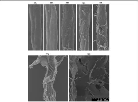
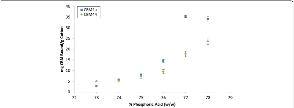
As protein-mediated amorphogenesis is still an evolving concept, we initially assessed the sensitivity and reproduci-bility of a CBM-mediated method for quantifying changes in cellulose accessibility by using concentrated phosphoric acid to disrupt cotton fibres to varying degrees of disassoci-ation [67]. The use of harsh acid treatments was intended to provide an exaggerated range of disrupted substrates, and was not intended to be representative of milder, bio-logical treatments. The disruptive effect of the acid was ini-tially qualitatively assessed using SEM (Figure 2) followed by a quantitative assessment where the adsorption of each of the substructure-specific CBMs [61] was determined (Figure 3).
for crystalline regions [61,71]. As CBM2a is thought to interact with 2–3 chains in the ordered crystal lattice [70], in the work reported here, we have defined a crys-talline region as one with 2–3 adjacent crystalline cellu-lose chains. In contrast, the amorphous-cellulose binding CBM used in this work, CBM44, is a Type B CBM with a binding site comprised of a narrow groove lined with hydrophobic aromatic residues [72]. This groove confers binding specificity to free polysaccharide chains, such as those present in amorphous cellulose, but does not enable binding to the tightly packed chains found within crystalline cellulose [72].
To try to progressively disrupt cotton fibres in order to assess the ability of CBM adsorption to quantify cellulose accessibility a range of phosphoric acid concentrations
was used. As the concentration of phosphoric acid was increased, an increase in the degree of disruption of the sample, including the splitting, delaminating and roughen-ing of the fibres was apparent (Figure 2). An increase in disruption generally correlated with an increase in binding of both the amorphous-binding and crystalline-binding CBMs up to a concentration of 77% acid treat-ment (Figure 3). This increase in the combined binding of both CBMs provided a good indication that the overall (crystalline and amorphous) cellulose accessibility had increased. Surprisingly, the use of increasingly harsh acid treatments resulted in only a relatively small increase in the amount of CBM2a bound to the substrate when com-pared to the increase in CBM44 binding. It had been anticipated that the increasing disruption of the fibres
Figure 2SEM images of cotton fibres disrupted by phosphoric acid treatments.SEM micrographs of cotton fibres after treatment with a range ofo-phosphoric acid concentrations. After control treatment (nanopure water, 0% (w/w) acid), cotton fibres appear smooth, with few surface features. As the acid concentration was increased to near the point of cellulose dissolution (~73%-78%), manifestations of
would result in the amorphous-binding CBM44 being bound more than the crystalline-binding CBM2a. How-ever, as CBM2a recognizes only two to three adjacent chains as being‘crystalline’, the observed relative increase in CBM2a binding over CBM44 binding as the acid con-centration increased was likely due to the increased solv-ent exposure of small microcrystalline substructures within the acid-disrupted cotton fibres.
As the acid concentration was further increased from 77% to 78%, the SEM micrographs of the cotton showed that any residual fibre structure had been lost and that the substrate now had the form of amorphous cellulosic
‘mats’ (Figure 2). These SEM observations complemen-ted the CBM adsorption results, where increasing the acid concentration from 77% to 78% resulted in a large increase in the amount of adsorbed CBM44, without sig-nificantly altering the adsorption of CBM2a (Figure 3). It was apparent that the specificity of CBM adsorption was distinct enough that changes in the surface morphology of the cellulosic substrates could be readily differentiated.
After determining that CBM adsorption could be used to quantify acid-induced changes in cellulose accessibil-ity, we attempted to correlate the degree of substrate disruption (quantified by CBM adsorption) with enzym-atic hydrolyzability. Each of the phosphoric-acid dis-rupted cotton fibre samples was hydrolyzed using a commercial cellulase mixture (30 filter paper units/g cel-lulose of Celluclast 1.5 L, supplemented with 15 cello-biase units/g cellulose of Beta-glucosidase (Novozym 188, Novozymes A/S, Bagsværd, Denmark)). The initial hydrolysis rate (defined here as the hydrolysis rate over the first 30 minutes of the reaction) was plotted against the adsorption of each individual CBM, as well as the
sum of their adsorptions (Figure 4). The adsorption of each individual CBM, and particularly their summed adsorptions, was found to correlate well with the enhanced enzymatic hydrolyzability of the cotton fibres. The steeper slope of the curve for CBM44 when com-pared to CBM2a seemed to indicate that, at least for the initial stages of hydrolysis, enzymatic hydrolysis rates were influenced more by the amount of accessible amorphous cellulose than they were by the amount of accessible crystalline cellulose.
Interestingly, although the hydrolyzability and CBM44 adsorption increased with every incremental increase in phosphoric acid concentration, this was not the case for CBM2a. As the acid concentration was increased from 77% to 78%, the adsorption of CBM2a to the substrate was not significantly affected, even as the hydrolyzability continued to increase. This suggested that any attempts to correlate changes in substrate accessibility to hydro-lyzability using a specific mono-component cellulase may be problematic, as some cellulases contain CBMs specific for crystalline cellulose and might therefore underestimate the accessibility of the highly amorphous regions of the substrate.
This seemed to indicate that substructure specific CBMs could be used to quantify acid-induced changes in cellu-lose accessibility and that increases in accessibility (as determined by CBM adsorption) can provide a good pre-dictor of initial rates of enzymatic hydrolysis.
Quantification of Swollenin-induced increases in cellulose accessibility
As Swollenin had previously been shown to disrupt mercer-ized cotton fibres [16], we next tried to quantify any changes in cellulose accessibility and surface morphology of
mercerized cotton fibres treated with Swollenin by looking at the degree of adsorption of substructure-specific CBMs to the treated fibres. Although mercerization is known to cause a significant reduction in the crystallinity of cellulosic substrates, mercerized cellulose has been shown to retain some adsorptive capacity for crystalline binding CBMs [73]. After incubation with Swollenin, binding of both the crys-talline and amorphous-specific CBMs to the mercerized cotton fibres increased (Figure 5).
The increase in binding was more pronounced for-CBM2a than for CBM44 after Swollenin treatment, indicat-ing that the increase in accessibility was not simply due to Swollenin-mediated decrystallization of the cellulose at the microfibril surface. This suggested that Swollenin might act by promoting the delamination or fibrillation of the sub-strate, or by promoting the “splitting” of microfibrils, thereby exposing new crystalline regions of cellulose to the CBMs.
Subsequent SEM micrographs of Swollenin-treated mer-cerized cotton fibres indicated that Swollenin treatments resulted in a smoothing of the roughened patches produced during mercerization (Figure 6). This smoothing effect was in contrast to the buffer- and BSA-treated mercerized cot-ton fibres, which retained their roughened surface. After Swollenin treatment, the roughened patches at the surface of the fibres appeared to have been sloughed off, revealing the smooth, well ordered surface of the underlying cotton fibre. The observed increase in turbidity in the supernatant after Swollenin treatment (data not shown) was also indica-tive of the release of small particles into solution. It is pos-sible that the roughened patches at the surface of the
mercerized cotton fibres will contain a higher proportion of amorphous cellulose than the underlying fibre, as these protruding rough regions were more exposed to the NaOH used for mercerization. This treatment has been shown to promote the conversion of crystalline cellulose I into amorphous cellulose and crystalline cellulose II [74]. It is possible that the release of these roughened particles from the surface of the fibre resulted in an increase in both the amount of exposed amorphous cellulose (primarily on the released particles) and the amount of exposed crystalline cellulose (primarily on the newly exposed surface of the underlying cotton fibre). However, it should be noted that the small roughened particles that are released from the surface of the cotton fibres appear to be approximately 100 nm in the shorter direction, and up to 1000 nm in the longer direction (estimated from Figure 6). Since the cellu-losic cores of cotton microfibrils have diameters of only 3– 5 nm and lengths of 100 s to 1000 s of nm [5,48,49], it is possible that the small, roughened particles released from the surface of the cotton fibres still contained significant amounts of crystalline cellulose.
Although it was not evident by which specific mechanism the Swollenin resulted in this“smoothing”effect, it is pos-sible that Swollenin acts in a similar manner to the Expan-sin family of proteins, which have been shown to weaken plant cell walls through disruption of the hydrogen bonding network between plant cell wall polymers [39]. If Swollenin disrupts hydrogen bonding in a similar mode to Expansins this might also explain how Swollenin appears to both dis-rupt the cell wall structure of Whatman filter paper No. 1 fibres and result in the swelling of cotton fibres [16,17].
R² = 0.9828 R² = 0.9728
R² = 0.9979
0 100 200 300 400 500 600 700 800
0 10 20 30 40 50 60 70
Initial Hydrolysis Rate
ug/mL glucose/hour
mg CBM Bound/mg Cotton
CBM2a
CBM44
Sum CBMs
These results indicated that substructure-specific CBMs could not only be used to track changes in cellulose acces-sibility after harsh acid treatments, but could also be used to track changes in surface morphology after the milder, Swollenin-induced, amorphogenesis. This technique has several advantages over current alternatives as it provided a direct, quantitative method able to consolidate changes in multiple substrate characteristics. Specifically, changes in the amounts of accessible amorphous cellulose, access-ible crystalline cellulose and the total (amorphous and crystalline) accessible cellulose can all be quantified. It was also apparent that this method could help better indicate the mode of action of non-hydrolytic, disruptive/amorpho-genesis-inducing proteins and has potential to yield novel insights into the mechanisms of glycosyl hydrolases and the other accessory enzymes involved in lignocellulose de-construction. Additionally, this technique has the potential
to facilitate comparisons of the disruptive capabilities of various non-hydrolytic proteins which might promote an increase in cellulose accessibility to the more traditional, hydrolytic components of the cellulase enzyme mixture.
It is also possible that CBMs with specificities for certain hemicelluloses, such as xylan or mannan, might be able to be used to track changes in the accessibility of these poly-mers during pretreatment, amorphogenesis and hydrolysis of softwoods, hardwoods and agricultural residues. In pre-vious work, Filonova et al., (2007) demonstrated the use of fluorescently-tagged mannan-specific CBM’s to quantify the accessibility of mannan in wood tissues and pulp, after applying a protein-based lignin-blocking technique to pre-vent non-specific adsorption of the CBMs to lignin [75]. The use of CBM-specific antibodies, or conjugation of CBMs to distinct fluorophores, have been used to provide direct visualization of the locations of the different 47.6%
22.1%
38.0%
0 5 10 15 20 25
Sum CBMs CBM44
CBM2a
mg CBM Bound/g Cotton
Control
Swollenin
Figure 5Adsorption of CBMs to Swollenin-treated cotton fibres.Swollenin-induced changes in the accessibility and surface morphology of mercerized cotton fibres quantified using CBM adsorption. The % values represent the % increase in adsorption of the CBMs after Swollenin treatment. A BSA negative protein control was found to have no significant effect on the extent of binding of either CBM. At least three replicates were performed for each sample. Error bars represent one standard deviation from the mean.
polymers or substructures at the substrate surface [75,76]. Thus, by utilizing a suite of different CBMs with specifici-ties for a range of structural features of the substrate, it might be possible to track changes in the morphology of the substrate during pretreatment and hydrolysis while better quantifying the role that enzyme access to the cellu-lose plays in limiting the rate and extent of enzymatic hydrolysis.
Conclusions
Previous attempts to try and quantify the “cellulose swel-ling/delamination” or the “amorphogenesis step” of cellu-lose hydrolysis have tended to make use of a suite of complementary qualitative and semi-quantitative techni-ques. While these techniques have provided some useful in-formation regarding the effects of amorphogenesis-inducing proteins on (ligno) cellulosic substrates, they have typically provided little insight into the mode of action of these proteins. A novel technique, using non-hydrolytic substructure-specific CBMs capable of quantitatively meas-uring changes in cellulose accessibility and surface morph-ology was successfully used to track changes in the cellulose during Swollenin-induced amorphogenesis. This novel method provided useful insights into how proteins such as Swollenin might increase cellulose accessibility by non-hydrolytic mechanisms such as the swelling or delam-ination of the cellulose substructures.
Methods Proteins
CBM44 from Clostridium thermocellum was purchased from NZYTech (Lisbon, Portugal, CR0049). CBM2a from
Cellulomonas fimi was provided by Dr. Douglas Kilburn from the University of British Columbia, Canada, after re-combinant expression and purification from E. coli. Re-combinant Swollenin (Swo1) was generously provided by VTT Technical Research Centre, Finland. Briefly, the Swollenin was expressed in Trichoderma reesei as a histidine-tagged protein and purified in two chromato-graphic steps.
Phosphoric acid-induced amorphogenesis
Phosphoric acid-disrupted cotton fibres were prepared fol-lowing a protocol similar to that described by Zhanget al.
(2006) [67]. Briefly, ice-coldo-phosphoric acid (Fisher Sci-entific, Canada, A242) solutions were produced at various concentrations and 14.5 mL was added to 50 mL centrifuge tubes containing 0.2 g cotton fibres (Sigma-Aldrich, St. Louis MO, USA, C6663) pre-wetted with 0.5 mL nanopure water to give finalo-phosphoric acid concentrations of 0– 78% w/w. Samples were incubated for one hour on ice with occasional mixing. Ice-cold nanopure water (35 mL) was slowly added to each sample, followed by centrifugation at 10,000 g for 15 minutes. The fibres were resuspended in
50 mL nanopure water and washed a further 4 times with 50 mL nanopure water, followed by one wash with 50 mL 20 mM Na2CO3 (Fisher Scientific, Pittsburg PA, USA,
S263) and 2 subsequent washes in 50 mL nanopure water. The cotton fibres were then lyophilized overnight.
Swollenin-induced amorphogenesis
Prior to Swollenin treatment, 200 mg cotton fibres (Sigma-Aldrich, St. Louis MO, USA, C6663) were mercer-ized in 50 mL 25% (w/w) ice-cold NaOH for 15 minutes. The mercerized fibres were washed thoroughly with nano-pure water then lyophilized overnight. Dried cotton fibres (50 mg) were weighed into 2 mL screwcap tubes. Swolle-nin or BSA (10 μg/mg cotton in 50 mM sodium acetate buffer, pH 5), or buffer alone was added and the samples were incubated overnight at 50°C in a FinepcrCombi SV12 hybridization incubator at 30 rpm. Protein was removed from the samples by extensive washing with nanopure water. Samples were lyophilized overnight prior to CBM adsorption and microscopy studies.
Turbidity measurements
Supernatants from the Swollenin- or control-treated mercerized cotton fibres were transferred to 1 mL plastic cuvettes, and the optical density at 600 nm was read on a Varian Cary 50 Bio Spectrophotometer. Sodium acetate buffer (50 mM, pH 5) was used as a blank. Samples were run in triplicate.
Scanning electron microscopy
Lyophilized cotton fibres were mounted on aluminum SEM stubs using double sided tape and sputter-coated with 10 nm Au/Pd (80:20 mix) then imaged on a Hitachi S-2600 VP-SEM (Tokyo, Japan).
CBM adsorption
Enzymatic Hydrolysis
Cotton samples were hydrolyzed using 5 mg substrate in 1 mL 50 mM sodium acetate buffer, pH 5, at 50°C for 30 minutes, with an enzyme loading of 30 filter paper units Celluclast (Novozyme, USA) per gram cotton and sup-plemental β-glucosidase (Novozymes 188, Novozymes, Bagsværd, Denmark) at 1:2 cellobiase units/filter paper unit. After hydrolysis, the enzymes were heat inactivated at 100°C for 10 minutes, samples were centrifuged at 16,000 g in a benchtop centrifuge for 10 minutes, and the glucose concentration of the supernatant was deter-mined using the glucose oxidase assay [46,47].
Abbreviations
CBM: Carbohydrate binding module; SEM: Scanning electron microscopy; BSA: Bovine serum albumin.
Competing interests
The authors declare that they have no competing interests.
Acknowledgements
The Natural Sciences and Engineering Research Council of Canada (NSERC), Natural Resources Canada (NRCan) and Genome BC are gratefully
acknowledged for the support of this work. We particularly want to thank Dr. Doug Kilburn and Emily Kwan at UBC for generously donating CBM2a, as well as Dr. Merja Penttilä and Martina Andberg at VTT Technical Research Centre of Finland for their generous donation of purified Swollenin.
Authors' contributions
KG designed and carried out the experiments, analyzed results and drafted the manuscript. VA and JNS coordinated the study, helped write and review the manuscript. All authors read and approved the final manuscript.
Received: 16 May 2012 Accepted: 2 July 2012 Published: 24 July 2012
References
1. Lynd L, Weimer P, van Zyl W, Pretorius I:Microbial cellulose utilization: fundamentals and biotechnology.Microbiol Mol Biol Rev2002, 66(3):506–577.
2. Zhang YH, Lynd LR:Toward an aggregated understanding of enzymatic hydrolysis of cellulose: noncomplexed cellulase systems.Biotechnol Bioeng2004,88(7):797–824.
3. Chundawat SP, Beckham GT, Himmel ME, Dale BE:Deconstruction of lignocellulosic biomass to fuels and chemicals.Annu Rev Chem Biomol Eng2011,2:121–145.
4. Somerville C, Bauer S, Brininstool G, Facette M, Hamann T, Milne J, Osborne E, Paredez A, Persson S, Raab T,et al:Toward a systems approach to understanding plant cell walls.Science2004,306(5705):2206–2211. 5. Himmel ME, Ding SY, Johnson DK, Adney WS, Nimlos MR, Brady JW, Foust
TD:Biomass recalcitrance: engineering plants and enzymes for biofuels production.Science2007,315(5813):804–807.
6. Scheller H, Ulvskov P:Hemicelluloses.Annu Rev Plant Biol2010,61:263–289. 7. Arantes V, Saddler JN:Cellulose accessibility limits the effectiveness of
minimum cellulase loading on the efficient hydrolysis of pretreated lignocellulosic substrates.Biotechnol Biofuels2011,4:3.
8. Arantes V, Saddler J:Access to cellulose limits the efficiency of enzymatic hydrolysis: the role of amorphogenesis.Biotechnol Biofuels2010,3:4. 9. Lynd LR, Laser MS, Bransby D, Dale BE, Davison B, Hamilton R, Himmel M,
Keller M, McMillan JD, Sheehan J,et al:How biotech can transform biofuels.Nat Biotechnol2008,26(2):169–172.
10. Service RF:Is there a road ahead for cellulosic ethanol?Science2010,329 (5993):784–785.
11. Klein-Marcuschamer D, Oleskowicz-Popiel P, Simmons BA, Blanch HW:The challenge of enzyme cost in the production of lignocellulosic biofuels. Biotechnol Bioeng2012,109(4):1083–1087.
12. Mansfield S, Mooney C, Saddler J:Substrate and enzyme characteristics that limit cellulose hydrolysis.Biotechnol Prog1999,15(5):804–816.
13. Chandra RP, Bura R, Mabee WE, Berlin A, Pan X, Saddler JN:Substrate pretreatment: the key to effective enzymatic hydrolysis of lignocellulosics?Adv Biochem Eng Biotechnol2007,108:67–93. 14. Chundawat SPS, Donohue B, Sousa L, Elder T, Agarwal U, Lu F, Ralph J,
Himmel MVB, Dale B:Multiscale visualization and characterization of lignocellulosic plant cell wall deconstruction during thermochemical pretreatment.EES2011,4:12.
15. Banka RR, Mishra S, Ghose TK:Fibril formation from cellulose by a novel protein fromTrichoderma reesei: A non-hydrolytic cellulolytic component?World J Microb Biot1998,14(4):551–560.
16. Saloheimo M, Paloheimo M, Hakola S, Pere J, Swanson B, Nyyssönen E, Bhatia A, Ward M, Penttilä M:Swollenin, aTrichoderma reeseiprotein with sequence similarity to the plant expansins, exhibits disruption activity on cellulosic materials.Eur J Biochem2002,269(17):4202–4211. 17. Jäger G, Girfoglio M, Dollo F, Rinaldi R, Bongard H, Commandeur U, Fischer
R, Spiess AC, Buchs J:How recombinant swollenin fromKluyveromyces lactisaffects cellulosic substrates and accelerates their hydrolysis. Biotechnol Biofuels2011,4:33.
18. Gilkes N, Kilburn D, Miller RJ, Warren R, Sugiyama J, Chanzy H, Henrissat B: Visualization of the adsorption of a bacterial endo-beta-1,4-glucanase and its isolated cellulose-binding domain to crystalline cellulose.Int J Biol Macromol1993,15(6):347–351.
19. Din N, Gilkes N, Tekant B, Miller R, Warren A, Kilburn D:Non-hydrolytic disruption of cellulose fibres by the binding domain of a bacterial cellulase.Nat Biotech1991,9:1096–1099.
20. Din N, Damude H, Gilkes N, Miller RJ, Warren R, Kilburn D:C1-Cx revisited: intramolecular synergism in a cellulase.Proc Natl Acad Sci U S A1994, 91(24):11383–11387.
21. Cosgrove D:Loosening of plant cell walls by expansins.Nature2000, 407(6802):321–326.
22. Pinto R, Moreira S, Mota M, Gama M:Studies on the cellulose-binding domains adsorption to cellulose.Langmuir2004,20(4):1409–1413. 23. Gao PJ, Chen GJ, Wang TH, Zhang YS, Liu J:Non-hydrolytic disruption of
crystalline structure of cellulose by cellulose binding domain and linker sequence of cellobiohydrolase I fromfrom Penicillium janthinellum.Sheng Wu HuaXue Yu Sheng Wu Wu Li XueBao(Shanghai)2001,33(1):13–18. 24. Chen XA, Ishida N, Todaka N, Nakamura R, Maruyama J, Takahashi H,
Kitamoto K:Promotion of efficient Saccharification of crystalline cellulose byAspergillus fumigatusSwo1.Appl Environ Microbiol2010,76(8): 2556–2561.
25. Lee H, Lee S, Ko H, Kim K, Choi I:An expansin-like protein fromHahella chejuensisbinds cellulose and enhances cellulase activity.Mol Cells2010,29 (4):379–385.
26. Ciolacu D, Kovac J, Kokol V:The effect of the cellulose-binding domain fromClostridium cellulovoranson the supramolecular structure of cellulose fibers.Carbohydr Res2010,345(5):621–630.
27. Quiroz-Castañeda RE, Martínez-Anaya C, Cuervo-Soto LI, Segovia L, Folch-Mallol JL:Loosenin, a novel protein with cellulose-disrupting activity fromBjerkandera adusta.Microb Cell Fact2011,10:8.
28. Lee I, Evans B, Woodward J:The mechanism of cellulase action on cotton fibers: evidence from atomic force microscopy.Ultramicroscopy2000, 82(1–4):121–213.
29. Hall M, Bansal P, Lee J, Realff M, Bommarius A:Cellulose crystallinity–a key predictor of the enzymatic hydrolysis rate.FEBS J2010,277(6):1571–1582. 30. Wang L, Zhang Y, Gao P:A novel function for the cellulose binding
module of cellobiohydrolase I.Sci China C Life Sci2008,51(7):620–629. 31. Tabuchi A, Li LC, Cosgrove DJ:Matrix solubilization and cell wall
weakening byβ-expansin (group-1 allergen) from maize pollen.Plant J 2011,68(3):546–559.
32. Harris P, Welner D, McFarland K, Re E, Navarro Poulsen J, Brown K, Salbo R, Ding H, Vlasenko E, Merino S,et al:Stimulation of lignocellulosic biomass hydrolysis by proteins of glycoside hydrolase family 61: structure and function of a large, enigmatic family.Biochemistry2010, 49(15):3305–3316.
33. Langston JA, Shaghasi T, Abbate E, Xu F, Vlasenko E, Sweeney MD: Oxidoreductive cellulose depolymerization by the enzymes cellobiose dehydrogenase and glycoside hydrolase 61.Appl Environ Microbiol2011, 77(19):7007–7015.
exploits biomass components.Proc Natl Acad Sci U S A2011, 108(37):15079–15084.
35. Westereng B, Ishida T, Vaaje-Kolstad G, Wu M, Eijsink VG, Igarashi K, Samejima M, Ståhlberg J, Horn SJ, Sandgren M:The putative
endoglucanase PcGH61D fromPhanerochaete chrysosporiumis a metal-dependent oxidative enzyme that cleaves cellulose.PLoS One2011, 6(11):e27807.
36. Beeson WT, Phillips CM, Cate JH, Marletta MA:Oxidative cleavage of cellulose by fungal copper-dependent polysaccharide monooxygenases. J Am Chem Soc2012,134(2):890–892.
37. Vaaje-Kolstad G, Westereng B, Horn S, Liu Z, Zhai H, Sørlie M, Eijsink V:An oxidative enzyme boosting the enzymatic conversion of recalcitrant polysaccharides.Science2010,330(6001):219–222.
38. Forsberg Z, Vaaje-Kolstad G, Westereng B, Bunæs AC, Stenstrøm Y, MacKenzie A, Sørlie M, Horn SJ, Eijsink VG:Cleavage of cellulose by a CBM33 protein.Protein Sci2011,20(9):1479–1483.
39. McQueen-Mason S, Cosgrove D:Disruption of hydrogen bonding between plant cell wall polymers by proteins that induce wall extension. Proc Natl Acad Sci U S A1994,91(14):6574–6578.
40. Lemos MA, Teixeira JA, Domingues MRM, Mota M, Gama FM:The enhancement of the cellulolytic activity of cellobiohydrolase I and endoglucanase by the addition of cellulose binding domains derived fromTrichoderma reesei.Enzyme MicrobTech2003,32(1):35–40.
41. Baker J, King M, Adney W, Decker S, Vinzant T, Lantz S, Nieves R, Thomas S, Li L, Cosgrove D,et al:Investigation of the cell-wall loosening protein expansin as a possible additive in the enzymatic saccharification of lignocellulosic biomass.Appl Biochem Biotechnol2000,84–86:217–223. 42. Kim E, Lee H, Bang W, Choi I, Kim K:Functional characterization of a
bacterial expansin fromBacillus subtilisfor enhanced enzymatic hydrolysis of cellulose.Biotechnol Bioeng2009,102(5):1342–1353. 43. Wang Y, Tang R, Tao J, Gao G, Wang X, Mu Y, Feng Y:Quantitative
investigation of non-hydrolytic disruptive activity on crystalline cellulose and application to recombinant swollenin.Appl Microbiol Biotechnol2011, 91(5):1353–1363.
44. Hong J, Ye X, Zhang Y:Quantitative determination of cellulose accessibility to cellulase based on adsorption of a nonhydrolytic fusion protein containing CBM and GFP with its applications.Langmuir2007,23 (25):12535–12540.
45. Miller G:Use of dinitrosalicylic acid reagent for determination of reducing sugar.Anal Chem1959,31:3.
46. Berezin IV, Rabinovich ML, Sinitsyn AP:Applicability of quantitative kinetic spectrophotometric method for glucose determination.Biokhimiia1977, 42(9):1631–1636.
47. Berlin A, Maximenko V, Bura R, Kang K-Y, Gilkes N, Saddler J:A rapid microassay to evaluate enzymatic hydrolysis of lignocellulosic substrates. Biotechnol Bioeng2005,93(5):7.
48. Sjöström E:Wood chemistry, fundamentals and applications. 2nd edition. San Diego: Academic; 1993.
49. Beckham GT, Matthews JF, Peters B, Bomble YJ, Himmel ME, Crowley MF: Molecular-level origins of biomass recalcitrance: decrystallization free energies for four common cellulose polymorphs.J Phys Chem B2011, 115(14):4118–4127.
50. Zhou Q, Lv X, Zhang X, Meng X, Chen G, Liu W:Evaluation of swollenin fromTrichoderma pseudokoningiias a potential synergistic factor in the enzymatic hydrolysis of cellulose with low cellulase loadings.World J Microbiol Biotechnol2011,27(8):6.
51. Berlin A, Gilkes N, Kurabi A, Bura R, Tu M, Kilburn D, Saddler J:Weak lignin-binding enzymes: a novel approach to improve activity of cellulases for hydrolysis of lignocellulosics.Appl Biochem Biotechnol2005,121–124: 163–170.
52. Yang B, Wyman CE:BSA treatment to enhance enzymatic hydrolysis of cellulose in lignin containing substrates.Biotechnol Bioeng2006, 94(4):611–617.
53. Esteghlalian A, Srivastava V, Gilkes N, Kilburn D, Warren R, Saddle J:Do cellulose binding domains increase substrate accessibility?Appl Biochem Biotechnol2001,91–93:575–592.
54. Chandra RP, Esteghlalian A, Saddler J:Assessing substrate accessibility to enzymatic hydrolysis by cellulases.InCharacteristics of lignocellulosic materials. Edited by Hu TQ. Oxford: Blackwell; 2008:60–80.
55. Nelson R:The determination of moisture transition in cellulosic materials using differential scanning calorimetry.J Appl Polym Sci1977,21:10.
56. Nakamura K, Hatakeyama T, Hatakeyama H:Studies on bound water of cellulose by differential scanning calorimetry.Textile Res J1981,72(9):7. 57. Felby C, Thygesen LG, Kristensen JB, Jørgensen H, Elder T:Cellulose–water
interactions during enzymatic hydrolysis as studied by time domain NMR.Cellulose2008,15(5):8.
58. Jeoh T, Ishizawa CI, Davis MF, Himmel ME, Adney WS, Johnson DK: Cellulase digestibility of pretreated biomass is limited by cellulose accessibility.Biotechnol Bioeng2007,98(1):112–122.
59. Palonen H, Tjerneld F, Zacchi G, Tenkanen M:Adsorption ofTrichoderma reeseiCBH I and EG II and their catalytic domains on steam pretreated softwood and isolated lignin.J Biotechnol2004,107(1):65–72. 60. Rollin J, Zhu Z, Sathitsuksanoh N, Zhang Y:Increasing cellulose
accessibility is more important than removing lignin: A comparison of cellulose solvent-based lignocellulose fractionation and soaking in aqueous ammonia.Biotechnol Bioeng2011,108(1):22–30.
61. McLean B, Boraston A, Brouwer D, Sanaie N, Fyfe C, Warren R, Kilburn D, Haynes C:Carbohydrate-binding modules recognize fine substructures of cellulose.J Biol Chem2002,277(52):50245–50254.
62. Daniel G, Filonova L, Kallas Å, Teeri T:Morphological and chemical characterisation of the G-layer in tension wood fibres ofPopulus tremula andBetula ver rucosa: Labelling with cellulose-binding module CBM1HjCel7Aand fluorescence and FE-SEM microscopy.Holzforschung 2006,60:7.
63. Shultz M, Lassig J, Gooch M, Evans B, Woodward J:Palladium–a new inhibitor of cellulase activities.Biochem Biophys Res Commun1995, 209(3):1046–1052.
64. Lassig J, Shultz M, Gooch M, Evans B, Woodward J:Inhibition of cellobiohydrolase I fromTrichoderma reeseiby palladium.Arch Biochem Biophys1995,322(1):119–126.
65. Chandra R, Ewanick S, Hsieh C, Saddler J:The characterization of pretreated lignocellulosic substrates prior to enzymatic hydrolysis, part 1: a modified Simons' staining technique.Biotechnol Prog2008, 24(5):1178–1185.
66. Drnovšek T, Perdih A:Selective staining as a tool for wood fibre characterization.Dyes Pigments2005,67(3):10.
67. Zhang YH, Cui J, Lynd LR, Kuang LR:A transition from cellulose swelling to cellulose dissolution by o-phosphoric acid: evidence from enzymatic hydrolysis and supramolecular structure.Biomacromolecules2006, 7(2):644–648.
68. Xu GY, Ong E, Gilkes NR, Kilburn DG, Muhandiram DR, Harris-Brandts M, Carver JP, Kay LE, Harvey TS:Solution structure of a cellulose-binding domain fromCellulomonas fimiby nuclear magnetic resonance spectroscopy.Biochemistry1995,34:6993–7009.
69. Boraston AB, Bolam DN, Gilbert HJ, Davies GJ:Carbohydrate-binding modules: Fine-tuning polysaccharide recognition.Biochemical J2004, 382:769–781.
70. Bolam DN, Ciruela A, McQueen-Mason S, Simpson P, Williamson MP, Rixon JE, Boraston A, Hazlewood GP, Gilbert HJ:Pseudomonas cellulose-binding domains mediate their effects by increasing enzyme substrate proximity. Biochem J1998,331(3):775–781.
71. McLean BW, Bray MR, Boraston AB, Gilkes NR, Haynes CA, Kilburn DG:Analysis of binding of the family 2a carbohydrate-binding module fromCellulomonas fimixylanase 10A to cellulose: specificity and identification of functionally important amino acid residues.Protein Eng2000,13:801–809.
72. Najmudin S, Guerreiro CI, Carvalho AL, Prates JA, Correia MA, Alves VD, Ferreira LM, Romão MJ, Gilbert HJ, Bolam DN, Fontes CM:Xyloglucan is recognized by carbohydrate-binding modules that interact with beta-glucan chains.J Biol Chem2006,281:8815–8828.
73. Široký J, Benians T, Russell S, Bechtold T, Knox JP, Blackburn R:Analysis of crystallinity changes in cellulose II polymers using carbohydrate-binding modules.Carbohyd Polym2012,89(1):213–221.
74. Borysiak S, Garbarczyk J:Applying the WAXS method to estimate the supermolecular structure of cellulose fibres after mercerisation.Fibres Text East Eur2003,11(5):104–106.
75. Filonova L, Kallas AM, Greffe L, Johansson G, Teeri TT, Daniel G:Analysis of the surfaces of wood tissues and pulp fibers using carbohydrate-binding modules specific for crystalline cellulose and mannan.Biomacromolecules 2007,8(1):91–97.
77. Pace CN, Vajdos F, Fee L, Grimsley G, Gray T:How to measure and predict the molar absorption coefficient of a protein.Protein Sci1995,4(11): 2411–2423.
78. Kerff F, Amoroso A, Herman R, Sauvage E, Petrella S, Filée P, Charlier P, Joris B, Tabuchi A, Nikolaidis N,et al:Crystal structure and activity of Bacillus subtilis YoaJ (EXLX1), a bacterial expansin that promotes root colonization.Proc Natl Acad Sci U S A2008,105(44):16876–16881.
doi:10.1186/1754-6834-5-51
Cite this article as:Gourlayet al.:Use of substructure-specific carbohydrate binding modules to track changes in cellulose accessibility and surface morphology during the amorphogenesis step of enzymatic hydrolysis.Biotechnology for Biofuels20125:51.
Submit your next manuscript to BioMed Central and take full advantage of:
• Convenient online submission
• Thorough peer review
• No space constraints or color figure charges
• Immediate publication on acceptance
• Inclusion in PubMed, CAS, Scopus and Google Scholar
• Research which is freely available for redistribution