1Sai Kumar .N 2Akshata Akalwadi 3Vinod Babu .K 4Zubair Rafiq Wani
CORRESPONDING AUTHOR
*1 Sai Kumar .N, MPT
Professor & Dean,
K.T.G. College of Physiotherapy and KTG Multi Speciality Hospital, Bangalore-560 091, India.
Int J Physiother. Vol 2(4), 667-675, August (2015) ISSN: 2348 - 8336
ABSTRACT
Background: Sacroiliac joint dysfunction (SIJD) is a common problem that causes pain and disability.
Adductor pull back exercise is widely used for treating sacroiliac joint dysfunction. No yet research has been directly examined the efficacy of adductor pull back exercise for sacroiliac joint dysfunction. The purpose of the study to find the efficacy of adductor pull back exercise on pain and functional disability for subjects with sacroiliac joint dysfunction.
Method: An experimental study design, 40 subjects with unilateral Sacroiliac joint dysfunction were
randomized into two groups: study group (n=20), and control group (n=20). Subjects in study group received adductor pull back exercise along with conventional exercise and Subjects in control group received conventional exercise. The duration of treatment was given for two weeks, three times a day, total six days per week. Outcome measures such as pain was measured using Visual analog scale (VAS), and functional disability was measured using Oswestry Disability Index questionnaire (ODI) before and after 2 weeks of the treatment in both the groups.
Results: When means were analyzed using Independent ‘t’ test as a parametric and Mann Whitney U
test as a non-parametric test, there is a statistically significant improvements in means of VAS, and ODI within the groups. When means were compared using Independent ‘t’ and Mann Whitney U test, there is a significant difference in post-means of VAS and ODI between the groups.
Conclusion: The present study concludes that the 2 weeks of adductor pull back exercise along with
conventional exercise found statistically and clinically significant effect on improving pain, functional disability for subjects with sacroiliac joint dysfunction. Adductors pull back exercise along with conventional exercise techniques shown to have greater percentage of improvement in improving pain and functional disability for subjects with sacroiliac joint dysfunction.
Key words: sacroiliac joint dysfunction, adductor pull back exercise, conventional back exercise, Pain,
functional disability.
DOI: 10.15621/ijphy/2015/v2i4/67748
2,3Associate Professor in Physiotherapy,
4MPT, K.T.G. College of Physiotherapy and
KTG Multi Speciality Hospital. Bangalore. India. Affiliated to Rajiv Gandhi University of Health Sciences, Karnataka. India.
Received 15th June 2015, revised 27th July 2015, accepted 02nd August 2015
INTRODUCTION
Sacroiliac joint dysfunction (SIJD) generally refers to pain in the sacroiliac joint region that is caused
by abnormal motion in the sacroiliac joint1. SIJD
occurs in 13 to 30% of patients who seen in
outpatient departments.2,3 SIJD typically results in
inflammation of the sacroiliac joint, and can be
debilitating.4 Individuals with severe and
long-standing sacroiliac joint dysfunction can develop muscle deconditioning and atrophy throughout the body due to limitation of activities and exercise that
bring about pain in the low back.5
The causes of SI joint dysfunction could be due to hypomobility, hypermobility and sublaxation. There are three types of primary SI joint dysfunction a) Innominate shears, superior and inferior; b) Innominate rotations, anterior and posterior; and c) Innominate in-flare and out-flare. There are other types of dysfunction such as sacral torsions, flexion and extension and unilateral sacral lesions, flexion and extension. Impairments in these types of dysfunction results in unilateral
muscle weakness, muscle over activity,
ligament/capsule shortness, decreased passive joint ROM, a functional leg length discrepancy leads to asymmetrical postural pattern where individuals have a tendency to shift their body weight or center of gravity on the pelvis and lumbar vertebrae which is rotated towards unaffected side, and thoracic vertebrae, pubic
symphysis rotate to the affected side.6-9 This leads
to asymmetry of bone and joint position, muscle imbalance and possible patterns of compensation
which may lead to ligamentous laxity.5,10 This
pattern has been described as the Anterior Interior Chain (AIC) pattern in order to place focus on a polyarticular chain of muscles that can become imbalanced and that may respond to interventions and postural pattern such as left Anterior Interior Chain (LAIC) and Right anterior Interior chain
(RAIC).6 The word anterior is used because the
muscles are anterior to the spine. Interior is used because the muscles are posterior and deep to the rib cage and are not easily palpated. Chain represents muscles that have no break in continuity and therefore can functionally be
considered one muscle.7
Adductor pull back exercise is designed to correct this postural symmetrical pattern developed by the postural restoration institute to “reposition” the lumbopelvic femoral region of the body. The reposition is defined as, “The return of something, such as a bone, to its proper position. Repositioning of the body is believed to occur when a patient performs a non-manual technique for the purpose
of changing the body from a suboptimal position towards a more optimal position in order to reduce
impairments and improve function.10
Adductor Pull Back is used for a variety of musculoskeletal dysfunctions, to address postural asymmetry related to the AIC pattern and attempts to restore proper bony and soft tissue position of the trunk and pelvis would seem to be desirable for
patients.9,11 Patients with subjective complaints and
objective examination findings associated with asymmetry would perform the exercise with the intent to reduce pain and functional disability. Furthermore, it has been suggested that Adductor Pull Back exercise has a beneficial effect in reducing pain and functional impairments due to
sacroiliac joint dysfunction. 11,12
Kyndall L. Boyle et.,al. studied the efficacy of adductor pull back exercise on postural asymmetry
and is used to reposition the lumbopelvic‐femoral
region of the body. Repositioning of the body is
believed to occur when subject performs a non‐
manual technique for the purpose of changing the body from a suboptimal position towards a more optimal position in order to reduce impairments
and improve function.11,12
Therefore, the Study with research question, Whether there is any effect of adductor pull back exercise on pain and functional disability for subjects with sacroiliac joint dysfunction? There are no studies found on effect of adductor pull back exercise on pain and functional disability in sacroiliac joint dysfunction. Hence the purpose of study is to find the efficacy of adductor pull back exercise on pain intensity and functional disability for subjects with sacroiliac joint dysfunction. It was null hypothesized that there will be no significant difference in effect of adductor pull back exercise on improving pain and functional disability for subjects with sacroiliac joint dysfunction.
METHODOLOGY
An experimental study design with two groups- study and control groups. As this study involved human subjects the Ethical Clearance was obtained from the Ethical Committee of KTG College of Physiotherapy and K.T.G. Hospital, Bangalore as per the ethical guidelines of Bio-medical research on human subjects. This study was registered under Rajiv Gandhi University of Health Sciences for subjects for registration for dissertation with registration number 09__47247. Subjects included in the study were age group between 20 to 50 years, both males and females, subjects diagnosed with unilateral SI joint dysfunction and referred to physiotherapy, Positive provocation test, Ober’s
excluded were with history of Spinal injuries,
post-surgical condition from past 6 months,
Neurological Conditions, Cardiac Conditions, Spondylolisthesis, TB Spine, Metabolic Disorders, History of fractures in Pelvis or Spine.
The subjects were recruited and study was conducted at KTG Hospital, Bangalore. Subjects who meet inclusion criteria were randomized by Simple random sampling method using closed envelops, randomly allocated subjects into two groups. Subjects who meet inclusion criteria were informed about the study and a written informed consent was taken. Total 40 Subject (n=40), 20 in each group completed the study. The duration of treatment was given for two weeks, three times a day total six days per week.
Procedure of intervention for Study Group: In this group the subjects, received adductor pull back exercise along with conventional exercise.
Adductor pull back exercise:11,12
The subject was instructed to lie down on side lying
with their hips and knees in approximately 900
flexion and the lumbar spine in flexion (relative posterior pelvic tilt). Then the subject was asked to touch the wall with their feet for proximal movement stability. They were instructed to keep their neck muscles in a relaxed position using one or two pillows.
The patient was asked to lie on unaffected side than the affected side hand rested on the plinth mat/towel/pillow or blanket and pillows are placed between the knees so that it should be large enough which made the affected side foot to be higher than affected side (Upper knee) knee allowing the passive left hip femoral acetabular internal rotation (FAIR). The subject was than instructed to inhale through their nose while simultaneously moving their affected side (Upper side) femur back into affected side hip internal rotation (IR) and adduction. This motion helps to approximate the affected side femoral head into the acetabulum which may help to correct a functionally long leg as a result of loose anterior hip ligaments so that their affected side knee moves behind their unaffected side knee, without changing the sagittal position of the hip.
The next step was instructed the subject to exhale through their mouth and squeeze down into the towel for three seconds. This promotes more hip adduction where the affected side femur moves on the affected side acetabulum in an internal rotation direction. Then the patient was instructed to repeat the process of pulling their affected side femur back even more upon inhalation followed by
exhalation until they reach their ROM endpoint which is usually three cycles.
Figure-1: Right side lying position. (Starting position for Left Adductor Pull Back exercise)
Figure-2: Right side lying (Performing Left Adductor Pull Back exercise)
Conventional exercise:16
The conventional exercises: One session under supervision and another two session advised to perform at home. Conventional back exercise regimen which consisted of mobility exercises (such as supine static back exercises) with ten repetitions.
The subject was asked to lie on their stomach with arms above the head. Keeping their knee and elbow straight, slowly lift the opposite arm and leg tightening the lower back and bottom muscles. Hold for 2 seconds and then return to the starting position. Repeat this process with the other arm and leg. Perform 10 times on each side.
The subject was asked slowly pull their belly button in "away from your belt line" and breathe normally. The rib cage should remain relaxed and should not elevate during this process. The subject able to hold and feel the muscle contracting if they press deeply 2 cm in from the bony process at the front of their pelvis. The subject holds this muscle at one third of a maximal contraction for as long as possible during the activity.
Procedure of intervention for control group:
Outcome Measurements:
The measurements such as pain was measured using VAS scale and functional disability was measured using Oswestry Disability Index and
measurements measured pre and post
Intervention for the subjects in both the groups.
Visual Analog Scale (VAS): Pain intensity was assessed using a visual analogue scale (VAS). Subjects placed a mark on a continuous 100mm horizontal line to indicate pain intensity, ranging from no pain or discomfort to the worst pain they could possibly feel.
Visual Analog scale (VAS) has high reliability and concurrent validity to measure intensity of
pain.17,18
Oswestry Disability Index (ODI)
The detailed explanation was given to subjects about the modified Oswestry Disability Index. It consists of 10 multiple-choice questions of sacroiliac joint dysfunction included disability in daily function and leisure time activities, for each questions the patient select one sentence out of six that best describe his/her disability. For each section of six statements the total score is 5, if the first statement is marked, the score is zero, if the last is marked the score is 5.The final score = (5*number of questions answered)*100%. The test-retest reliability of the modified ODI has been shown to be high. The ODI is a self-administered questionnaire that requires 5 minutes to complete and 1 minute to score. Scores are associated with degree of disability ranging from minimal to bedbound. ODI is a valid, reliable, and responsive condition-specific assessment tool that is suited for use in clinical practice. It is easy to administer and score, objectifies client’s complaints, and monitors
effects of therapy.19,20
Statistical Methods
Descriptive statistical analysis was carried out in the present study. Out Come measurements
analyzed are presented as mean SD. Significance
is assessed at 5 % level of significance with p value was set at 0.05 less than this is considered as statistically significant difference. Paired ‘t’ test as
a parametric and Wilcoxon signed rank test as a non-parametric test have been used to analysis the variables pre-intervention to post-intervention with calculation of percentage of change. Independent‘t’ test as a parametric and Mann Whitney U test as a non-parametric test have been used to compare the means of variables between two groups with calculation of percentage of difference between the means. The Statistical software namely SPSS 16.0, Stata 8.0, MedCalc 9.0.1 and Systat 11.0 were used for the analysis of the data and Microsoft word and Excel have been used to generate graphs, tables etc.
RESULTS
The study was carried on total 40 subjects (Table-1) in study Group there were 20 subjects with mean age 39.60 years and there were 7 males and 13 females were included in the study. In control group there were 20 subjects with mean age 42.05 years and were 8 males 12 females were included in the study. There is no significant difference in mean ages between the groups. In study group the mean duration of the symptoms is 5.90 weeks and in control group the mean duration of the symptoms was 5.60 weeks.
When means of pain and ODI was analyzed within the groups from pre intervention to post intervention (Table-2) it was found that there is a statistically significant change in means of VAS and ODI score in percentage with p<0.000 with negative percentage of change showing that there is decrease in the post means. There is clinical significant improvement with large effect size in both the groups.
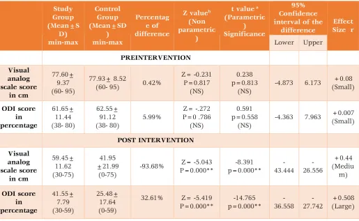
Comparative analysis between the groups (Table-3) shows that there is no statistically significant difference in pre-intervention means of Visual analogue score for pain and ODI score between groups. There is a statistically significant difference in post-intervention means of VAS and ODI score when compared between the groups. There is a clinical significant difference in post means with medium and large effect size.
Table-1: Basic Characteristics of the subjects studied
Basic Characteristics of the subjects
studied Study Group
Control Group
Between the groups Significance
Number of subjects studied (n) 20 20 --
Age in years (Mean± SD)
39.60± 7.91 (15-50)
42.05± 5.01
(31-50) p= 0.316 (NS)
Gender Males 7 8 --
Females 13 12 --
Duration in weeks 5.90± 4.68
(1-16)
5.60± 5.19
Table-2: Analysis of pain and functional disability within study and control Groups (Pre to post test analysis)
** Statistically Significant difference p<0.05; NS- Not significant; a. Pared t test. b. Wilcoxon Signed Ranks Test
Table -3: Comparison of means of pain and functional disability between study and control Groups (PRE AND POST INTERVENTION COMPARISION)
Study Group (Mean±S D) min-max Control Group (Mean±SD ) min-max Percentag e of difference
Z valueb
(Non parametric
)
t value a
(Parametric ) Significance
95% Confidence interval of the
difference
Effect Size r
Lower Upper
PREINTERVENTION Visual analog scale score in cm 77.60± 9.37 (60- 95) 77.93± 8.52
(60- 95) 0.42%
Z= -0.231 P=0.817 (NS) 0.238 p=0.813 (NS)
-4.873 6.173 +0.08
(Small) ODI score in percentage 61.65± 11.44 (38- 80) 62.55± 91.12 (38- 80) 5.99% Z= -.272 P=0 .786 (NS) 0.591 p=0.558 (NS)
-4.363 7.963 +0.007
(Small) POST INTERVENTION Visual analog scale score in cm 59.45± 11.62 (30-75) 41.95 ±21.99 (0-75)
-93.68% Z= -5.043
P=0.000** -8.391 p=0.000** -43.444 -26.556 +0.44 (Mediu m) ODI score in percentage 41.55± 7.79 (30-59) 25.48± 17.64 (0-59)
32.61% Z= -5.419
P=0.000** -14.765 p=0.000** -36.558 -27.742 +0.508 (Large)
** Statistically Significant difference p<0.05; NS- Not significant a. Independent t test b. Mann-Whitney Test Pre intervention (Mean±SD) min-max Post intervention (Mean±SD) min-max Perecntage change
Z valueb
(Non parametric significance)
t valuea
(Parametric) Parametric Significance
P value
95%Confidence interval of the
difference
Effect Size
(r)
Lower Upper
Study Group
Visual analog scale score
in mm
77.60± 9.37 (60- 95)
59.45± 11.62
(30-75) -23.38%
-3.924 p <0.000**
15.973
P <0.000** 46.75 60.85
+0.65 (Large)
ODI score in percentage
61.65± 11.44 (38- 80)
41.55± 7.79
(30-59) -32.60%
-3.924 p <0.000**
26.499
P <0.000** 49.78 58.31
+0.71 (Large) Control Group Visual analog scale score in mm 77.93± 8.52 (60- 95) 41.95±21.99
(0-75) -46.16%
-3.784 p <0.000**
6.246
P <0.000** 12.06 24.23
+0.73 (Large) ODI score in percentage 62.55± 91.12 (38- 80) 25.48± 17.64
(0-59) -59.26%
-3.827 p<0.000**
7.929
P <0.000** 14.79 25.40
Graph 1: Comparison of means of VAS between Study and Control Groups
The above graph shows that there is no statistically significant difference in means of Visual analogue score for pain when pre interventionmeans were compared between study and control groups.
There is a statistically significant difference when post-intervention VAS score means were compared between the groups.
Graph 2: Comparison of means of ODI between Study and control Groups
The above graph shows that there is no statistically significant difference in means of ODI score when pre-intervention means were compared between study and control groups. There is a statistically significant difference when post-intervention ODI means were compared between the groups
DISCUSSION
The present study found that the subjects who received adductor pull back exercise and along with conventional exercise and subjects who received conventional exercise found a statistically significant improvement in means of VAS, and ODI within the groups. When compared the effect there is a statistically significant difference in improvements between the groups. However the subjects in Study group shown greater percentage of improvements than control group
In study group, the improvment in pain and functional disability could be because of adductor pull back exercise technique and conventional exercise that shown to have several effects. In SIJD there is poorly approximated left hip and left hip capsule restriction, weak and long adductors, internal obliques (IO) and transverse abdominus (TA), short and over acting paraspinals which can lead pain and disability. Adductor Pull back exercise techniques brings the position and alignment of SIJ and on left anterior gluteus medius and ischiocondylar adductor magnus activation due to neuro-physiological factors based on activating and shortening of adductors, internal oblique, transverse abdominus and inhibiting and lengthening of paraspinal muscles and thereby facilitating the elongation of this muscle-tendon unit. The Reciprocal inhibition describes the
0 10 20 30 40 50 60 70 80
Pre-intervention:VAS Post-intervention:VAS
77.60
59.45 77.93
41.95
Mea
ns
o
f V
AS
in
mm
Study Group Control Group
0% 10% 20% 30% 40% 50% 60% 70%
Pre-itervention:ODI Post-intervention:ODI
61.65%
41.55% 62.55%
25.48%
M
ea
ns of
OD
I in pe
rce
ntage
phenomenon that when a muscle group is activated, its antagonist is inhibited. During inhalation phase of adductor pull back exercises, the left hip moves into internal rotation and adduction that put the left posterior hip capsule and ischiofemoral ligament in a lengthened position. The ischiofemoral ligament is a spiral ligament, and thus is affected by rotational movements. When the hip is flexed, internal rotation is limited by tension in the capsule and ischiofemoral ligament. The left hip motion lengthens and inhibits the muscles of the right anterior outlet (adductors, levatorani, obturator internus) and muscles of the left anterior inlet (rectus femoris, Sartorius). The left posterior hip capsule may become tight and short as a result of compensation for a Left AIC. This motion requires the muscles that adduct and internally rotate the hip leads to activation of left adductor Magnus and left medial hamstrings (semimembranosus and semitendinosus). The Recruitment and activation of these muscles may help to shorten and strengthen them if they are long or weak. During the inhalation, the diaphragm contracts, which forces the left pelvic floor fulcrum to open the left posterior pelvic outlet so that upright left hip (internal rotation and adduction) more easily obtained and are not be limited by the pelvis. Thus, application of adductor pull back exercise technique cause improvement in function and
reducing the pain in subjects with SIJD.11,12,21
Michael T Cibulkaet al, in a study that examined the effects of postural restoration exercise technique on the sacroiliac joint pain and found the significant improvement in pain followed by an
exercise program with restoration technique.21
Kyndall L. Boyle et al in their study examined the effects of right side lying adductor pull back exercise intervention they found reduction in pain
received in the group.22
The improvements in study group are also attributed due to the conventional exercises they received. Therefore, in the present study found the significant improvement in outcome measures following application of adductor pull back exercise on pain and functional disability.
In control group, there is a significant improvement in means of VAS and ODI. It can be speculated due to conventional exercise. The static exercise has been used to control lumbar stabilization, motor control training. The static exercise leads to increased metabolic activity and blood flow to the muscles and increase the co-ordination. The improved co-ordination lead to antagonistic pairs of muscles work together even more effectively; when the prime mover contracts
more rapidly the antagonist muscle relax as quickly and relieve inflammation associated with
muscular pain.7
Comparative analysis between the groups found that there is no statistically significant difference in means of VAS and ODI when pre-intervention means were compared between the groups. When means of post intervention were compared between the groups, there is a significant difference in means of VAS and ODI between the groups. Even though there is significant improvement in the both technique, however, study group found greater percentage of improvement in pain and functional disability than control group; this could be an added effect of adductor pull back exercises may be due to effect of increase in muscle strengthening, endurance and flexibility that influenced on SI joint dysfunction. The added effect of adductor pull back exercises along with conventional exercise might have shown improvement due to counter-irritant effects, or a spinal reflex mechanism for the relief of muscles spasm. Hence, there is no statistically significance difference in improvement of pain and functional disability obtained between the groups, based on the finding in this study found that there is a significant effect of adductor pull back exercise on pain and functional disability for SIJD. Therefore study rejects null hypothesis.
Limitation of the study
1. Subject with wide range group between 20 to 50
year of age were considered for the study, thus result cannot be generalized to individual age.
2. Duration of the symptoms and dosage of
exercises performance was not considered specific to the individual subjects that may influence on the performance of the outcome measures.
3. Only pain, functional disability parameters
were studied. Measures such as ROM, functional length discrepancy and quality of life were not studied.
Further study recommendations:
1. Further studies are needed to find the effect of
these techniques on follow-up to find the recurrence of SIJD.
2. Further studies can be carried out to find effect
of these techniques on other outcome measurements.
3. Further study can be carried out to find the
effect of these techniques in combination with other physiotherapy interventions.
Conclusion
The present study concludes that the 2 weeks of
conventional exercise found statistically and clinically significant effect on improving pain, functional disability for subjects with sacroiliac joint dysfunction. Adductors pull back exercise along with conventional exercise techniques shown to have greater percentage of improvement in improving pain and functional disability than only conventional exercises for subjects with sacroiliac joint dysfunction.
Acknowledgement
Authors were expressing their sense of gratitude’s to the people who helped and encouraged them for the guidance and completion of this study
Conflicts of interest: None
REFERENCES
1. Sherman, Andrew; Gotlin, Robert; et al.
"Sacroiliac Joint Injury". Retrieved 18 January 2011.
2. Maigne JY, Aivaliklis A, Prefer F. Results of
sacroiliac joint double block and value of sacroiliac pain provocation tests in 54 patients with low back pain. Spine. 1996; 21:1889–92.
3. O'Sullivan PB. Altered motor control strategies
in subjects with sacroiliac joint pain during the active straight-leg-raise test. Spine. 2002; 27(1): E1–8.
4. John A. McCulloch: Macnab’s Backache,
Lesions of the sacroiliac joints. 3rd edition, Williams and Wilkins: 180.
5. Foley, BS; Buschbacher, RM. "Sacroiliac joint
pain: anatomy, biomechanics, diagnosis, and treatment". Am J Phys Med Rehabil. 2006; 85 (12): 997–1006.
6. J, Kyncl M, et al. Postural function of the
diaphragm in persons with and without chronic low back pain. J Orthop Sports PhysTher. 2012; 52(4):352–362.
7. Robey J, Boyle K. Bilateral Functional Thoracic
Outlet Syndrome in a College Football Player. N Am J Sports PhysTher Boyle K. Ethnography of the postural restoration subculture: a posture
based approach to patient/client
management [Dissertation]. Fort Lauderdale, FL, Nova Southeastern University; 2006.
8. Mezieres F. MethodesOrthopediques and La
Fonction du Sympathique. Cahiers de la MethodeNaturelle. 1973:52–53.
9. Hansen H, Manchikanti L A systematic
evaluation of the therapeutic effectiveness of sacroiliac joint interventions. 2012;15(3):E247-78.
10.Boyle K. Management of a Female with Left
Low Back Pain and Sacroiliac Joint Pain with
therapeutic Exercise: A Case
Report. Physiother Can. 2011;63 (2):154–163.
11.Kyndall L. Boyle. Clinical application of right
side lying adductor pull back exercise .2013; 8(3): 349–358.
12.Holly Spence, postural restoration: an effective
physical therapy approach to patient
treatment. 2008 ;12 (2) 102-104.
13.Simopoulos TT, ManchikantiL A systematic
evaluation of prevalence and diagnostic accuracy of sacroiliac joint interventions. Pain Physician. 2012; 15(3): E305-E344.
14.Rupert MP, Evaluation of sacroiliac joint
interventions: a systematic appraisal of the literature. 2009;12(2):399-418.
15.Jerry Hesch, Henderson, Nevada: Evalution
and treatment of the most common patterns of sacroiliac joint dysfunction; Body Mechanics & Gainesille PT / SI DYSFUNCTION.COM
16.Boyle K. Conservative Management for
Patients with Sacroiliac Joint Dysfunction. In: Norasteh AA, editor. Low Back Pain. Rijeka, Croatia: InTech; 2012:293–332.
17.Ngoc Quan Phan, Christine Biome, Fleur Fritz,
Joachim Gress, Adam Reich, Toshi Ebata, Matthias Augustine, Jacek C. Szepietowski and Sonja Stander. Assessment of Pruritis Intensity: prospective Study on Validity and Reliability Scale of the Visual Analog Scale, Numerical Rating Scale and Verbal Rating Scale in 471 patients with Chronic Pruritus: clinical report: Acta Derm Venereol.2012; (92): 502-507.
18.Leighann Litcher-Kelly, Sharon A.
Martino, Joan E. Broderick, Arthur A Stone. A systematic review of measures used to assess chronic musculoskeletal pain in clinical and randomized controlled clinical trials. Journal of pain. Dec 2007; 8(12): 906–913.
19.Jeremy C. T. Fairbank, Paul B. Pynsent. The
Oswestry Disability Index. Spine 2000;25: 2940– 2953.
20.Michael Vianin DC. Psychometric properties
and clinical usefulness of the Oswestry Disability Index. Journal of Chiropractic Medicine 2008; 7: 161–163.
21.Michael T Cibulka MT.Anatomy of the
sacroiliac joints.2013 Mar; 222(3).
22.Kyndall L. Boyle Managing a Female Patient
Citation
Sai Kumar .N, Akshata Akalwadi, Vinod Babu .K & Zubair Rafiq Wani
. (2015).
EFFICACY OFADDUCTOR PULL BACK EXERCISE ON PAIN AND FUNCTIONAL DISABILITY FOR SACROILIAC