143
Original article
Computational structure prediction and analyze active ligand binding site
of defense and lytic enzymes of Trichoderma harzianum
Ved Ratan, Supriya Dixit, Mukesh Srivastava*, Shubha Trivedi, Abhishek Mishra, Y.K. Srivastava and D .K. Srivastava**
Biocontrol Lab, Department of Plant Pathology, Chandra Shekhar Azad University of Agriculture and Technology, Kanpur-208002, Uttar Pradesh, India
*Registrar, Rani Laxmi Bai Central Agricultural University, Jhansi-284003, Uttar Pradesh, India
**Joint Director, Council of Science and Technology, Bans Mandi, Qaiserbagh, Lucknow-226018, Uttar Pradesh, India
Received June 13, 2018: Revised July 30, 2018: Accepted August 5, 2018: Published online December 30, 2018
Abstract
Trichoderma spp. are considered as efficient biocontrol agent that can significantly reduces the growth of several soil borne and plant pathogens such as Rhizoctonia solani, Sclerotium rolfsii,
Phythium aphanidermatium, Fusarium oxysporum in several mode of action. Trichoderma harzianum is one the important species in genus Trichoderma, which is capable of producing several effective lytic enzymes and antifungal antibiotics that compete to other fungal pathogens and promotes plant growth. The aim of present study is to predict and analyze the tertiary structure and their potential binding sites through bioinformatic tools and techniques. The protein sequences of enzymes were retrieved from UniProt database, followed by modelling of tertiary structure by Swiss-model Workspace and further validated using PROCHECK server which showed that from 75% to 90% residues are in favored region of Ramchandran Plot. These different validation steps proved that the predicted models are stable and it will provide an insight to its functional aspect which is based on tertiary structures. Furthermore, functional and conserved motifs were predicted through PROSITE database. These findings allow us to determine the protein families and domain which remain conserved throughout the evolution which may act as inducing or suppressing the biological activity of protein. Ligand binding site of enzymes has been predicted using SiteHound server by using four different chemical probes which allow us studying different ligand binding site. Thus, this study supported a scientific base for 3D structure modelling of lytic and defence enzymes and opens the new opportunities for further investigations in biological control of phytopathogens.
Key words: Physicochemical properties, signal peptides, transmembrane region, comparative modelling, ligand binding site
Copyright @ 2018 Ukaaz Publications. All rights reserved. Email: [email protected]; Website: www.ukaazpublications.com
1. Introduction
The modern agriculture approaches increases the use of genetically modified crop plants, fertilizer, pesticides, various biotechnolo-gically advanced equipment and water to produce large quantity of single modified crop which causes increase in disease susceptibility which will lead to destructive crop diseases caused by microorganism (Waard et al., 1993). Therefore, nowadays, various researches have been aimed to control plant diseases by replacing the pesticides with eco-friendly biological control agen ts wh ich could be rhizopheric fungi and bacteria, having antagonistic activities towards pathogens. Biological control agents should have ability to survive under unfavorable conditions, efficient to utilize nutrient and increases the quality of soil (Haran et al., 1996).
From decades, Trichoderma species are well known saprophytic fungi habitat in the rhizosphere, which have received significant
Author for correspondence: Mrs. Supriya Dixit
Biocontrol Lab, Department of Plant Pathology, Chandra Shekhar Azad University of Agriculture and Technology, Kanpur-208002, Uttar Pradesh, India
E-mail: [email protected] Tel.: +91-7522090030
Annals of Phytomedicine 7(2): 143-160, 2018
DOI: 10.21276/ap.2018.7.2.23; Print ISSN : 2278-9839 and Online ISSN : 2393-9885 7(2):143-160 (2018) Ann. Phytomed.,
observation as potential fungal antagonist that could be used as effective biopesticides. Among all known Trichoderma spp (100), Trichoderma harzianum was found to be very efficacious bioagent against soil borne phytopathogens (Green et al., 1999).
brinjal plants during T. harzianum and F. solani interaction to find any correlation that may exist in host defense mechanism and they noticed that T. harzianum was significantly interacts with peroxidase activity which is increased in the period of infection and lasts till the end of incubation period which also indicates that there is a correlation with resistance of plant against infection. Thus, the aim of this study is to explore more about the lytic and defence enzymes through various computational software and techniques. This goal requires the knowledge of three dimensional structures of proteins obtained through X-ray crystallography and NMR spectroscopy. The crystal structures of defense and lytic enzymes are not available in protein data bank (PDB), but these experimental techniques are very expensive, time consuming and complex process. Hence, by using computational technique, the 3D structure of defense and lytic proteins has been modeled through comparative or homology modelling which predict model based on experimental 3D structure of related homologous protein (serve as template). The Swiss model server is used to construct the three dimensional structure of proteins and, further validated through Web server software by plotting Ramachandran Plot.
However, the studies suggested that approximate by one third of proteins are associated with small probes and their presence and absence directly affects the catalytic mechanism of proteins and it is also helpful to stabilize the tertiary structure of proteins (Shi and Chance, 2008). So, in order to identify the most potential probe with their putative ligand binding site, the SITEHOUND -Web server (http://sitehound.sanchezlab.org) is used.
2. Materials and Methods
The protein sequences of defense and lytic enzymes produced by T. harzianum were retrieved from UniProt Database in fasta format and used further for the prediction of physicochemical properties liketheoretical pI, molecular weight, half-life, instability index and grand average of hydropathicitythrough ExPASy (Expert Protein Analysis System) proteomics server of the Swiss Institute of Bioinformatics (Wilkins et al., 1999). The presence of signal peptides in defence and lytic proteins was analysed by using SignalP 4.1 server (Petersen et al., 2011). The protein sequences were also used for the prediction of conserved motif which plays an important
role for inducing defense in plants and also performs catalytic activity. Thus, ScanProsite program (De Castro et al., 2006) was used which allows scanning of protein sequence for matches against
the PROSITE collection of motifs with a meaningful signature or pattern and then similarity searching was done for validation of the identified motif in other Trichoderma species in order to check wh ether th e identified motif is presen t in other strain s of Trichoderma or not. For comparative modelling, Swiss model Automated Workspace (Arnold et al., 2006) was used to determine the 3D structure. The predicted models were further evaluated in order to know the quality of structures. PROCHECK server was used to validate predicted structure. The PROCHECK server generates ‘Ramachandran Plot’ on the basis of steriochemical configuration of amino acids (Laskowski et al., 1993). Identification of binding site with favorable interaction with four different ligands (Methyl Carbon, Phosphate Oxygen, Hydroxyl and Aromatic) were done using SITEHOUND Web server (Hernandez et al., 2009).
3. Results and Discussion
The present computational studies focused on sequence and structural analysis of lytic and defence enzymes which participates in fungal cell wall degradation includes chitinases, glucanases, protease and peroxidase of T. harzianum. Sequences of lytic enzymes were retrieved from UniProt Database and are varied in length from 300 to 1040 amino acid (Table 1) and used for further analysis.
Table 1:The pathogenic proteins name and their accession numbers E nz y me s Ac ces sio n number
(Prot ein s equenc e) Alkaline Proteinase A0A0F9X8B4
Beta-1,3 exoglucanase O 14 40 2
Beta-1,3-glucan-binding A0A0F9Y010
Beta-1,6-glucanase B9VQ17
Catalase-peroxidase A0 A0F9X3 Z8
Endochitinase 33 Q 12 71 3
Endochitinase 37 Q8NJQ5
Endochitinase 42 P4 8 8 2 7
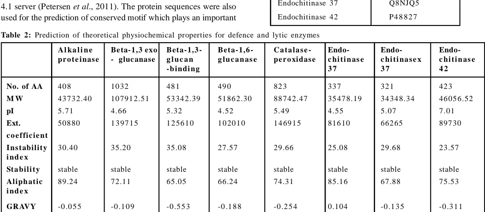
Table 2: Prediction of theoretical physiochemical properties for defence and lytic enzymes
A l ka l i n e Be ta- 1,3 exo Bet a- 1,3 - Bet a- 1,6 - C a t a la s e - Endo- Endo- Endo-pr o t e inas e - gluc anase g l u c a n g lu c a n as e pe r o xidas e c hi t i n as e c hi t i na s e x c hi t i n as e
- b indi ng 3 7 3 7 4 2
No. of AA 40 8 10 32 48 1 49 0 82 3 33 7 32 1 42 3
M W 43 73 2.40 10 791 2.51 53 34 2.39 51 86 2.30 88 74 2.47 35 47 8.19 34 34 8.34 46 05 6.52
pI 5.71 4.66 5.32 4.52 5.49 4.55 5.07 7.01
Ext. 508 80 1 3 97 1 5 1 2 56 1 0 1 0 20 1 0 1 4 69 1 5 816 10 662 65 897 30 c o e f fic ie nt
I ns t abilit y 30.40 35.20 35.08 27.57 29.66 25.08 29.68 23.57
i nd e x
St a bili t y stable stable stable stable stable stable stable stable
Aliph at ic 89.24 72.11 65.05 66.24 74.31 85.16 67.88 75.53
i nd e x
Another important property is grand averages of hydropathy (GRAVY) which indicate the hydrophilic and soluble behavior of proteins is ranging from – 0.055 to – 0.553 as this score indicating that all enzymes are hydrophilic in nature except endochitinase 37 as its predicted score is positive which means it is hydrophobic. The aliphatic index defines the relative volume occupied by aliphatic chains of proteins and the increased positive score shows the increased thermostability. Here, all enzymes showed high positive score but alkaline protease is potentially stable over a high temperature range.
3.2 Signal peptide prediction
Signal peptide plays important role in targeting the translocation of integral membrane proteins and secretory proteins. For the prediction of signal peptide in all enzymes, SignalP program was used. The SignalP results are summarized in Table 3. SignalP gives output in graphical and numerical values which comprises three different scores, C, S and Y.
3.1 Physicochemical analysis
By using the ProtParam tools, several physicochemical properties were computed for alkaline proteinase, 1,3 exoglucanase, Beta-1, 3 -glucan-bin ding protein, Beta-1 ,6-glu canase, Catalase-peroxidase, Endochitinase 33, Endochitinase 37 and Endochitinase 42 (Table 2).
The predicted instability index for T. harzianum enzymes are below 40 which indicated that all enzymes are in stable form. A protein with an instability index lower than 40 is predicted as stable, while a value greater than 40 predicts that the protein may be unstable. The significance for computation of instability index is to store proteins in correct solvent. Grasso et al. (2016) predicted instability index for mouse caltrin I, bovine caltrin and human insulin is 51.99, 24.86 and 43.05, respectively, indicating that while storing in aqueous solution, the mouse caltrin I and human insulin turns in aggregated form with loss of biological activity. Hence, these unstable proteins are not suitable for long term storage in aqueous solution. In our prediction, it is clearly visible that all defence and lytic enzymes are in stable form while storing in aqueous solution.
Table 3: Prediction of signal peptides in defense and lytic enzymes through SignalP
between pos. 20th serine and 21th
serine 0.671 1-20 0.760 1-20 0.566 21 0.416 21 Endo-chitinase 42
between pos. 19th alanine and 20th
glycine 0.901 1-19 0.925 1-19 0.872 20 0.821 20 Endo-chitinase 33
between pos. 25st alanine and 26th
glutamine 0.835 1-25 0.847 1-25 0.820 26 0.799 26 Endo-chitinase 37 between pos. 22ndalanine and
23rd threonine 0.634 1-22 0.788 1-22 0.452 23 0.312 29 Beta-1,3 exo-glucanase
between pos. 20st alanine and 21st
leucine 0.906 1-20 0.933 1-20 0.875 21 0.820 21 Alkaline proteinase Value Position Value Position Value Position Value Position Cleavage site D score Mean S Max. Y Max. C SignalP measure
The first predicted score is C-score defines “cleavage site” score
reported for each position which is significantly high at the cleavage site. For alkaline proteinase enzyme, the reported C-score position indicating first cleavage residue is at 21 position of mature protein which means cleavage site between amino acids 20-21 corresponds to the mature protein starting at position 21 including position 20.
The second predicted score is Y-max which is derivative of C-score combined with S-score resulting better cleavage site prediction rather than raw C-score alone. In a graph, multiple high peaks of C-score is found in one sequence where only one is true cleavage site.
Another predicted score is S-mean and D-score which is for discrimination of secretory and non-secretory proteins. All scores given by SignalP should be very low in case of non-secretory proteins.
3.3 Transmembrane region prediction
Transmembrane region prediction is a part of functional study. Predictions were made through SOSUI server which distinguishes
between membrane and soluble protein and predicts membrane helix. The results are tabulated in Table 4. It is clearly visible that alkaline proteinase, beta-1,3-glucan-binding and endochitinase 37 were predicted as membrane proteins and other enzymes, Beta-1,3 exoglucan ase, Beta-1 ,6 -glucan ase, Catalase-peroxidase, Endochitinase 33 and Endochitinase 42 are soluble proteins. Transmembrane regions are rich in hydrophobic amino acids. 3.4 Motif prediction
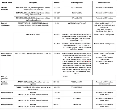
defence enzymes were used as an input for motif search against PROSITE database. Results are tabulated below in the Table 5. The hits were retrieved by matched pattern in case of alkaline proteinase, catalase-peroxidase, en doch itin ase 3 3, endoch itin ase 42 , endochitinase 37 and in case of Beta-1,3 exoglucanase,
Beta-1,3-glucan-binding Beta-1,6-glucanase hits were made by matched profile. No hits were found for Beta-1,6 glucanase enzymes. The predicted length of motifs is varied from 10-18 amino acids. It also predicts the conversed active site residues with their behavior.
Table 4: Prediction of transmembrane region through SOSUI server
Membrane protein 23 TRLLDASFLLLPVIASTLFGTAS Endochitinase 37 Membrane protein 23 TIMPLLGILLGLAISGFLIWDGM Beta-1,3-glucan-binding Membrane protein 23 MAWFKTLALFLFTVTPYVTALPL Alkaline proteinase
Type of protein Length Transmembrane region Enzyme name Membrane protein 23 TRLLDASFLLLPVIASTLFGTAS Endochitinase 37 Membrane protein 23 TIMPLLGILLGLAISGFLIWDGM Beta-1,3-glucan-binding Membrane protein 23 MAWFKTLALFLFTVTPYVTALPL Alkaline proteinase
Type of protein Length
Transmembrane region Enzyme name
Table 5: Prediction of motifs through PROSITE database
Active site at 171thposition (Proton donor) FDGIDIDwE
163 - 171
PS01095
CHITINASE_18 Chitinases family 18 active site
Endo-chitinase 42
Active site at 160thposition (Proton donor) FDGIDIDiE
152 - 160
PS01095
CHITINASE_18 Chitinases family 18 active site
Endo-chitinase 37
Active site at 167thposition (Proton donor) VDGFDFDfE
159 - 167
PS01095
CHITINASE_18 Chitinases family 18 active site
Endo-chitinase 33
TVALIAGGHAF 341 - 351
PS00435 PEROXIDASE_1 Peroxidases proximal heme-ligand signature
Active site at 174thposition (Proton acceptor) GPffIRLaWHNA
165–176
PS00436 PEROXIDASE_2 Peroxidases active site signature Catalase-peroxidase No Hits Beta-1,6-glucanase
Domain at 149th– 458thposition GH16
Active site at 293rdposition (Nucleophile) Active site at 298thposition
(Proton donor) EDFSNGLDPsiwtkevQVggFGNGeFEqttggdsn vfveNGHlmikatlqdanlveknnvinllkdgtCTskdyy SCVAATNTTNGNSSVVPPTL---SGRINTFKGAKIKYGRVEVTAKLPVGDW LWPAIWMLPVNDTYGPWPssGEIDIMESR GNNHTYsqggnniASSALHWGPDPANDAW WKTNNKrkalHTTYSAEFNTFGLEWSQKY LFTYinsrllqvtytnFNKPMWKRgafpdstangtrlV DIWSSTGRDNTPFDTEFYLILNLAvgGTNG WFEDgmsGKPWLDHSPNAKKDFWNARD TWYPT 149 - 458
PS51762 GH16_2 Glycosyl hydrolases family 16 (GH16)
Beta-1,3-glucan-binding protein
Domain at 927th - 1024th position GWNFRGCYTDSvNARALIAESVPNGPSsM
TIEACQSVCKGLGYTLAGLEYADECYCG NSLANGATIApdgNAGCNMNCAGNAAET
CGGPNRLDIYSYG 927-1024
Domain at 816th– 914thposition GWNFLGCYSDNvNGRTLANQVQVAGGA
saMSIEACETASESAGYTIAGVEYSGECWC DTKFQNGGGPAsdgSAQCTMTCSGAPQET
CGGPNRLDVYSLA 816–914
PS51212WSC domain
Signal peptide from 1st – 17th Lipid at 18thposition N-palmitoyl
cysteine site Lipid at 18thposition S-diacylglycerol cysteine site MGFIRSAVLSALTFAAAC
1 - 18
PS51257 PROKAR_LIPOPROTEIN Prokaryotic membrane lipoprotein lipid attachment site profile
Beta-1,3 exoglucanase
Active site at 353th position GTSmSaPhVAG
351 - 361
PS00138 SUBTILASE_SER Serine proteases, subtilase family, serine active site
Active site at 197thposition HGThVAGiIAG
197 - 207
PS00137 SUBTILASE_HIS Serine proteases, subtilase family, histidine active site
Active site at 166thposition AYVVDSGVftkH
162 - 173
PS00136SUBTILASE_ASP Serine proteases, subtilase family, aspartic acid active site
Alkaline Proteinase Predicted features Matched patterns Position Description Enzyme names
Active site at 171thposition (Proton donor) FDGIDIDwE
163 - 171
PS01095
CHITINASE_18 Chitinases family 18 active site
Endo-chitinase 42
Active site at 160thposition (Proton donor) FDGIDIDiE
152 - 160
PS01095
CHITINASE_18 Chitinases family 18 active site
Endo-chitinase 37
Active site at 167thposition (Proton donor) VDGFDFDfE
159 - 167
PS01095
CHITINASE_18 Chitinases family 18 active site
Endo-chitinase 33
TVALIAGGHAF 341 - 351
PS00435 PEROXIDASE_1 Peroxidases proximal heme-ligand signature
Active site at 174thposition (Proton acceptor) GPffIRLaWHNA
165–176
PS00436 PEROXIDASE_2 Peroxidases active site signature Catalase-peroxidase No Hits Beta-1,6-glucanase
Domain at 149th– 458thposition GH16
Active site at 293rdposition (Nucleophile) Active site at 298thposition
(Proton donor) EDFSNGLDPsiwtkevQVggFGNGeFEqttggdsn vfveNGHlmikatlqdanlveknnvinllkdgtCTskdyy SCVAATNTTNGNSSVVPPTL---SGRINTFKGAKIKYGRVEVTAKLPVGDW LWPAIWMLPVNDTYGPWPssGEIDIMESR GNNHTYsqggnniASSALHWGPDPANDAW WKTNNKrkalHTTYSAEFNTFGLEWSQKY LFTYinsrllqvtytnFNKPMWKRgafpdstangtrlV DIWSSTGRDNTPFDTEFYLILNLAvgGTNG WFEDgmsGKPWLDHSPNAKKDFWNARD TWYPT 149 - 458
PS51762 GH16_2 Glycosyl hydrolases family 16 (GH16)
Beta-1,3-glucan-binding protein
Domain at 927th - 1024th position GWNFRGCYTDSvNARALIAESVPNGPSsM
TIEACQSVCKGLGYTLAGLEYADECYCG NSLANGATIApdgNAGCNMNCAGNAAET
CGGPNRLDIYSYG 927-1024
Domain at 816th– 914thposition GWNFLGCYSDNvNGRTLANQVQVAGGA
saMSIEACETASESAGYTIAGVEYSGECWC DTKFQNGGGPAsdgSAQCTMTCSGAPQET
CGGPNRLDVYSLA 816–914
PS51212WSC domain
Signal peptide from 1st – 17th Lipid at 18thposition N-palmitoyl
cysteine site Lipid at 18thposition S-diacylglycerol cysteine site MGFIRSAVLSALTFAAAC
1 - 18
PS51257 PROKAR_LIPOPROTEIN Prokaryotic membrane lipoprotein lipid attachment site profile
Beta-1,3 exoglucanase
Active site at 353th position GTSmSaPhVAG
351 - 361
PS00138 SUBTILASE_SER Serine proteases, subtilase family, serine active site
Active site at 197thposition HGThVAGiIAG
197 - 207
PS00137 SUBTILASE_HIS Serine proteases, subtilase family, histidine active site
Active site at 166thposition AYVVDSGVftkH
162 - 173
PS00136SUBTILASE_ASP Serine proteases, subtilase family, aspartic acid active site
A
B
C
E
F
G
H
3.5 Template selection, molecular modelling and validation
BLAST search was performed in order to search of crystal structure of homologous proteins against protein data bank ( ). Homologues models having sequence identities between 68-25% were chosen as a template structure. Sequence identity percentage (total number of amino acids matched between two different sequences) correlated with coverage percentage (% of query sequence overlaps to template sequence) plays significant role in choosing reliable template structure for comparative modelling. Accuracy of modeled structure depends on when the selected template is having more than 50% of sequence similarity (Baker and Sali, 2001). So, in our work, the h ighest percen tage sequ en ce similarity was obtained for endochitinase 42 of the crystal structure of A chitinase crchi1 from the nematophagous fungus, Clonostachys rosea (PDB: 3G6L_A) with 68% sequence similarity and 96% coverage.
In case of beta-1,3 selected template was ofPDB ID: 5M5Z_A with 4 6% seq uence similarity an d 7 2% coverage, followed by endochitinase 33 (PDB: 2UY2_A with 45% sequence similarity and
89% coverage) and alkaline proteinase (PDB: 3F7O_A with 42% sequence similarity and 68% coverage). The selected PDB ID 5KZ6_A (33% sequence similarity and 89% coverage) and 5NGK_A (31% sequence similarity and 87% coverage) for endochitinase 37 and beta-1,6-glucanase, respectively. The lowest sequence identity (29%) template is of PDB: 3AZX_A with 57% coverage was found for beta-1,3-glucan-binding protein.
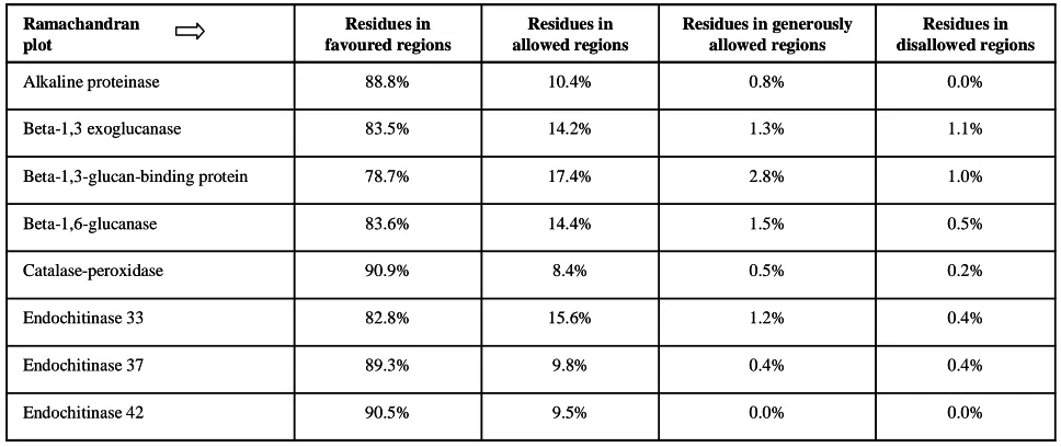
For further analysis, the models were developed by the Swiss-model Workspace. PROCHECK is a Web server, u sed for stru cture assessment. It gives Ramachandran Plot which observed residues in favoured, allowed and outer lined regions and it also allows studying steric hindrance. Ramachandran Plot of all enzymes are given below which showed that the residues present in the protein, if found in red colored region denotes that there is no steric hindrance, yellow color denotes some steric hindrance and white color denotes the forbidden zone, where only glycine “G” is found as it has no side chains (Figure 1). It also describes % residues found in favored region and rest of amino acid percentage in outer lined region (Table 6).
Table 6: Ramachandran Plot results through PROCHECK
0.0% 0.0%
9.5% 90.5%
Endochitinase 42
0.4% 0.4%
9.8% 89.3%
Endochitinase 37
0.4% 1.2%
15.6% 82.8%
Endochitinase 33
0.2% 0.5%
8.4% 90.9%
Catalase-peroxidase
0.5% 1.5%
14.4% 83.6%
Beta-1,6-glucanase
1.0% 2.8%
17.4% 78.7%
Beta-1,3-glucan-binding protein
1.1% 1.3%
14.2% 83.5%
Beta-1,3 exoglucanase
0.0% 0.8%
10.4% 88.8%
Alkaline proteinase
Residues in disallowed regions Residues in generously
allowed regions Residues in
allowed regions Residues in
favoured regions Ramachandran
plot
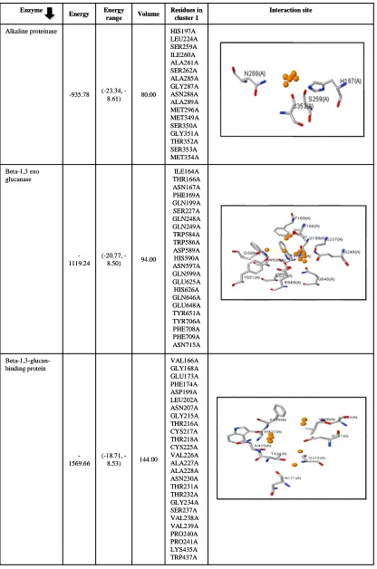
3.6 Structure ligand binding site identification
Active site prediction is most important aspect as the interaction behavior of proteins with their ligands and other small molecules reveals the cellular function and it also helps to characterized proteins in functional point of view (Singh and Chaube, 2014). Binding site prediction of lytic and defence enzymes of T. harzianum were predicted by using SiteHound Web server which identified ligand binding site by computing the interaction energy between protein structure and various chemical probe and carries out cluster analysis. All clusters are, further ranked up according to total interaction energy calculated by molecular interaction field (MIF) (Hernandez et al., 2009). The Web server uses four probes, i.e., methyl probe used to predict binding site for drug like molecules, hydroxyl probe used to characterized sugar binding site, phosphate probe used for the detection of phosphorylated ligands and an aromatic carbon probe used to characterize various other ligands. In a study done by Ghersi and Sanchez (2009), in which test was
conducted on 77 protein-ligand complexes containing drug-like molecule showed 95% correct binding site in case of using methyl probe while using phosphate probe in set 120 protein bound with phosphorylated ligands, it gives similar accuracy results as given in methyl probe.
In case of motif analysis, some features were predicted in case of, beta-1,3-glucan-binding protein, catalase-peroxidase, endochitinase 33, endochitinase 37 and endochitinase 42 which includes active site prediction at different amino acid position. The predicted three active sites of alkaline proteinase at 166, 197 and 353positions were found interacting with phosphate oxygen probe which suggests that binding of phosphorylated ligand with their identified putative binding site may increase or decrease the catalytic mechanism as proteases penetrates the fungal pathogenic cell wall and leads to lysis.
that other ligands such as various metal, ions and inorganic compound shall increase or decrease the catalytic mechanism of enzyme but it is important to check the other ligands interaction with particular enzymes because in recent studies; it was found that those small ligands causes harmful effect to T. harzianum which lead to deactivation of enzymes. A similar in silico study was conducted by Gavanji et al. (2013), in which they check the possible interaction of chitinase enzyme of T. harzianum with silver nitrate (AgNO3) particles as it has strong antimicrobial activity against pathogenic microbes through docking technique. Their observations suggested that chitinase and silver nitrate have interactions which will lead to
deactivation of enzymes. Another bioinformatics study was conducted by predicting copper, zinc and iron binding proteins with fungal pathogenic species Paracoccidioides and the obtained results suggested that these ions played important role in virulence (Tristão et al., 2015).
In case of catalase-peroxidase and endochitinase 37, all predicted active sites were found to be interacting with all four probes whereas endochitinase 33 is interacting with all probes except phosphate oxygen. Endochitinase 42 was found to be interacting with methyl carbon probe. Binding site and descriptors calculations are given in Tables 7-10 for lytic and defence enzymes of T. harzianum. Table 7: SiteHound results with methyl carbon (CMET) probe
ASP323A PRO324A ALA328A TRP330A LYS331A THR371A ARG382A GLY383A ALA384A PHE385A ASP387A SER388A THR389A ASP397A ILE398A SER400A SER401A ARG404A 112.00
(-21.62, -8.94) -1377.14
Beta-1,3-glucan-binding protein
ILE164A THR166A ASN167A PHE169A GLN199A SER227A GLN248A TRP584A TRP586A ASP589A HIS590A SER596A ASN597A GLN599A GLU625A HIS626A GLN646A GLU648A TYR651A TYR706A PHE708A PHE709A ASN715A 98.00
(-19.16, -8.93) -1184.14
Beta-1,3 exo glucanase
HIS132A ALA133A PRO134A TRP135A ALA138A ALA139A SER142A LYS144A PRO145A GLY146A ALA147A LYS148A SER312A TRP314A 52.00
(-17.79, -8.96) -589.70
Alkaline proteinase
Residues in cluster 1 Volume
Energy range Energy
Enzyme
ASP323A PRO324A ALA328A TRP330A LYS331A THR371A ARG382A GLY383A ALA384A PHE385A ASP387A SER388A THR389A ASP397A ILE398A SER400A SER401A ARG404A 112.00
(-21.62, -8.94) -1377.14
Beta-1,3-glucan-binding protein
ILE164A THR166A ASN167A PHE169A GLN199A SER227A GLN248A TRP584A TRP586A ASP589A HIS590A SER596A ASN597A GLN599A GLU625A HIS626A GLN646A GLU648A TYR651A TYR706A PHE708A PHE709A ASN715A 98.00
(-19.16, -8.93) -1184.14
Beta-1,3 exo glucanase
HIS132A ALA133A PRO134A TRP135A ALA138A ALA139A SER142A LYS144A PRO145A GLY146A ALA147A LYS148A SER312A TRP314A 52.00
(-17.79, -8.96) -589.70
Alkaline proteinase
Residues in cluster 1 Volume
Energy range Energy
TYR32A SER37A ALA38A ASN39A SER40A PHE63A ASP117A ASP165A PHE166A GLU167A ALA197A PRO198A GLN199A ASP204A GLN222A TYR224A ASN225A ALA272A MET299A TRP301A 84.00
(19.07, -8.96) -1002.82
Endo chitinase 33
PRO166A PHE167A ILE169A ARG170A
TRP173A HIS174A ASP203A ILE307A TYR308A VAL309A PRO311A ILE327A PHE331A LEU344A ILE345A GLY348A
HIS349A PHE351A LYS353A THR354A HIS355A THR393A SER394A TRP400A LEU456A SER458A TRP490A 150.00
(20.89, -8.91) -1831.63
Catalase-peroxidase
SER37A ASN38A GLN39A ALA40A GLY41A TYR43A LYS44A THR345A GLY346A ALA347A TRP348A ASN349A GLN350A THR390A PHE402A GLN403A THR404A TYR407A ASN480A VAL482A 114.00
(19.34, -8.91) -1353.49
Beta-1,6-glucanase
TYR32A SER37A ALA38A ASN39A SER40A PHE63A ASP117A ASP165A PHE166A GLU167A ALA197A PRO198A GLN199A ASP204A GLN222A TYR224A ASN225A ALA272A MET299A TRP301A 84.00
(19.07, -8.96) -1002.82
Endo chitinase 33
PRO166A PHE167A ILE169A ARG170A
TRP173A HIS174A ASP203A ILE307A TYR308A VAL309A PRO311A ILE327A PHE331A LEU344A ILE345A GLY348A
HIS349A PHE351A LYS353A THR354A HIS355A THR393A SER394A TRP400A LEU456A SER458A TRP490A 150.00
(20.89, -8.91) -1831.63
Catalase-peroxidase
SER37A ASN38A GLN39A ALA40A GLY41A TYR43A LYS44A THR345A GLY346A ALA347A TRP348A ASN349A GLN350A THR390A PHE402A GLN403A THR404A TYR407A ASN480A VAL482A 114.00
(19.34, -8.91) -1353.49
TYR43A PHE71A TRP131A ASP169A GLU171A TYR172A ALA211A MET237A TYR239A ASP240A TYR293A ARG295A ILE319A TRP378A 80.00
(-20.20, -8.90) -967.93
Endo chitinase 42
TYR43A PHE80A GLY123A
ASP158A ILE159A GLU160A ALA199A PRO200A GLU201A TYR204A TYR219A GLN238A TYR239A TYR240A ASN241A GLY288A LEU289A PRO290A ALA295A GLY296A GLY297A MET320A TRP322A 77.00
(-21.56, -8.97) -939.54
Endo chitinase 37
Table 8: SiteHound results with phosphate oxygen (OP) probe
ASP166A
SER167A GLU195A
HIS197A
LEU224A SER227A VAL228A SER259A ILE260A ALA261A ALA285A GLY287A ASN288A ALA289A MET296A MET349A SER350A GLY351A THR352A
SER353A
MET354A 139.00
(29.96, -8.51) -1913.38
Alkaline proteinase
Interaction site Residues in
cluster 1 Volume
Energy range Energy
Enzyme
ASP166A
SER167A GLU195A
HIS197A
LEU224A SER227A VAL228A SER259A ILE260A ALA261A ALA285A GLY287A ASN288A ALA289A MET296A MET349A SER350A GLY351A THR352A
SER353A
MET354A 139.00
(29.96, -8.51) -1913.38
Alkaline proteinase
Interaction site Residues in
cluster 1 Volume
Energy range Energy
ASP127A TRP180A ASN242A GLU243A ASN246A GLN248A TYR251A HIS287A ASN288A HIS312A TYR314A GLU339A TRP341A THR342A PRO343A THR345A TRP369A GLY384A GLY385A CYS386A GLY387A THR388A 191.00
(25.40, -8.50) -2438.03
Beta-1,6-glucanase
VAL166A GLY167A GLY168A GLY172A GLU173A PHE174A LEU197A ASP199A LEU202A ASN207A GLY215A THR216A CYS217A THR218A SER219A VAL226A ALA227A ALA228A ASN230A THR231A THR232A GLY234A SER237A VAL238A VAL239A PRO240A PRO241A TRP271A PRO436A TRP437A 213.00
(27.83, -8.54) -2715.94
Beta-1,3- glucan-binding protein
PRO129A ASN175A GLN204A ASN205A ASP233A MET234A THR235A ARG253A ASN254A THR392A ARG393A SER394A LYS395A PRO396A GLN397A GLU399A 120.00
(23.33, -8.53) -1558.93
Beta-1,3 exo glucanase
ASP127A TRP180A ASN242A GLU243A ASN246A GLN248A TYR251A HIS287A ASN288A HIS312A TYR314A GLU339A TRP341A THR342A PRO343A THR345A TRP369A GLY384A GLY385A CYS386A GLY387A THR388A 191.00
(25.40, -8.50) -2438.03
Beta-1,6-glucanase
VAL166A GLY167A GLY168A GLY172A GLU173A PHE174A LEU197A ASP199A LEU202A ASN207A GLY215A THR216A CYS217A THR218A SER219A VAL226A ALA227A ALA228A ASN230A THR231A THR232A GLY234A SER237A VAL238A VAL239A PRO240A PRO241A TRP271A PRO436A TRP437A 213.00
(27.83, -8.54) -2715.94
Beta-1,3- glucan-binding protein
PRO129A ASN175A GLN204A ASN205A ASP233A MET234A THR235A ARG253A ASN254A THR392A ARG393A SER394A LYS395A PRO396A GLN397A GLU399A 120.00
(23.33, -8.53) -1558.93
THR45A TRP47A GLY48A TYR50A ASP51A ARG52A ILE292A TYR293A SER314A TRP315A GLU316A ILE319A TRP320A ASP321A TRP378A GLU379A SER381A ALA382A 165.00
(34.51, -8.51) -2398.85
Endo chitinase 42
PHE80A ILE83A GLY122A GLY123A ALA124A THR125A ALA126A GLY127A ILE128A ILE157A ASP158A ILE159A GLU160A THR161A ALA199A PRO200A GLU201A TYR204A TYR219A GLN238A TRP322A 84.00
(30.24, -8.57) -1189.43
Endo chitinase 37
GLN35A ASN36A SER37A ALA38A PHE63A ASN65A GLY66A MET71A THR72A ASN73A ALA75A GLY115A GLY116A ASP117A SER118A GLN121A 145.00
(30.18, -8.54) -2030.74
Endo chitinase 33
PRO166A ILE169A ARG170A TRP173A HIS174A ASP203A LEU306A ILE307A TYR308A VAL309A ASP310A PRO311A ILE327A THR330A PHE331A LEU344A ILE345A GLY348A HIS349A PHE351A GLY352A LYS353A THR354A HIS355A THR393A SER394A LEU396A TRP400A LEU456A SER458A ASP459A 320.00
(29.05, -8.52) - 4496.18
Catalase-peroxidase
THR45A TRP47A GLY48A TYR50A ASP51A ARG52A ILE292A TYR293A SER314A TRP315A GLU316A ILE319A TRP320A ASP321A TRP378A GLU379A SER381A ALA382A 165.00
(34.51, -8.51) -2398.85
Endo chitinase 42
PHE80A ILE83A GLY122A GLY123A ALA124A THR125A ALA126A GLY127A ILE128A ILE157A ASP158A ILE159A GLU160A THR161A ALA199A PRO200A GLU201A TYR204A TYR219A GLN238A TRP322A 84.00
(30.24, -8.57) -1189.43
Endo chitinase 37
GLN35A ASN36A SER37A ALA38A PHE63A ASN65A GLY66A MET71A THR72A ASN73A ALA75A GLY115A GLY116A ASP117A SER118A GLN121A 145.00
(30.18, -8.54) -2030.74
Endo chitinase 33
PRO166A ILE169A ARG170A TRP173A HIS174A ASP203A LEU306A ILE307A TYR308A VAL309A ASP310A PRO311A ILE327A THR330A PHE331A LEU344A ILE345A GLY348A HIS349A PHE351A GLY352A LYS353A THR354A HIS355A THR393A SER394A LEU396A TRP400A LEU456A SER458A ASP459A 320.00
(29.05, -8.52) - 4496.18
Table 9: SiteHound results with hydroxyl (OA) probe
VAL166A GLY168A GLU173A PHE174A ASP199A LEU202A ASN207A GLY215A THR216A CYS217A THR218A CYS225A VAL226A ALA227A ALA228A ASN230A THR231A THR232A GLY234A SER237A VAL238A VAL239A PRO240A PRO241A LYS435A TRP437A 144.00
(18.71, -8.53)
-1569.66
Beta-1,3-glucan-binding protein
ILE164A THR166A ASN167A PHE169A GLN199A SER227A GLN248A GLN249A TRP584A TRP586A ASP589A HIS590A ASN597A GLN599A GLU625A HIS626A GLN646A GLU648A TYR651A TYR706A PHE708A PHE709A ASN715A 94.00
(20.77, -8.50)
-1119.24 Beta-1,3 exo
glucanase
HIS197A LEU224A SER259A ILE260A ALA261A SER262A ALA285A GLY287A ASN288A ALA289A MET296A MET349A SER350A GLY351A THR352A SER353A MET354A 80.00
(23.34, -8.61) -935.78
Alkaline proteinase
Interaction site Residues in
cluster 1 Volume
Energy range Energy
Enzyme
VAL166A GLY168A GLU173A PHE174A ASP199A LEU202A ASN207A GLY215A THR216A CYS217A THR218A CYS225A VAL226A ALA227A ALA228A ASN230A THR231A THR232A GLY234A SER237A VAL238A VAL239A PRO240A PRO241A LYS435A TRP437A 144.00
(18.71, -8.53)
-1569.66
Beta-1,3-glucan-binding protein
ILE164A THR166A ASN167A PHE169A GLN199A SER227A GLN248A GLN249A TRP584A TRP586A ASP589A HIS590A ASN597A GLN599A GLU625A HIS626A GLN646A GLU648A TYR651A TYR706A PHE708A PHE709A ASN715A 94.00
(20.77, -8.50)
-1119.24 Beta-1,3 exo
glucanase
HIS197A LEU224A SER259A ILE260A ALA261A SER262A ALA285A GLY287A ASN288A ALA289A MET296A MET349A SER350A GLY351A THR352A SER353A MET354A 80.00
(23.34, -8.61) -935.78
Alkaline proteinase
Interaction site Residues in
cluster 1 Volume
Energy range Energy
TYR32A ALA38A PHE63A GLY116A ASP117A ASP165A GLU167A ALA197A PRO198A GLN199A GLN222A TYR224A ASN225A ALA272A MET299A TRP301A 71.00
(19.05, -8.58) -804.44
Endo chitinase 33
PRO166A ILE169A ARG170A TRP173A HIS174A ASP203A ILE307A TYR308A VAL309A ASP310A PRO311A ILE327A THR330A PHE331A LEU344A ILE345A GLY348A HIS349A PHE351A LYS353A THR354A HIS355A THR393A SER394A LEU396A TRP400A LEU456A SER458A ASP459A PHE486A TRP490A 241.00
(24.15, -8.51)
-2780.23
Catalase-peroxidase
ASP127A TRP180A ASN242A GLU243A ASN246A GLN248A TYR251A TYR314A GLU339A TRP341A THR342A PRO343A TRP369A GLY384A GLY385A CYS386A THR388A 85.00
(18.81, -8.57) -941.82
Beta-1,6-glucanase
TYR32A ALA38A PHE63A GLY116A ASP117A ASP165A GLU167A ALA197A PRO198A GLN199A GLN222A TYR224A ASN225A ALA272A MET299A TRP301A 71.00
(19.05, -8.58) -804.44
Endo chitinase 33
PRO166A ILE169A ARG170A TRP173A HIS174A ASP203A ILE307A TYR308A VAL309A ASP310A PRO311A ILE327A THR330A PHE331A LEU344A ILE345A GLY348A HIS349A PHE351A LYS353A THR354A HIS355A THR393A SER394A LEU396A TRP400A LEU456A SER458A ASP459A PHE486A TRP490A 241.00
(24.15, -8.51)
-2780.23
Catalase-peroxidase
ASP127A TRP180A ASN242A GLU243A ASN246A GLN248A TYR251A TYR314A GLU339A TRP341A THR342A PRO343A TRP369A GLY384A GLY385A CYS386A THR388A 85.00
(18.81, -8.57) -941.82
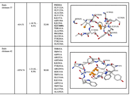
THR45A TRP47A ASP51A ARG52A ASP240A ILE292A TYR293A ARG295A SER314A TRP315A GLU316A ILE319A ASP321A TRP378A GLU379A ALA382A 90.00
(21.62, -8.58) -1074.74
Endo chitinase 42
PHE80A GLY122A GLY123A ALA126A GLY127A ILE157A ASP158A ILE159A GLU160A THR161A ALA199A PRO200A GLU201A TYR204A TYR219A GLN238A 52.00
(18.79, -8.55) -614.51
Endo chitinase 37
Table 10: SiteHound results with aromatic (CR1) probe
THR166A ASN167A PHE169A GLN199A SER227A GLN248A TRP584A TRP586A ASP589A HIS590A ASN597A GLN599A GLU625A HIS626A GLU648A TYR651A 41.00
(16.24, -8.67) -448.52
Beta-1,3 exo glucanase
HIS132A ALA133A TRP135A ALA138A ALA139A SER142A LYS144A PRO145A GLY146A ALA147A LYS148A SER312A TRP314A 18.00
(13.64, -8.71) -188.04
Alkaline proteinase
Interaction site Residues in
cluster 1 Volume
Energy range Energy
Enzyme
THR166A ASN167A PHE169A GLN199A SER227A GLN248A TRP584A TRP586A ASP589A HIS590A ASN597A GLN599A GLU625A HIS626A GLU648A TYR651A 41.00
(16.24, -8.67) -448.52
Beta-1,3 exo glucanase
HIS132A ALA133A TRP135A ALA138A ALA139A SER142A LYS144A PRO145A GLY146A ALA147A LYS148A SER312A TRP314A 18.00
(13.64, -8.71) -188.04
Alkaline proteinase
Interaction site Residues in
cluster 1 Volume
Energy range Energy
TRP173A HIS174A
ASP203A ILE307A
TYR308A
VAL309A LEU344A ILE345A
GLY348A HIS349A
SER458A ASP459A
TRP490A 50.00
(14.59, -8.51) -506.21
Catalase-peroxidase
SER37A ASN38A
GLN39A
GLY41A TYR43A TRP348A
ASN349A GLN350A
PHE402A GLN403A
THR404A
TYR407A ASN480A
VAL482A 49.00
(13.27, -8.55) -500.58
Beta-1,6-glucanase
PHE169A
GLY170A ASN171A
GLU173A ARG246A
TRP273A ALA275A
TRP277A LEU279A
TRP288A GLU293A
ASP295A
GLU298A HIS319A ASN326A
TRP329A ASN418A
ALA420A 62.00
(14.57, -8.51) -660.34
Beta-1,3- glucan-binding protein
TRP173A HIS174A
ASP203A ILE307A
TYR308A
VAL309A LEU344A ILE345A
GLY348A HIS349A
SER458A ASP459A
TRP490A 50.00
(14.59, -8.51) -506.21
Catalase-peroxidase
SER37A ASN38A
GLN39A
GLY41A TYR43A TRP348A
ASN349A GLN350A
PHE402A GLN403A
THR404A
TYR407A ASN480A
VAL482A 49.00
(13.27, -8.55) -500.58
Beta-1,6-glucanase
PHE169A
GLY170A ASN171A
GLU173A ARG246A
TRP273A ALA275A
TRP277A LEU279A
TRP288A GLU293A
ASP295A
GLU298A HIS319A ASN326A
TRP329A ASN418A
ALA420A 62.00
(14.57, -8.51) -660.34
TYR172A PRO213A ALA214A GLY215A ASN218A MET237A TYR239A ASP240A TYR241A ALA242A GLY243A PHE245A SER246A PHE267A ARG295A 42.00
(-13.64, -8.59) -430.47
Endo chitinase 42
TYR43A PHE80A GLY123A ASP158A ILE159A GLU160A ALA199A PRO200A GLN238A TYR239A TYR240A GLY288A LEU289A PRO290A ALA295A MET320A TRP322A 37.00
(-16.66, -8.56) -394.20
Endo chitinase 37
TYR32A ALA38A SER40A PHE63A ASP117A ASP165A GLU167A GLN222A TYR224A ALA272A MET299A TRP301A 38.00
(-14.01, -8.50) -398.27
Endo chitinase 33
TYR172A PRO213A ALA214A GLY215A ASN218A MET237A TYR239A ASP240A TYR241A ALA242A GLY243A PHE245A SER246A PHE267A ARG295A 42.00
(-13.64, -8.59) -430.47
Endo chitinase 42
TYR43A PHE80A GLY123A ASP158A ILE159A GLU160A ALA199A PRO200A GLN238A TYR239A TYR240A GLY288A LEU289A PRO290A ALA295A MET320A TRP322A 37.00
(-16.66, -8.56) -394.20
Endo chitinase 37
TYR32A ALA38A SER40A PHE63A ASP117A ASP165A GLU167A GLN222A TYR224A ALA272A MET299A TRP301A 38.00
(-14.01, -8.50) -398.27
The aim of using SiteHound Web server is to identify different ligand binding sites with the use of four different probes which corresponds to putative binding sites. The use of different chemical probes allows to study and characterize different potential binding site which helps to understand the fundamental mechanism of protein ligand interaction.
4. Conclusion
In Trichoderma, mycoparasitism is one of major mechanism involved in their antagonistic activity against phytopathogens. Multiple enzymes play crucial role in mycoparasitic interaction. In this study, lytic and defense enzymes were selected for in silico studies to improve their potential functional aspects. Due to lack of experimental structures of lytic and defence enzyme, in silico structural analysis was performed using homology modelling approach and further validated. Prediction of motifs in protein sequences allows studying conserved patterns throughout the evolutionary process. In silico identification and prediction of potential of binding site is very crucial feature of structure-based drug designing. But, here in this study, prediction of different ligand binding sites, u sin g chemical probes will provide to stu dy interaction between ligands and enzymes to improve the efficacy of biological experiment for the development of small compounds which increases the catalytic mechanism of enzymes, involves in mycoparasitism and will lead to destruction of phytopathogens.
Acknowledgements
The authors are grateful for the Financial Support given by the Council of Science and Technology, Lucknow.
Conflict of interest
We declare that we have no conflict of interest.
References
Arnold, K.; Bordoli, L.; Kopp, J. and Schwede, T. (2006). The SWISS-MODEL Workspace: A web-based environment for protein stru ctu re homology modelling. Bioinformatics, 22:195-201.
Baker, D. and Sali, A. (2001). Protein structure prediction and structural genomics. Science, 294:93-96.
Berman, H.; Westbrook, J.; Feng, Z.; Gilliland, G.; Bhat, T. N.; Weissig, I.; Shindyalov, I. and Bourne P. E. (2000). The Protein Data Bank. Nucleic Acids Res., 28:235-242.
Bindu, V. B. B.; Srinath, M.; Shailaja, A. and Giri, C. C. (2017). Comparative protein profile studies and in silico structural/functional analysis of HMGR (ApHMGR) in Andrographis paniculata (Burm.f.) Wall. ex Nees. Ann. Phytomed., 6(1):30-44.
Camacho, C.; Coulouris, G.; Avagyan, V.; Ma, N., Papadopoulos, J.; Bealer, K. and Madden, T. L. (2009).BLAST: Architecture and applications. BMC Bioinformatics, 10:421-430.
Chakraborty, M. R. and Chatterjee, N. C. (2007). Interaction of Trichoderma harzianum with Fusarium solani during its pathogenesis and the associated resistance of the host. Asian J. Exp. Sci., 21:353-357.
De Castro, E.; Sigrist, C. J. A.; Gattiker, A.; Bulliard, V.; Langendijk Genevaux, P. S.; Gasteiger, E.; Bairoch, A. and Hulo, N. (2006). ScanProsite: detection of PROSITE signature matches and ProRule-associated functional and structural residues in proteins. Nucleic Acids Res., 1: 34 (Web Server issue): W362-5.
Devi, N. and Azmi, W. (2018). Structural analysis and characterization of a clinically importa nt low molecula r weight natu ral dextr an synthesized by Leuconostoc lactis KU665298 dextransucrase. Ann. Phytomed., 7(1):5 2-62.
Fotoohiyan, Z.; Rezaee, S.; Shahidi Bonjar, H. Gh.; Mohammadi, H. A. and Moradi, M. (20 15). I ndu ction of systemic r esista nce by Tr ic hod er ma harzianum isolates in Pistachio plants infected with Verticillium dahlia. Journal of Nuts, 6(2):95-111.
Gavanji, S.; Aziz, A. H.; Larki, B. and Mojiri, A. (2013). Computational pr ediction and analysis of inter action of silver nitr ate with chitinase enzyme. IJSRES, 1(4):50-62.
Ghersi, D. and Sanchez, R. (2009). Easy MIFS and Site Hound: A toolkit for the identification of ligand-binding sites in protein structures. Bioinformatics, 25:3185-3186.
Grasso, J. E.; Sottile, E. A. and Coronel, E. C. (2016). Structural prediction and in silico physicochemical characterization for mouse caltrin I and bovine caltrin proteins. Bioinformatics and Biology Insights. 10:225-236 doi: 10.4137/BBI.S38191.
Green, H.; Larsen, J.; Olsson, A. P.; Jensen, F. D. and Jakobsen, I. (1999).
Suppression of the biocontrol agent Trichoderma harzianum by mycelium of the a rbu scular mycorr hizal fungus Glom us in tra radices in root-free soil. Applied and Envir onmental Microbiology, 65(4):1428-1434.
Haran, S.; Schickler, H. and Chet, I. (1996). Molecular mechanisms of lytic enzymes involved in the biocontrol activity of Tr icho der ma harzianum. Microbiology, 142:2321-2331.
Hernandez, M., Ghersi, D. and Sanchez, R. (2009).SITEHOUND-web: A server for ligand binding site identification in protein structures. Nucleic Acids Research, 37:W413-W416 doi:10.1093/nar/gkp281.
Hirokawa, T.; Boon-Chieng, S., and Mitaku, S. (1998).SOSUI: Classification and secondary structure prediction system for membrane proteins. Bioinformatics, 14(4):378-379.
Khaled, L. B.; P´erez-Gilabert, M.; Dreyer, B.; Oihabi, A.; Honrubia, M. and Morte, A. (20 08). Peroxidase changes in Ph oen ix dactylifer a pa lms inoculated with mycorrhizal and biocontrol fungi. Agron. Sustain. Dev., 28(3):411-418.
Laskowski, R. A.; MacArthur, M. W.; Moss, D. S. and Thornton J. M. (1993).
PROCHECK: A program to check the stereochemical quality of protein structures. J. Appl. Cryst., 26:283-291.
Petersen, N. T.; Brunak, S.; Heijne, V. G. and Nielsen, H. (2011). SignalP 4.0: discriminating signal peptides from transmembrane regions. Nature Methods: doi:10.1038/nmeth.1701.
Shi, W. and Chance, M. R. (2008). Metallomics and metalloproteomics. Cell.Mol. LifeSci, 65:3040-3048. doi:10.1007/s00018-008-8189-9.
Singh, A. and Chaube, R. (2014). Bioinformatic analysis, structure modeling and a ctive site pr ediction of aqu aporin pr otein from ca tfish heteropneustes fossilis. IJRITCC, 2(10):3208-3215.
Strakowska, J.; Blaszyk, L. and Chelkowski, J. (2014). The significance of cellu lolytic enzymes pr odu ced by Tr ich od erm a in the
oppor-tunistic lifestyle of this fungus. J. Basic Microb., 54:S2-S13.
The Un iProt Cons ortium (20 17). UniProt: The universal pr otein knowledgebase. Nucleic Acids Res., 45:D158-D169.
Tristão, G. B.; Assunção, Ldo. P.; Dos Santos, L. P.; Borges, C. L.; Silva-Bailão, M. G.; Soares, C. M.; Cavallaro, G. and Bailão, A. M. (2015). Predicting copper, iron, and zinc-binding proteins in pathogenic species of the Para coccidioides genus. Front. Microbiol., 9(5) :761 . doi: 10.3389/fmicb.2014.00761.
Waard, M. A. D.; Georgopoulos, G.; Hollomon, D. W.; Ishii, H.; Leroux, P.; Ragsdale, N. N. and Schwinn, F. J. (1993). Chemical control of plant diseases: Problems and prospects. Anm. Rev. Phtopathol., 34 :403-42 1.
Wilkins, M. R.; Gasteiger, E. and Bairoch, A. (1999). Protein identification and analysis tools in the ExPASy server. Methods Mol Biol.,