AMELOBLASTIC CARCINOMA –
REVIEW AND HISTOPATHOLOGY OF 5 CASES
1 2 3 4 5 6
Maya Ramesh B. Sekar S. Murali Saramma Mathew James Chacko George Paul
1 2 3 4
Senior Lecturer Reader Professor and Head, Ex Professor Emeritus, Department Of Oral Pathology, Vinayaka Missions Sankarachariyar Dental College,
Vinayaka Missions University, Ariyanoor, Salem, Tamil Nadu 5
Oral Surgeon, Penang International college, Malaysia, 6 Consultant Oral Surgeon, Sharon hospital, Salem )
Corresponding Author: Maya Ramesh,144, Andal Street, Thirumal Nagar, Alagapuram, Salem-636004. 0091 427 2444886 ®, 0091 9600918804(Mob), Email:
Abstract
Ameloblastic carcinoma is a rare malignant lesion that occurs primarily in the mandible in a wide range of age groups. No sex or race predilection has been noted. Ameloblastic carcinomas have characteristic histopathologic features and dictate more aggressive surgical approach than that of ameloblastoma. It is difficult to differentiate between a benign and malignant odontogenic lesion. Only less than 60 cases have been published in the English literature between the years 1984-2007. We are presenting the review and histopathologic features of 5 ameloblastic carcinoma cases here which were reported to our institution in a short span of 5 years in order to throw more light on the clinical features, histopathology, treatment and prognosis of the rare lesion.
Keywords : Ameloblastic carcinoma, Ameloblastoma
mayaramesh96@gmail.com
novo, ex-ameloblastoma, or ex-odontogenic
Introduction:
cyst. Most ameloblastic carcinomas are
1 presumed to have arisen de novo, with very few
O d o n t o g e n i c c a r c i n o m a s a r e
cases of malignant transformation of malignant odontogenic epithelial tumours that
a m e l o b l a s t o m a b e i n g a p p a r e n t . are of odontogenic origin and that bear little or
Dedifferentiation tends to occur in no resemblance to odontogenic apparatus.
2 ameloblastoma spontaneously or due to
The term ameloblastic carcinoma was first used
repeated surgical procedures or therapeutic by Shafer in 1974. The term ameloblastic
radiation. Peripheral ameloblastic carcinoma carcinoma is currently defined as 'a malignant
arising from gingiva or alveolar mucosal epithelial odontogenic tumour that has retained
epithelium is very rare. the features of ameloblastic differentiation, yet
also exhibits cytologic features of malignancy.'
Little information is available on
3 ameloblastic carcinoma, as only less than 65
Ameloblastic carcinomas seem to be
cases have been published in the English more common compared to malignant
literature between the years 1984-2007. This ameloblastoma in the ratio 2:1. Ameloblastic
article reports five additional cases of carcinomas have been reported to metastasize
ameloblastic carcinoma and outlines the clinical to the lungs and to distant sites. Male to female
features, biological behaviour, histopathology, ratio is almost 1.4:1. Lesions are located in the
treatment protocol and the prognosis of this mandible and the maxilla in 66% and 34% of the
rare tumour. patients, respectively.
3
Case Reports
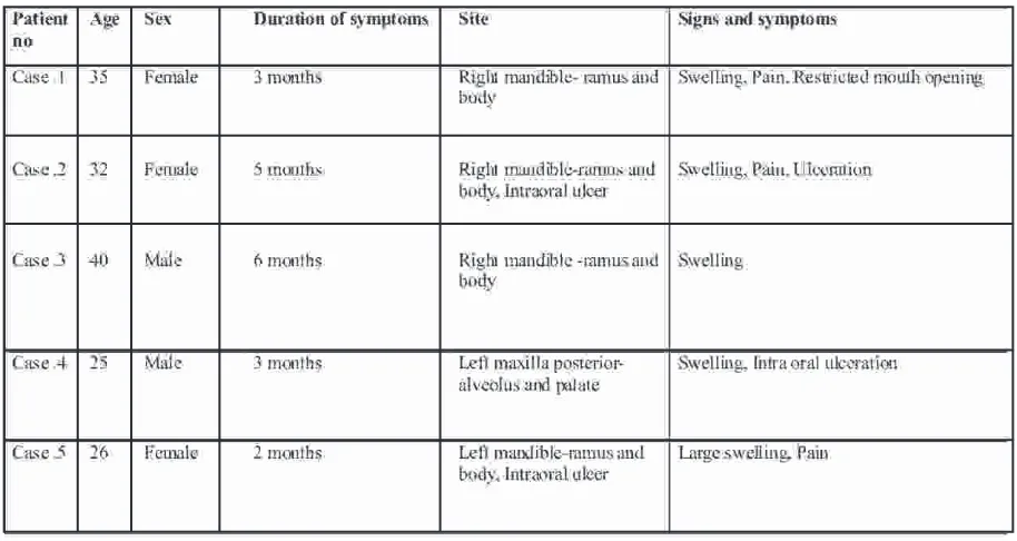
Our series of patients with ameloblastic carcinoma consisted of three females and two males who visited our institution in the past six years. Two patients sought treatment in 2002, another in 2005 and the other two in 2007. All were in the age group of 25-40 years. All patients reported with swelling and two patients showed intra-oral ulceration. One 40 year old
The Orthopantomograms revealed male patient was HIV positive. Three of the
solitary large radiolucency in three of the cases, cases were provisionally diagnosed as
bilocular radiolucency in one case and an ill odontogenic tumours, one as sarcoma and the
defined radiolucency in one case. Impacted other as a HIV / AIDS related tumour.
teeth were present in three of the lesions which were displaced by the growing tumour. All the patients were treated in the Department of Oral and Maxillofacial Surgery of two major hospitals. The case history is given in Table No I. All the five were diagnosed as ameloblastic carcinomas by hematoxylin and eosin sections. The malignancy was confirmed by proliferation marker PCNA which showed almost 5-6 times the value of ameloblastomas.
Discussion
Some of the reported cases of ameloblastic carcinoma in the literature are shown in Table II. The review by various authors showed more number of cases in the mandible ( 60-80 %) especially in the posterior region. Four of my cases were also in the mandibular posterior area. Only 19 cases are reported in maxilla till date and one of my case
1
was in maxilla. Corio et al reported 1:1 male : female ratio while my cases showed 3:2 ratio. The mean age of my patients was 32 years while
1
Corio et al reported a mean age of 30 years . Only very less number of reported cases showed metastasis. All these five cases were diagnosed before metastases. All our cases were in dentulous arch like most of the reported cases. The patients showed the features of rapid
15
growth and pain of the swelling. S Akrish in his review reported multilocularity and ill defined borders radiographically. The radiographic features of my cases included destructive radiolucency with multilocularity in one case and impacted teeth in three cases.
differentiated, usually non keratinizing form of The histopathologic features are shown ameloblastic carcinoma, both lesions being in Table no III. A predominantly follicular derived basically from odontogenic epithelial growth pattern was seen in three cases and remnants". In our series, there was no ambiguity plexiform growth pattern was seen in two cases. as all lesions showed clear evidence of Stellate reticulum like cells were minimal in all ameloblastic carcinoma.
the five cases. Squamous metaplasia and keratin
Among the differential diagnosis, pearls were zresent in two cases, but three cases
carcinomas metastazing from distant primaries showed only squamous metaplasia. No osteoid,
mimicking ameloblastic carcinoma and primary dentinoid or calcified tissues were present in any
intra alveolar carcinoma are the important ones. of the cases. Necrosis was present in three cases
Treatment and Prognosis and cystic degeneration in two cases. Peripheral
cells were multilayered in all cases with cuboidal
Surgical resection was the primary in three cases, squamous in one case and
modality of treatment in all the five cases. cuboidal and columnar in one case. Reversal of
Hemimandibulectomy was performed in the polarity was absent in four cases and present in
four mandibular lesions and maxillectomy was one case. Cells in the central areas were
done for the maxillary lesion. No neck predominantly spindle shaped in one, spindle
dissection was performed in any of the cases. and squamous in two cases, squamous in one
Surgical treatment decisions were made on the case and undifferentiated in one case.
clinical background that all lesions were N0 with Hyperchromatism and pleomorphism with
no evidence of metastasis. The patients showed increased nuclear cytoplasmic ratio were
good healing in the post operative period, present in all the 5 cases. In other areas the nuclei
justifying the treatment plan of oral surgeon. were vesicular. Spindle cells were present in all
the cases with two cases showing abundance.
The rarity and unusual behaviour make Clear cells were seen in two cases. Vascular the prognosis of the ameloblastic carcinoma invasion was present in all the five cases with
difficult. Recurrences and metastatic spread can only one case showing neural invasion.
be expected with inadequate treatment. Clear Infiltration into the surrounding connective cell type of ameloblastic carcinoma is seen to tissue was seen in all the five cases, but
have the worst prognosis. None of our series infiltration of the malignant cells into the
were of the clear cell type. Maxillary surface epithelium was present only in two
ameloblastic carcinomas have been stated to cases. have more unfavourable prognosis.
3
Bruce and Jackson stated the difficulty
The two patients treated in 2002 were in differentiating the histologic appearance of
followed up for a year and were then lost to primary intra alveolar carcinoma and the
follow up. The patient treated in 2005 was HIV
1
ameloblastic carcinoma. Corio et al stated:
positive and has not reported after his first post "Although the primary intra-alveolar carcinoma
operative visit. The two of the patients treated in and the ameloblastic carcinoma exhibit some
2007 are currently being followed up with no clinical differences, their histologic features are
signs of recurrence. Survival of ameloblastic similar enough to suggest a histogenetic
carcinomas should be evaluated over a long relationship. It is possible that the primary
Table no: I Clinical Features of the 5 patients with Ameloblastic Carcinoma
5. Nuclear details Pleomorphic with increased N/C ratio, Vesicular in some areas and Hyperchromatic in other areas
Pleomorphic with increased N/C ratio, Vesicular in some areas and Hyperchromatic in other areas
Pleomorphic with increased N/C ratio, Vesicular in some areas and Hyperchromatic in other areas
Pleomorphic with increased N/C ratio, Vesicular in some areas and Hyperchromatic in other areas
Pleomorphic with increased N/C ratio, Vesicular in some areas and Hyperchromatic in other areas 6. Necrosis / Cystic
Degeneration
Necrosis Present Necrosis Present Absent Cystic degeneration Present
Both Present
7. Infiltration of malignant cells into epithelium
Absent Present Absent Present Absent
8. Connective Tissue
Infiltration of malignant cells into
connective tissue Present Present Present Present Present
9. Osteoid /Dentinoid / Calcification
- - - - -
10. Squamous metaplasia / Keratin pearls
Both present Both present Squamous metaplasia present
Squamous metaplasia present
Squamous metaplasia present
11. Vascular / Neural invasion
Vascular invasion present
Vascular invasion present
Vascular and neural invasion present
Vascular invasion present
Vascular invasion present
regional recurrences and metastases.
12. Shigeto Kawauchi, Yoshikazu Hayatsu, Mutsuo Takahashi, Tomoko Furuya, Atsunori Oga, Shin Reference
Ichiro Niwa, Kohsuke Sasaki. Spindle cell ameloblastic carcinoma: a case report with 1. Corio R L, Goldblatt L I, Edwards PA, Hartman
immunohistochemical ultrastructural and KS, Ameloblastic Carcinoma: a clinicopathologic
comparative genomic hybridisation analyses. study and assessment of eight cases, Oral Surg Oral
Oncology Reports 10:31-34, 2003 Med Oral Pathol 1987; 64: 570-6
13. Anni Suomalaien, Jarkko Heitanen, Soraya 2. Shafer- Text Book of Pathology, Fifth Edition,
Robinson, Jaakko Sakari Peltola, Helsinki . 2006, Elsevier, A Division of Reed Elsevier India
Ameloblastic carcinoma of the mandible resembling Private Limited, ISBN-10: 81-8147-847-9
odontogenic cyst in a panoramic radiograph, Oral Surgery, Oral Medicine, Oral Pathology, Oral 3. Reichart. P.A, Philipsen .H.P- Odontogenic
Radiology and Endodontology 2006; 101: 5 :638-Tumours and Allied Lesions, 2004 Quintessence
42 Publishing Co Ltd,London, ISBN 1-85097-059-9
14. James M.Hall, Dwight R. Weathers and K. 4. Nagai N,
Krishnan Unni. Ameloblastic Carcinoma: An analysis of 14 cases Oral Surgery, Oral Medicine, Oral Pathology Oral Radiology and Endodontology 2007;6 103: 799-807.
5. Lolachi C M, Madan SK, Jacobs JR. Ameloblastic
15. S J Akrish, Amos Buchner, Yitzhak Shoshani, Carcinoma of Maxilla J Laryngol Otol 1995;109:
Marilena Vered, Dan Dayan. Ameloblastic 1019-22
carcinoma : report of a new case , Literature review and comparison to ameloblastoma. J Oral 6. Khoo Suan Phaik, Ong Siew Tin. Ameloblastic
Maxillofac Surg, 2007; 65: 777-783. Carcinoma: A Case with cervical node And
pulmonary metastasis, Annals Dent Univ Malaya
16. Brent B.Ward, Steven Edlund, James Scuibba, 1998;5:49-52
Joseph I. Helman Ameloblastic Carcinoma (primary type) Isolated to the anterior maxilla ; case 7. DP Cox, Muller S, Carlson GW, Murray D.
report with review of literature, J OralMaxillofac Ameloblastic Carcinoma ex ameloblastoma of the
Surg 2007:65;9,1800-1803. mandible with malignancy
17. Adil Benlyazid, Magali Lacroix – Triki, Richard 8. Jesus Sastre, Munoz M, Naval L, Adranos M.
Aziza, Anni Gomez-Brouchet, Maryalis Ameloblastic Carcinoma of Maxilla: report of a case.
Guichard, Jerome Sarini, Toulouse. Ameloblastic J Oral Maxillofac Surg 2002;60:102-4
carcinoma of maxilla: case report and review of literature, Oral Surg Oral Med Oral Pathol Oral 9. Karan
Radiol Endod 2007;104:e17-e24
10. J Hall, K Unni, D Weathers, Ameloblastic carcinoma: Analysis of 11 cases. Oral Surgery, Oral Medicine and Oral Pathology, Vol 96, No 3, 2003
11. Rajiv Datta, Winston J S, Diaz - Reyes G, Loree T R, Myers L, Kuriakose MA et al. Ameloblastic carcinoma : Report of an aggressive case with multiple bony metastases. Am J Otolaryngol 2003; 24:64-9
Takeshita N, Nagatsuka H, Inoue M Nishijima K, Nojima T et al: Ameloblastic Carcinoma- Case report and review, J Oral Pathol and Med 1991; 20:460-3.
Dhir , Sciubba J, Tufano R P: Ameloblastic Carcinoma of the Maxilla, Oral Oncol 2003; 39:736 -41
Source of Support - Nil