1. Introduction
espite a gradual decline in overall stroke death in many countries, stroke remains the third leading cause of death; stroke is also the leading cause of disability in adults.
The human and financial costs of stroke are
immense. Approximately 30% of survivors require sistance with activities of daily living, 20% require as-sistance with ambulation, and 16% require institutional care (Hunckey and Warlaw, 2000).
Routine empirical measurement of C-reactive protein (CRP) is a valuable aid in patient’s management across
Seyed Abbas Hasani 1, Seyed Ali Ziai2, Masoud Mehrpour 3,*, Mandana Amiri1, Mohamad R Motamed 3
1. Department of Emergency Medicine, Tehran University of Medical Sciences, Iran. 2. Department of Pharmacology, Shahid Beheshti University, Tehran, Iran. 3. Department of Neurology, Tehran University of Medical Sciences, Iran.
D
* Corresponding Author:
Masoud Mehrpour, MD.
Department of Neurology, Firoozgar Clinical Research Development Center (FCRDC), Firoozgar General Hospital, Tehran University of Medical Sciences. Fax: +98-21-88942622
Introduction: Elevated levels of CRP are present among patients at risk for further
first-ever myocardial infarction and stroke. It has been shown that after ischemic
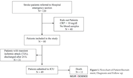
stroke, increased levels of CRP are associated with unfavorable outcomes. Methods: From 120 patients admitted to the emergency unit of our hospital with the diagnosis of stroke; CRP, D-dimer and ferritin level was measured and the patients were followed until discharge or death.
Results: CRP level was significantly different between the patients with TIA and stroke. D-Dimer level was also significantly different between the TIA & the
admitted groups. Ferritin was not different between the prognosis groups. There was a correlation between CRP and D-Dimer (r = 0.381, p = 0.001), and also between CRP and ferritin (r = 0.478, p= 0.000).
Discussion: CRP is a useful adjuvant marker to determine the prognosis of patients with cerebro-vascular events admitted to the hospital, in both patients
with stroke positive history and first-ever stroke.
A B S T R A C T
Article info:
Received: 20 August 2011 First Revision: 28 August 2011 Accepted: 23 September 2011
Key Words: Stroke, Prognosis,
Acute Phase Reactant, C Reactive Protein, Transient Ischemic Attack.
a broad range of clinical practices. Sensitive CRP assay may become a new risk assessment marker for cardio-vascular diseases, and guidelines for its application are under discussion (Pepys, 2001). Elevated levels of CRP
are present among patients at risk for further first-ever
myocardial infarction and stroke (Koenig, 2001).
It has been shown that after ischemic stroke, increased levels of CRP are associated with unfavorable outcomes (Sander, 2002). In another study, survival in patients
with CRP levels >1.01 mg/dl was significantly worse
Autumn 2011, Volume 3, Number 1 Basic and Clinical
By determining the CRP level we could better under-stand the prognosis of patients and could make better decisions about their hospitalization and implementa-tion of more aggressive treatments.
2. Methods
From 120 patients admitted to the emergency unit of the Rasool-e-Akram hospital in Tehran in spring of 2008 with the diagnosis of cerebrovascular disease, eighty patients were included in the study. All patients were informed about the study and consents were ob-tained from them. The inclusion criteria were
diagno-sis of stroke, either ischemic or hemorrhagic (with first
ever and previous stroke history), and TIA (transient ischemic attack). Exclusion criteria were related mainly to the conditions associated with increased levels of
in-flammation markers, including infection like pneumo
-nia, sepsis or other inflammatory diseases like vasculitis
(initial CRP level >10 mg/dl). CT scan of the brain was performed at enrollment within 24 hours after stroke
onset to confirm the diagnosis of ischemic and hemor -rhagic stroke. A full neurological examination,
includ-ing screeninclud-ing with Modified Rankin Scale, was also car -ried out. All the patients were re-examined by the same neurologist, 24 hours after cerebrovascular attack.
Several laboratory parameters and cardiovascular risk factors such as smoking, hypertension, diabetes mellitus, and previous myocardial infarction were
de-termined. Based on prognosis, patients were separated into three groups: 1) TIA: transient ischemic attack with symptoms lasting less than 24 hours, 2) admitted: pa-tients who had ischemic or hemorrhagic stroke and need to be admitted, 3) death: patients who died after admis-sion. We also categorized patients based on their CT
scan into three groups: 1) patients with non-significant
CT scan, 2) patients with ischemic lesions and 3) pa-tients with hemorrhagic pattern.
A 902 Roche/Hitachi auto analyzer was used to de-termine the levels of CRP, D-Dimer, and Ferritin. In all patients, these were measured immediately after admission. Levels of CRP were determined with a commercially available, high-sensitivity, immunoneph-elometric, latex enhanced assay (Tina quant CRP Ul-tra sensitive, Roche diagnostic). D-Dimer and Ferritin were also determined by immunonephelometric method (Tina quant D-Dimer and Tina quant Ferritin, Roche diagnostic). Statistical analysis was performed with a SPSS v11 package.
3. Results
Eighty patients were included in the study, 37 male and 43 female. The patient’s mean age was 67.1 ± 13.1 years (mean ± SD). The group distribution has been de-picted in chart I. Patient’s characteristics on admission
Aspirin 6 9 2
MI history 5 5 3
Stroke history 7 11 4
Diabetes mellitus 10 13 5
Hypertension 21 28 9
Smoking 5 6 4
Table 2. CRP, D-Dimer, and ferritin in three prognosis groups (mean ± SD).
TIA Admitted Death P value TIA vs. admitted P value TIA vs. Death P value Admit-ted vs. Death
CRP 0.23 ± 0.21 1.64 ± 1.80 1.47 ± 1.97 0.000*** 0.016* 0.73
D- Dimer 0.91 ± 1.05 3.64 ± 5.83 3.81 ± 5.77 0.024* 0.119 0.929
Ferritin 117.6 ± 137.3 158.9 ± 136.2 122.4 ± 83.1 0.519 0.999 0.632
Table 3. Distribution of diagnosis groups between prognosis groups.
TIA Admitted Death
Non-significant CT 16 15 0
Ischemic CT 2 29 7
Hemorrhagic CT 0 7 4
are listed in table 1. Individual CRP, D-Dimer and ferri-tin values in the three groups are shown in table 2. CRP
was significantly different between prognosis groups (table 2). D-Dimer was also significantly different be -tween TIA & admitted groups (table 2). Ferritin was not different between prognosis groups. There was a corre-lation between CRP and D-Dimer (r = 0.381, p = 0.001), and also between CRP and ferritin (r = 0.478, p< 0.001).
We found that CRP is significantly different between
prognosis groups in patients with previous stroke
his-tory (p= 0.05) as well as first ever stroke patients (p =
0.012).
Categorizing the patients based on diagnosis into the
following three groups: non-significant (n = 18, %22.5),
ischemic (n = 51, %63.8), and hemorrhagic (n = 11,
%13.8), we found a significant difference in CRP level between them (non-significant & ischemic patients: p = 0.001; non-significant & hemorrhagic patients: p =
0.04). Table 3 shows the distribution of CT scan diag-nosis groups between progdiag-nosis groups. There is a good correlation between the diagnosis groups and the prog-nosis groups (r = 0.563, p<0.001).
4. Discussion
In this study the CRP concentrations increased (> 5 mg/l) in about %65 of patients within 24h after ischemic stroke. We found that CRP measurement on admission for acute-cerebro-vascular events predicts the further
-Autumn 2011, Volume 3, Number 1 Basic and Clinical
ference between TIA and admitted or death patients. CRP level was less than 5 mg/l in about 93% of TIA group. In a previous work, researchers had not found any prediction value for the patient’s groups (Canova et al., 1999). In our study we did not found any difference in CRP between admitted and death groups. But we
found a significant correlation between CRP, D-Dimer,
and Ferritin, which corresponds with previous works (Di Napoli et al. 2002). Categorizing patients based on diagnosis showed that there is a good correlation be-tween the prognosis and the diagnosis groups. There was a correlation between CRP and prognosis groups (r = 0.358, p = 0.001), but there was no correlation be-tween CRP and the diagnosis groups. This pattern was also repeated for D-Dimer (r = 0.262, p = 0.31). It may be concluded that CRP and D-Dimer can predict the outcome independent of the CT scan’s results.
High CRP in healthy individuals indicates increased risk of coronary and cereberovascular events (Ridker et al., 1997), and a worse prognosis in myocardial in-farction (Pietila et al., 1996), and ischemic stroke. This indicates that CRP should be considered as a marker of cardiovascular risk (Pepys, 2001). In patients with acute myocardial infarction or ischemic stroke the extent of necrosis is the main but not the only determinant of prognosis (Di Napoli et al, .2001). Response to the ne-crotic insult are probably multiple and may need inde-pendent additional determinants of prognosis (Canova et al., 1999).
in order to be in accordance with other authors who
studied on first ever stroke patients (Di Napoli et al.
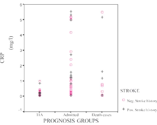
Figure 2. Distribution of CRP between prognosis groups (Transient Ischemic Attack (TIA), admitted, and death cases) categorized based on stroke history. Only 2 of TIA prognosis group (6.7%) had CRP > 5 mg/l. But, 25 cases of the admitted group (66%) and 7 of the death group (64%) had CRP > 5 mg/l.
2000), we also divided patients into two major groups,
with previous stroke history and first ever stroke, corre -lation between CRP, D-Dimer and Ferritin was stronger in patients with previous stroke history (Di Napoli et al., 2002). So we can conclude that CRP is a useful adju-vant marker to determine the prognosis of patients with cerebro-vascular events admitted to the hospital, in both
patients with stroke positive history and first-ever stroke
patients. Unfortunately it is impossible to measure the CRP level before stroke and we could not determine its value as a diagnostic factor. More studies are needed to determine any difference between different sources of stroke.
Acknowledgment
The authors thank Dr. S. M. Blourchi, manager of Daneshafzar Eng. Co., and Miss. Pakdel for providing the CRP kit.
References
Canova CR, Courtin C, Reinhart WH (1999). C-reactive protein (CRP) in cerebro- vascular events. Atherosclerosis; 147:49-53.Di Napoli M. (2001). C reactive protein and acute phase of ischaemic stroke. BMJ; 322(7302): 1605-6.
Di Napoli M, Di Gianfilippo G, Sollecito A, Bocola V (2000). C-reactive protein and outcome after first-ever ischemic stroke.
tigators. Inflammation, hemostatic markers, and antithrom -botic agents in relation to long-term risk of new
cardiovascu-lar events in first-ever ischemic stroke patients. Stroke, 33(7):
1763-71.
Hunkey GJ, and Warlow CP (2000). Transient ischemic attacks of the brain and eye. London: WB Saunders/ Ballier Tindall.
Koenig W (2001). Inflammation and coronary heart disease: an
overview. Cardiol. Rev., 9: 31-35.
Muir KW, Weir CJ, Alwan W, Squire IB, Lees KR, (1999). C-reactive protein and outcome after ischemic stroke, Stroke, 30: 981-5.
Pepys MB, (2001).The renaissance of C reactive protein, BMJ, 322: 4-5.
Pietila KO, Harmoinen AP, Jokiniitty J, Pasternack AI (1996). Serum C-reactive protein concentration in acute myocardial infarction and its relationship to mortality during 24 months of follow-up in patients under thrombolytic treatment. Eur Heart J, 9:1345-9.
Ridker PM, Cushman M, Stampfer MJ, Tracy RP, Hennekens CH (1997).
Inflammation, aspirin, and the risk of cardiovascular disease in
apparently healthy men. N Engl J Med, 336(14): 973-9.
Sander D, (2002) Prognostic relevance of early serial C-reactive
protein measurements after first ischemic stroke. Stroke, 33: