the visual function and thus the visual field as well. Therefore, early detection of these defects is crucial to starting the treatment in time. It has been not-ed that substantial retinal ganglion cell damage can happen long before its detection using standard ex-aminations assessing RGC damage [8]. Some au-thors, however, claim that M-cell damage does not affect the results of visual field examinations [9].
Due to its mostly symptomless beginnings, glaucoma is hard to diagnose. That is why pre-ventive treatment and performing periodic health tests are crucial. The most important examination assessing optic nerve function is a visual field test.
Visual Field
Visual field is a term for the area one sees while the eye is still. Another, more intuitive definition would be ‘the vision island’ surrounded by the ‘sea of darkness’ [10]. Measurements of the visual field Glaucoma is an optic nerve neuropathy
as-sociated with progressive visual field loss. One of the most frequent eye diseases these days, it is believed to have affected 60 m people worldwide in 2014 [1]. It has also been estimated that over 20 m patients will have lost their sight due to glau-coma by 2020 [2].
Nowadays it is said that glaucoma damage doesn’t only affect optic nerve fibers, but also retinal ganglion cells [RGC] [3], which become damaged at an even earlier stage [4]. The type and timing of cel-lular changes leading to RGC loss in glaucoma re-main incompletely understood, including whether specific RGC subtypes are preferentially impact-ed at early stages of the disease [5]. It seems that magnocellular retinal cells (M-cells) are more sen-sitive than parvocellular retinal cells (P-cells) and become some of the first to be damaged in glauco-ma [6]. Since these cells are very sensitive to glau-coma damage, they suffer complete loss at an ear-ly stage of glaucoma [7], which in turn influences
Kamil Kaczorowski
B–D, Małgorzata Mulak
A, D, E, Dorota Szumny
C–E,
Marta Misiuk-Hojło
FHeidelberg Edge Perimeter:
The New Method of Perimetry
Department and Clinic of Ophthalmology, Wroclaw Medical University, Poland
A – research concept and design; B – collection and/or assembly of data; C – data analysis and interpretation;
D – writing the article; E – critical revision of the article; F – final approval of article
Abstract
Glaucoma is an optic nerve neuropathy associated with progressive visual field loss. One of the most frequent eye diseases these days, it is believed to have affected 60 million people worldwide in 2014. Various visual field exami-nation methods are known, from the confrontational test to kinetic and static perimetry. The latest device to access the visual field is the Heidelberg Edge Perimeter (HEP). It is a flicker perimeter, but, unlike others of its kind, it uses a unique stimulus called FDF (Flicker Defined Form). A 5-grade round stimulus is created by reversing the phase of flickering black and white dots, thereby forming illusory outlines. The test uses randomly flickering points in medium illumination (50 cd/m2). The background remains the same during the whole test. Background luminance is 50 cd/m2, the marker showing time is 400 ms, and the frequency is 15 Hz. Current studies show that HEP can detect early visual field loss which remains invisible during a standard visual field test with standard automated perimetry. HEP might also prove useful in the early detection of other diseases connected with visual field loss, for example in neurology (Adv Clin Exp Med 2015, 24, 6, 1105–1112).
Key words: glaucoma, visual field, Heidelberg Edge Perimeter (HEP), flicker defined form.
REVIEWS
Adv Clin Exp Med 2015, 24, 6, 1105–1112
can be taken with a perimeter. The visual field was first measured by Thomas Young, then, in 1825, Purkinje modified Young’s ranges, changing them to 60° nasal, 100° temporal, 60° in the upper and 80° in the lower field [10]. It was Albrecht von Gräfe that first introduced perimetry into clinical practice [11].
In the course of glaucoma development, the vi-sual field decreases in a characteristic way, related to the arrangement of nerve fibers in the retina and the way they come from its specific parts. The fi-bers from the spot and the nasal part of the retina go immediately to the optic nerve disc, whilst those from the retina’s temporal segment make bends. The latter are also the most sensitive to glaucoma-tous damage. Visual field tests in glaucomaglaucoma-tous pa-tients prove the existence of bends in which no vi-sual information is perceived [12].
Visual Field Test
Various visual field examination methods are known, from the confrontational test to kinetic and static perimetry.
Kinetic perimetry is the oldest measurement technique known. It consists of showing the pa-tient a marker whose illumination is changing. The clinician moves the marker at a speed varying from 2º/s to 5º/s from the area in which the patient
cannot see it to where it becomes noticeable [13]. The patient informs the doctor when they can see the marker, owing to which isopters (lines con-necting the visibility areas of a marker given) are obtained.
Currently, the most common visual field ex-amination method is static perimetry. It presents many markers of changing illumination in the same area in such a way that the smallest degree of marker brightness in that place can be deter-mined [14]. This way a three-dimensional vision island of the patient is established, using which one can state where exactly the patient’s response to light stimuli is weaker than it should be based on the patient’s age. Following the invention of auto-matic perimeters, this technique has become much easier and faster than kinetic perimetry.
Markers may vary in size and illumination as well as showing time. Their size is normally given using the 5-grade Goldmann scale, whose mark-ers are 1/16 to 64 mm2 big. The marker’s showing
time varies according to the device. The illumina-tion, measured in apostilbs or decibels in perime-try, is an indication of stimulus intensity.
Static perimeters measure the patient’s re-sponse in the central and peripheral visual fields, whose width is usually 10°, 24°, 30° and 60°. Tests
are marked as, for example, 24-2 or 30-1, with the first digit indicating the range of the visual field test (given in degrees) and the latter (either 1 or 2) – the marker arrangement [15].
The most frequent Standard Automated Pe-rimeters (SAP) are Humphrey and Octopus, rec-ommended by the European Glaucoma Society as the most useful for routine glaucoma diag-nostics [16]. The basic element of a perimeter is a hemisphere-shaped cap, within which markers are shown at any place given. A change in marker intensity can be achieved through an alteration in its size or illumination.
Another perimeter type is Flicker Defined Pe-rimetry (whose one example is the Heidelberg Edge Perimeter – HEP), which uses static perime-ters using a different kind of stimulation – a flick-ering marker of periodically changing illumina-tion [17, 18]. It is more difficult to detect, which is believed to be related to the lesser sensitivity of magnocellular retinal cells [18, 19]. These perime-ters are particularly useful in detecting early glau-comatous changes.
The frequency-doubling technology (FDT) perimeter is similar to Flicker Defined Perime-try in terms of usage. The eye observes a sinusoi-dal net of a low spatial frequency, but flickering with a high frequency and perceives it as an el-ement of twice as big spatial frequency, which is an optical illusion [15, 20]. As in flicker perim-etry, M-cells do react to this kind of stimulus, which provides information about the early stage of glaucoma [21].
Heidelberg Edge Perimeter
HEP is a flicker perimeter, but, unlike others of this kind, it uses a unique stimulus called Flick-er Defined Form (FDF). A 5-grade round stim-ulus is created by reversing the phase of flicker-ing black and white dots, thereby formflicker-ing illusory outlines [22, 23]. The test uses randomly flickering points in medium illumination (50 cd/m2). Thebackground remains the same during the whole test. Background luminance is 50 cd/m2, with
a marker showing time of 400 ms, whereas the fre-quency is 15 Hz.
HEP can be used for testing peripheral as well as central visual field, 10°, 24°, 30° or 60° wide. An SAP examination can also be performed the same way as on a standard perimeter.
The following examinations can be carried out using the HEP field analyzer: S-30 (FDF), 10-3 (FDF), 10-2 (SAP III), 24-2 (FDF), 24-2 (SAP III and V), 30-2 (FDF) and 30-2 (SAP III).
Also, the HEP perimeter uses different test strategies: Adaptive Staircase Thresholding Algo-rithm (ASTA) was elaborated by prof. John Fla-nagan. His technique uses the step function up or down, modifying it to suit the database. The results are presented as a sensitivity measure expressed in decibels (dB). Three ASTA strategies are operated by HEP: Standard, Follow-up and Fast.
ASTA Standard is based on a probability es-timate in order to ensure optimum test effective-ness. This strategy ought to be performed on all new patients as it provides satisfying results and enables the assessment of possible defect progres-sion. First the 4 : 2 : 2 strategy is used to determine loss in a segment given, then, if the patient can-not see the stimulus, the power is increased first by 4 dB, which makes the stimulus more notice-able, and by an additional 2 dB should the stim-ulus still not be seen. This sensitivity is a starting point for the neighboring areas, where the strate-gy used is 2 : 2. Should these segments differ a lot from the rest or be very limited, they ought to be tested again.
ASTA Follow-up is a test that can be per-formed following the others. It compares the re-sults of previous examinations with the current ones; this strategy uses the 2 : 2 step function and is faster than ASTA Standard.
The ASTA Fast algorithm uses the 4 : 2 func-tion, thereby making the examination faster. It is recommended for the monitoring of ‘within the normal range’ patients or those who have previous-ly proven tired of the examination methods. The results are less comparable, but the test is shorter.
HEP also has different screening test strategies at its disposal. Since the examination time is short-er, it is particularly advisable in the case of patients who cannot take a long test.
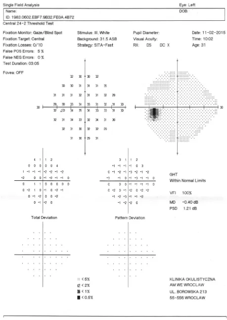
Reliability measures such as fixation loss-es (FL), false positive errors (FP) or false nega-tive errors (FN) are taken into account while in-terpreting the test results [15]. Fixation losses
imply the patient having wrongly looked sideways, while false positive errors indicate the patient has pressed the ‘stimulus’ button automatically, with-out having really seen it. False negative errors take place when the patient doesn’t notice a marker of greater brightness than that seen in the same loca-tion before. The results of the test can be analyzed from a graphical printout.
Heidelberg Retina Tomograph (HRT) can be connected to the HEP so that examinations from both can be compared and analyzed by a special program. HRT is a device used for measuring op-tic nerve fiber thickness right by the edge of the optic disc. The measurement once taken is then compared to the apparatus’ database, which con-tains the range of ‘normal’ results for each pa-tient age. The possibility of connecting HEP and HRT makes it possible to assess existing visual field loss.
HEP Examinations so Far
HEP is a new perimeter whose advantages and drawbacks are being intensely examined all over the world. It has only been used for patient exam-ination for a few years now, therefore, rather few works on its effectiveness in visual field testing are known so far.The introduction of a new perimeter has made it necessary to compare it to those already in use. As a logarithmic scale measuring the sensitivi-ty to a stimulus, a decibel range is used for evalu-ating glaucoma defect. ‘Zero’ on the decibel scale equals a complete lack of reaction to the brightest stimulus. The greater the decibel value, the dark-er the stimuli that wdark-ere noticed. The decibel scale, however, is not the same for different parameters. ‘0’ on the Octopus device doesn’t denote the same as ‘0’ for Humphrey [24]. The comparison has been shown in Table 1.
The range of the HEP scale is 0–25 dB and it is not easily compared to the Humphrey results. Nevertheless, the device itself has a program which converts its measurements to the generally recog-nized SAP perimeter scale – HEP software release
Table 1. The comparison of decibel scales in SAP (Humphrey) and FDF (HEP)
SAP FDF
Size of the stimulus Goldman III and V 5 × 5° for 24°, 30° and 60° visual field, 3 × 3° for 10°
Luminance of the stimulus 40–0 dB 28–0 dB
Brightness of the background 10 cd/m2 50 cd/m2
2.2 which includes: HEP Acquisition Module (AQM) v. 2.2, HEP Viewing Module (VWM) v. 2.2 and Heidelberg Eye Explorer (HEYEX) v. 1.7. Ow-ing to that, a comparison of the results obtained is possible. Differences in the decibel values are crucial to assessing the stage of glaucoma defect. Thus far the Hodapp differentiation method has been used to indicate it in SAP perimeters: 0–6 dB means small glaucoma damage, 6–12 dB indicates medium changes, whereas above 12 dB means seri-ous damage [26]. It hasn’t been stated whether the same criteria can be applied to HEP yet.
The differences between HEP and standard perimeters are only now being established. Some of the first studies have already demonstrated the HEP visual field test can detect changes at an earli-er stage than standard pearli-erimetry. In 2012, Haslearli-er S. and Stürmer J. claimed that the Heidelberg Edge perimetry seemed to be more sensitive than con-ventional static perimetry in the early detection of visual field alterations in patients with ocular hy-pertension and incipient glaucoma [27].
Despite normal visual field findings in the Oc-topus perimetry (MD < 2.0 dB), pathological visual fields have been detected in 48 out of 90 eyes with the HEP examination (53%).
Examinations with flicker defined form (FDF) and frequency doubling technology (FDT) perim-etry have been carried out to determine their abil-ity to detect glaucoma at an earlier stage than stan-dard automated perimetry (SAP) [27]. The purpose of the study was to examine the structure-function relationship between FDF, FDT, SAP, and confo-cal scanning laser ophthalmoscopy (cSLO) in glau-coma patients. FDF perimetry presented with the strongest structure-function relationship (global correlation with the rim area was 0.44, whereas the range of significant sectoral FDF values was 0.23– –0.69), followed by FDT (whose global correlation with the rim area was 0.35 and the range of sig-nificant sectoral FDT values: 0.25–0.60). SAP fea-tured the weakest structure-function relationship and fewer statistically relevant results (global cor-relation: 0.32; the range of significant sectoral SAP values: 0.23–0.58). The correlation between struc-ture and function was stronger for FDF and FDT than in SAP. The connections appeared strongest in temporal areas – usually the first to suffer glau-comatous damage.
While examining patients who were clinically suspicious for glaucoma due to optic nerve head (ONH) or retinal nerve fiber layer (RNFL) dam-age, it was noted that in more than half of them FDF managed to detect visual field loss while SAP still gave ‘normal’ results [23]. The compari-son of HEP and SAP results in patients with pri-mary open-angle glaucoma proved that out of
the total of 42 open angle glaucoma (OAG) pa-tients with abnormal SAP MD, 38 also had path-ological FDF MD (mean deviation). Neverthe-less, FDF MD was aberrant in 28 out of 55 OAG patients with normal SAP MD. The FDF MD proved more correlated with RNFL thickness than SAP MD. To conclude, FDF perimetry may be useful in early glaucomatous nerve atrophy detection [21].
Performing HEP is particularly important in the case of glaucoma suspicion. Horn et al. have pointed out that the FDF stimulus is able to detect early glaucoma damage [22]. Patients with SAP MD values exceeding 5 dB should be monitored with conventional perimetry because of its larger dynamic range.
The usefulness of HEP in early glaucoma de-tection has also been emphasized by other au-thors. Prokosch et al. [28] have compared white-on-white standard automated perimetry (SAP), matrix frequency doubling technology (FDT), and flicker-defined form perimetry (FDF) in their use for detecting nerve fiber layer loss in patients at an early stage of glaucoma. They concluded that the sensitivity to RNFL loss detection in early glaucoma appeared higher in FDF and FDT ma-trix than in SAP perimetry, much as the specifici-ty was highest in SAP. The sensitivispecifici-ty was highest for FDF perimetry (87%), followed by FDT ma-trix (62.5%) and then SAP (40%). MD and pattern standard deviation (PSD) in FDF and FDT ma-trix were significantly different between patients with RNFL loss and those without it (p < 0.05), while no difference had been found in SAP. That is why simultaneous use of FDF/FDT matrix and SAP perimetry is recommended for fully accurate results.
Studies aiming to improve the understanding of perimeter work mechanisms are also being car-ried out. Shahidi et al. have pointed out the differ-ences in HEP-FDF and FDT matrix stimuli, based on the findings from their own study: the visu-al sensitivity increase noted was only significant for HEP-FDF stimuli and not for the other device examined [29].
Octopus. As in the case of other perimeters, the patient has to learn to undergo this examination. Usually, the first two or three results are not as re-liable as the later ones, which has to do with the marker notice learning effect, correct eye position and general examination learning.
Another relevant issue is contrast sensitivi-ty in the patient, which may heavily influence the results. In the case of reduced contrast sensitivi-ty, patients examined using the HEP perimeter can experience trouble seeing the stimulus, which is dark grey and shown against a flickering grey background.
Conclusions
The crucial element of glaucoma treatment is an early diagnosis, when only few nerve fibers have been damaged and the patient can undergo treat-ment for many years to come, using the slowdown methods available and continuing to have good vi-sual field. Current studies show that the HEP can detect early visual field loss which remains invisi-ble during a standard visual field test with the SAP perimeter. HEP might also prove useful in the ear-ly detection of other diseases connected with visual field loss, for example in neurology.
References
[1] Weinreb R: Glaucoma Worldwide: A Growing Concern (February 2015). Available from: http://www.glaucoma. org/gleams/glaucoma-worldwide-a-growing-concern.php.
[2] Facts about Glaucoma, National Eye Institute (February 2015). Available from: http://www.nei.nih.gov/health/ glaucoma/glaucoma_facts.asp.
[3] Chen YS, Green CR, Danesh-Meyer HV, Rupenthal ID: Neuroprotection in the treatment of glaucoma – A focus on connexin43 gap junction channel blockers. Eur J Pharm Biopharm 2015.
[4] Park JW, Jung HH, Heo H, Park SW: Validity of the temporal-to-nasal macular ganglion cell-inner plexiform layer thickness ratio as a diagnostic parameter in early glaucoma. Acta Ophthalmol 2015.
[5] El-Danaf RN, Huberman AD: Characteristic patterns of dendritic remodeling in early-stage glaucoma: evidence from genetically identified retinal ganglion cell types. J Neurosc 2015, 35, 2329–2343.
[6] Kerrigan-Baumrind LA, Quigley HA, Pease ME, Kerrigan DF, Mitchell RS: Number of ganglion cells in glau-coma eyes compared with threshold visual field tests in the same persons. Invest Ophthalmol Vis Sci 2000, 41, 741–748.
[7] Maddess T, Hemmi JM, James AC: Evidence for spatial aliasing effects in the Y-like cells of the magnocellular visual pathway. Vision Res 1998, 38, 1843–1859.
[8] Hood DC, Kardon RH: A framework for comparing structural and functional measures of glaucomatous damage. Prog Ret Eye Res 2007, 26, 688–710.
[9] White AJ, Sun H, Swanson WH, Lee BB: An examination of physiological mechanisms underlying the frequency-doubling illusion. Invest Ophthalmol Vis Sci 2002, 43, 3590–3599.
[10] Simpson DA, Crompton JL: The Visual Fields: An Interdisciplinary History II. Neurosurgeons and Quantitative Perimetry. J Clin Neurosc 2008, 15, 229–236.
[11] Lanska DJ, von Gräfe A: In: Aminoff MJ, Daroff RB, editors. Encyclopedia of the Neurological Sciences (Second Edition). Oxford: Academic Press 2014, 475–476.
[12] Fankhauser F, Spahr J, Bebie H: Some aspects of the automation of perimetry. Survey of Ophthalmology 1977, 22, 131–141.
[13] Mills R: Perimetry With and Without Automation. Am J Ophthalmol 1987, 104, 197.
[14] Schiefer U, Patzold J, Wabbels B, Dannheim F: Conventional techniques of visual field examination: part 4 Static perimetry: interpretation – perimetric indices – follow-up – perimetry in childhood. Ophthalmologe 2006, 103, 235–254.
[15] Henson DB: Perimetry. In: Dartt DA, editor. Encyclopedia of the Eye. Oxford: Academic Press 2010, 300–306.
[16] Hitchings R: Terminology and Guidelines for Glaucoma (February 2015). Available from: http://www.eugs.org/ eng/EGS_guidelines.asp.
[17] Dannheim F: Flicker and conventional perimetry in comparison with structural changes in glaucoma. Ophthalmologe 2013, 110, 131–140.
[18] Yoshiyama KK, Johnson CA: Which method of flicker perimetry is most effective for detection of glaucomatous visual field loss? Invest Ophthalmol Vis Sci 1997, 38, 2270–2277.
[19] Comer GW: Chapter 15 – Visual-Field Screening and Analysis. In: BenjaminConsultant WJ, Borish IM, editors. Borish’s Clinical Refraction (Second Edition). Saint Louis: Butterworth-Heinemann 2006, 544–618.
[20] Kelly DH: Frequency Doubling in Visual Responses. J Opt Soc Am 1966, 56, 1628–1632.
[21] Horn FK, Tornow RP, Junemann AG, Laemmer R, Kremers J: Perimetric measurements with flicker-defined form stimulation in comparison with conventional perimetry and retinal nerve fiber measurements. Invest Ophthalmol Vis Sci 2014, 55, 2317.
[22] Horn FK, Kremers J, Mardin CY, Junemann AG, Adler W, Tornow RP: Flicker-defined form perimetry in glau-coma patients. Graefe’s Arch Clin Exp Ophthalmol 2014.
[24] Sharma P, Sample PA, Zangwill LM, Schuman JS: Diagnostic Tools for Glaucoma Detection and Management. Surv Ophthalmol 2008, 53, Suppl 6, 17–32.
[25] Mills RP, Hopp RH, Drance SM: Comparison of Quantitative Testing with the Octopus, Humphrey, and Tübingen Perimeters. Am J Ophthalmol 1986, 102, 496–504.
[26] Susanna R Jr, Vessani RM: Staging Glaucoma Patient: Why and How? Open Ophthalmol J 2009, 3, 59–64.
[27] Hasler S, Stürmer J: First experience with the Heidelberg Edge Perimeter® on patients with ocular hypertension
and preperimetric glaucoma. Klin Monbl Augenheilkd 2012, 229, 319–322. DOI: 10.1055/s-0031-1299210. Epub 2012 Apr 11.
[28] Prokosch V, Eter N: Correlation between early retinal nerve fiber layer loss and visual field loss determined by three different perimetric strategies: white-on-white, frequency-doubling, or flicker-defined form perimetry. Graefe’s Arch Clin Exp Ophthalmol 2014, 252, 1599–1606.
[29] Shahidi AM, Hudson C, Patel SR, Flanagan JG: The effect of hypercapnia on the sensitivity to flicker defined stimuli. Br J Ophthalmol 2015, 99, 323–328.
[30] Mulak M, Szumny D, Sieja-Bujewska A, Kubrak M: Heidelberg edge perimeter employment in glaucoma diag-nosis – preliminary report. Adv Clin Exp Med 2012, 21, 665–670.
Address for correspondence:
Kamil KaczorowskiDepartment and Clinic of Ophthalmology Wroclaw Medical University
Borowska 213 50-556 Wrocław Poland
Tel: +48 534 834 355
E-mail: drkamilkaczorowski@gmail.com
Conflict of interest: None declared