J
ERZYG
OSK, R
OMANR
UTOWSKIThe Injuries of the Bone−Joint System Accompanying
the Perinatal Brachial Plexus Palsy
Obrażenia układu kostno−stawowego
towarzyszące okołoporodowym uszkodzeniom splotu ramiennego
Department of Trauma and Hand Surgery, Silesian Piasts University of Medicine in Wrocław, Poland Adv Clin Exp Med 2006, 15, 2, 297–301
ISSN 1230−025X
ORIGINAL PAPERS
Abstract
Background. The obstetrical brachial plexus palsy requires differentiating from bone−joint system injuries. The simultaneous occurrence of the perinatal brachial plexus injuries with bone fractures and joint dislocations is rather rare in medical practice.
Objectives. Analysis of the clinical data in cases with co−existent injuries of the brachial plexus and bone−joint system.
Material and Methods. Clinical material consisted of 9 children with co−existent lesions of the brachial plexus and skeleton. This group was chosen from 83 children with obstetrical brachial plexus palsy treated at the Department of Trauma and Hand Surgery in the period 1994–2003. The following parameters were analysed statistically: duration of pregnancy, duration of the II stage of delivery, age of mother, birth weight, body length, head and chest circumfer− ence, Apgar scale at 1 min in scheme: control group – children with lesions of the brachial plexus without bone−joint injuries – children with co−existent injuries of the brachial plexus and bone−joint system. The control group consisted of 56 healthy born children. The other parameters including: presentation, shoulder dystocia, type of brachial plexus palsy and side affected, severity of injuries, kind of treatment and localisation of skeleton lesions were also analysed.
Results. Co−existent injuries of the brachial plexus and skeleton made−up 10.8% of all cases (9 from 83 cases). In own material the authors found 7 cases of clavicle fracture, 1 case of humeral shaft fracture and 1 case of glenohumeral joint dislocation. Only in 1 case the breech presentation was observed. Shoulder dystocia was found in 4 deliveries. In these cases during the surgical treatment the authors observed injuries with discontinuity of the neural elements of the brachial plexus. There were no statistically important differences in the analysed parameters between group with isolated injuries of the brachial plexus and group with co−existent injuries of brachial plexus and skeleton.
Conclusions. Injuries of the bone−joint system may co−exist with perinatal brachial plexus palsy with different local− isation and degree of injury severity. Classical risk factors of perinatal brachial plexus palsy do not influence signifi− cantly on the possibility of appearance of bone−joint system injury (Adv Clin Exp Med 2006, 15, 2, 297–301).
Key words: obstetrical brachial plexus palsy, bone fractures, joint dislocations.
Streszczenie
Wprowadzenie. Okołoporodowe uszkodzenia splotu ramiennego wymagają diagnostyki różnicowej z obrażenia− mi układu kostno−stawowego. Jednoczesne występowanie okołoporodowych uszkodzeń splotu ramiennego ze zła− maniami kości i zwichnięciami stawu jest rzadko spotykane w praktyce klinicznej.
Cel pracy. Analiza danych klinicznych w przypadkach jednoczesnego uszkodzenia splotu ramiennego i układu kostno−stawowego.
Materiał i metody. Analizą objęto 9 dzieci ze współistniejącymi objawami uszkodzenia splotu ramiennego i układu kostno−stawowego. Grupę tę wydzielono spośród 83 dzieci leczonych w Klinice Chirurgii Urazowej i Chirurgii Ręki w latach 1994–2003. Analizowano statystycznie następujące parametry: czas trwania ciąży, czas trwania II okresu po− rodu, wiek matki, urodzeniową masę ciała dziecka, długość ciała, obwód głowy i klatki piersiowej, skalę Apgar w pierwszej minucie w układzie: grupa kontrolna – dzieci z uszkodzeniami splotu bez uszkodzeń układu kostno−sta− wowego – dzieci z uszkodzeniami splotu i układu kostno−stawowego. Grupę kontrolną stanowiło 56 zdrowo urodzo− nych dzieci. Pozostałe oceniane parametry zawierały: położenie płodu, zaklinowanie barku, rodzaj i stronę uszkodze− nia splotu, ciężkość uszkodzenia, rodzaj leczenia oraz umiejscowienie uszkodzenia układu kostno−stawowego.
The perinatal brachial plexus palsy requires differentiating from bone−joint system injuries which may appear in the early period after deliv− ery with similar clinical manifestation [1, 2]. They usually include: clavicle fracture, humeral shaft fracture, proximal epiphyseolysis of humerus and glenohumeral joint dislocation [3]. X−ray exami− nation which can be performed directly after a perinatal injury and, if needed, repeated in the 2nd−
3rdweek of child’s life is useful in differentiating
[4]. Co−existing perinatal brachial plexus palsy and bone fractures are seldom observed [5–7].
Material and Methods
Clinical material consisted of 9 children (5 girls, 4 boys) with perinatal brachial plexus palsy and co−existent injuries of bone−joint system. This group was chosen from 83 children with peri− natal brachial plexus palsy treated at the De− partment of Trauma and Hand Surgery in the peri− od 1994−2003. The analysed children were born by mothers between 20 and 41 years of age, from uni− foetal pregnancies, delivery through natural pas− sages, at term (38th−41st week), without caesarean
sections. The most essential clinical data are pre− sented in Table 1. The following parameters were analysed statistically with program ANO− VA/MANOVA (STATISTICA v. 4.5.): duration of pregnancy, duration of the II stage of delivery, age of mother, birth weight and body length, head and chest circumference, Apgar scale at I min in scheme: control group − children with brachial plexus palsy without bone−joint injuries −children with co−existent injuries of the brachial plexus and bone−joint system. The control group consisted of 56 healthy children born at term (37th−42nd week)
from unifoetal pregnancies, spontaneous labour, by mothers between 21 and 40 years of age.
Results
The evaluation of the statistical importance of the analysed parameters is presented in Table 2.
Discussion
Injuries of the bone−joint system had the form of clavicle fractures (7 cases), humeral shaft frac− tures (1 case) and glenohumeral joint dislocation (1 case). It is the most frequent localisation of peri− natal injuries of the bone−joint system [1–3]. In 6 cases a clavicle fracture appeared on the side of brachial plexus palsy, and in 1 case −on the oppo− site site (case 2). The child was born in delivery with breech complete presentation which was complicated with injury of the superior part of brachial plexus on the right side and clavicular fracture on the left side. In consequence of conser− vative treatment the symptoms of brachial plexus palsy disappeared and superior right extremity function was returned. In the remaining cases cephalic presentation was observed (8 cases). Shoulder dystocia was observed in 4 deliveries (cases 6–9) and the remaining 4 deliveries were without dystocia (cases 1, 3–5). Assistance was used in 3 deliveries: forceps (case 5), vacuum (case 6), manual assistance (case 9). According to Gherman’s analyses, plexus palsy as complication of deliveries without shoulder dystocia is less favourable because it has a weak tendency to spontaneous return of function and clavicular frac− tures more often co−exist with it [8]. Observations of the authors of that report are different. The cases without shoulder dystocia were prognosti− cally favourable and in 3 of them (cases 1, 3, 4) complete return of the upper extremity function was practically achieved in conservative treat− ment, and in 1 case (case 5) neurolysis of the brachial plexus was used. The cases with shoulder dystocia were complicated with brachial plexus injuries with discontinuity of neural trunks and avulsion of spinal nerves roots (cases 6–9). Co− existing injuries of neural and bone−joint systems constituted 10.8% of the amount of cases of peri− natal brachial plexus palsy (9 from 83 cases). Other authors observed a similar frequency of such complications. Nehme analysing material including 30 children, in 3 cases observed clavic− ular fracture, and in 1 case fracture of humeral shaft [5]. Bisinella and co−workers observed nej i 1 przypadek zwichnięcia stawu ramiennego. Tylko w jednym przypadku obserwowano położenie pośladko− we płodu. Niewspółmierność barkowa występowała w przebiegu 4 porodów. W tych przypadkach podczas lecze− nia chirurgicznego zaobserwowano uszkodzenia z przerwaniem ciągłości elementów nerwowych splotu ramienne− go. Nie stwierdzono istotnych statystycznie różnic analizowanych parametrów między grupą z izolowanymi uszko− dzeniami splotu a grupą ze współistniejącymi uszkodzeniami splotu i układu kostno−stawowego.
Wnioski. Obrażenia układu kostno−stawowego mogą towarzyszyć okołoporodowym uszkodzeniom splotu ramien− nego o różnym umiejscowieniu i stopniu ciężkości uszkodzenia. Klasyczne czynniki ryzyka wystąpienia okołopo− rodowych uszkodzeń splotu ramiennego nie wpływają w sposób zasadniczy na możliwość wystąpienia uszkodze− nia układu kostno−stawowego (Adv Clin Exp Med 2006, 15, 2, 297–301).
T
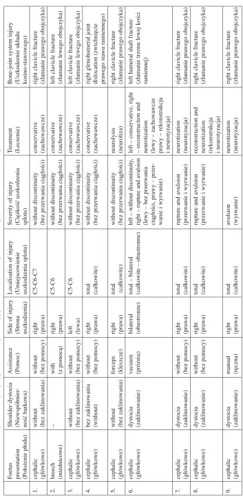
able 1.
The clinical data of the cases with co−existent injuries of the brachial plexus and bone−joint system
T
abela 1.
Zestawienie danych klinicznych przypadków ze współistniejącymi uszkodzeniami splotu ramiennego i układu kostno−stawowego
Foetus
Shoulder dystocia
Assistance
Side of injury
Localisation of injury
Severity of injury
T
reatment
Bone−joint system injury
presentation (Niewspółmier − (Pomoc) (Strona (Umiejscowienie (Ciężkość uszkodzenia (Leczenie) (Uszkodzenie układu (Położenie płodu) ność barkowa) uszkodzenia) uszkodzenia splotu) splotu) kostno−stawowego) 1. cephalic without without right C5−C6−C7 without discontinuity conservative
right clavicle fracture
(główkowe)
(bez zaklinowania)
(bez pomocy)
(prawa)
(bez przerwania ciągłości)
(zachowawcze)
(złamanie prawego obojczyka)
2. breech – with right C5−C6 w ithout discontinuity conservative
left clavicle fracture
(miednicowe)
(z pomocą)
(prawa)
(bez przerwania ciągłości)
(zachowawcze)
(złamanie lewego obojczyka)
3. cephalic without without left C5−C6 w ithout discontinuity conservative
left clavicle fracture
(główkowe)
(bez zaklinowania)
(bez pomocy)
(lewa)
(bez przerwania ciągłości)
(zachowawcze)
(złamanie lewego obojczyka)
4. cephalic bez zaklinowania without right total without discontinuity conservative
right glenohumeral joint
(główkowe)
(without)
(bez pomocy)
(prawa)
(całkowite)
(bez przerwania ciągłości)
(zachowawcze)
dislocation (zwichnięcie prawego stawu ramiennego)
5. cephalic without forceps right total without discontinuity neurolysis
right clavicle fracture
(główkowe)
(bez zaklinowania)
(kleszcze)
(prawa)
(całkowite)
(bez przerwania ciągłości)
(neuroliza)
(złamanie prawego obojczyka)
6.
cephalic
dystocia
vacuum
bilateral
total – bilateral
left – without discontinuity
,
left – conservative, right
left humeral shaft fracture
(główkowe)
(zaklinowanie)
(próżnia)
(obustronne)
(całkowite – obustronne)
right – rupture and avulsion
–
reconstruction and
(złamanie trzonu lewej kości
(lewy – bez przerwania
neurotization
ramiennej)
ciągłości, prawy – przer
−
(lewy – zachowawcze
wanie i wyrwanie)
prawy – rekonstrukcja i neurotyzacja)
7. cephalic dystocia without right total
rupture and avulsion
neurotization
right clavicle fracture
(główkowe)
(zaklinowanie)
(bez pomocy)
(prawa)
(całkowite)
(przerwanie i wyrwanie)
(neurotyzacja)
(złamanie prawego obojczyka)
8. cephalic dystocia without right total
rupture and avulsion
reconstruction and
right clavicle fracture
(główkowe)
(zaklinowanie)
(bez pomocy)
(prawa)
(całkowite)
(przerwanie i wyrwanie)
neurotization
(złamanie prawego obojczyka)
(rekonstrukcja i neurotyzacja)
9. cephalic dystocia manual right total avulsion neurotization
right clavicle fracture
(główkowe) (zaklinowanie) (ręczna) (prawa) (całkowite) (wyrwanie) (neurotyzacja)
5 cases of clavicular fracture and 3 fractures of humerus in a group including 74 children with perinatal brachial plexus palsy [6]. Gherman observed 16 cases of co−existing injuries of brachial plexus and bone in a group of 285 deliv− eries with shoulder dystocia [7]. Statistical differ− ence in all parameters evaluating the size of child between the control group and group with brachial plexus palsy with fractures and without fractures was observed with analyses of classical risk fac− tors. There were no statistically important differ− ences, however, between the group of isolated brachial plexus palsy and the group with addition−
al injury of the bone−joint system. In cases of motor functions disorders of an upper extremity, diagnosis of the bone−joint system injury should not relieve from the duty of neurological supervi− sion of the child because of the possibility of coex− istence of both systems injuries [9–11].
The authors conclude that injuries of the bone− joint system may co−exist with perinatal brachial plexus palsy with different localisation and degree of injury severity. Classical risk factors of perina− tal brachial plexus palsy do not influence signifi− cantly on the possibility of appearance of bone− joint system injury.
Table 2.The evaluation of the statistical importance of the analysed parameters in scheme: control group – children with lesions of the brachial plexus without bone−joint system injuries – children with co−existent injuries of the brachial plexus and bone−joint system
Tabela 2. Ocena istotności statystycznej badanych parametrów w układzie: grupa kontrolna – dzieci z uszkodzeniami splotu bez uszkodzeń układu kostno−stawowego – dzieci z uszkodzeniami splotu i układu kostno−stawowego
Examined parameter Control group Brachial plexus injuries Brachial plexus in− Statistical importance (Badany parametr) (Grupa kontrolna) without fractures juries with fractures (Istotność statystyczna)
C (Uszkodzenia splotu (Uszkodzenia splotu p n = 56 bez złamań) ze złamaniami)
WF F
n = 74 n = 9
Birth weight 3107 ± 399 4477 ± 648 4255 ± 649 C/WF – 0.000000
(Waga urodzeniowa) C/F – 0.000054
g F/WF – ns.
Body length 52.1 ± 1.9 58.7 ± 3.8 58.1 ± 3.7 C/WF – 0.000000
(Długość ciała) C/F – 0.000284
cm F/WF – ns.
Head circumference 32.7 ± 1.6 35.7 35.4 C/WF – 0.000000
(Obwód głowy) C/F – 0.006784
cm F/WF – ns.
Chest circumference 32.4 ± 1.5 36.6 ± 1.8 36.0 ± 2.2 C/WF – 0.000000
(Obwód klatki piersiowej) C/F – 0.002972
cm F/WF – ns.
Apgar scale at 1 min – points 9.4 ± 0.9 4.5 ± 3.1 5.4 ± 3.3 C/WF – 0.000000
(Skala Apgar w 1 min C/F – 0.000204
– punkty) F/WF – ns.
Mother’s age – years 27.2 ± 2.1 30.1 ± 5.6 31.4 ± 7.6 C/WF – 0.011361
(Wiek matki – lata) C/F – ns.
F/WF – ns.
Duration of the II stage 24.1 ± 17.8 22.1 ± 13.4 27.5 ± 10.6 C/WF – ns.
of labour C/F – ns..
(Czas trwania II okresu porodu) F/WF – ns
min
Pregnancy duration – weeks 39.0 ± 1.9 39.5 ± 1.4 39.7 ± 1.0 C/WF – ns.
(Czas trwania ciąży – tyg.) C/F – ns.
F/WF – ns.
References
[1] Birch R: Obstetric brachial plexus palsy. J Hand Surg 2002, 27, 3–8.
[2] Hoffer HM: Assessment and natural history of brachial plexus injury in children. In: Operative nerve repair and reconstruction. Eds.: Gelberman RH, JB Lippincot Company, Philadelphia 1991, 1361–1368.
[3] Koszla MM:Złamania i zwichnięcia u dzieci. PZWL, Warszawa 1986, wyd. 3, 314–321.
[5] Nehme A, Kany J, Sales−de−Gauzy J, Charlet JP, Dautel G, Cahuzac JP:Obstetrical brachial plexus palsy. Prediction of outcome in upper root injuries. J Hand Surg 2002, 27, 9–12.
[6] Bisinella GL, Birch R: Obstetric brachial plexus lesions: a study of 74 children registered with the British Paediatric Surveillance Unit. J Hand Surg 2003, 28, 40–45.
[7] Gherman RB, Ouzounian JG, Goodwin TM: Obstetric maneuvers for shoulder dystocia and associated fetal morbility. Am J Obstet Gynecol 1988, 178, 1126–130.
[8] Gherman RB, Ouzoniann JG, Miller DA, Kwok L, Goodwin TM: Spontaneous vaginal delivery: a risk factor for Erb’s palsy? Am J Obstet Gynecol 1988, 178, 423–427.
[9] Gosk J, Rutowski R: Analiza czynników ryzyka okołoporodowych uszkodzeń splotu ramiennego. Gin Pol 2005, 76, 270–276.
[10] Alfonso I, Papazian O, Grossman JA: Clinical presentations, differential diagnosis and management of obstet− ric brachial palsy. Rev Neurol 1998, 27, 258–263.
[11] Dutkowsky JP, Kasser JR:Nerve injury associated with fractures in children. In: Operative nerve repair and reconstruction. Eds.: Gelberman RH, JB Lippincot Company, Philadelphia 1991, 635–640.
Address for correspondence:
Jerzy Gosk
Department of Trauma and Hand Surgery, Silesian Piasts University of Medicine R. Traugutta 57/59
50−417 Wrocław Poland
tel.: +48 071 370 02 12
e−mail: [email protected]
Conflict of interest: None declared
Received: 3.06.2005 Revised: 30.06.2005 Accepted: 19.07.2005
Praca wpłynęła do Redakcji: 3.06.2005 r. Po recenzji: 30.06.2005 r.