inflammation, regulation of blood vessel contrac-tility and proper brain and eye retina function-ing. Thus, they are necessary not only for adequate growth and development of the human body but al-so for the functioning of the adult organism. The re-sults of studies on omega-3 fatty acids have aroused interest in the possibility of increasing their level in the human body through proper nutrition as well as through modified food and diet supplements.
The intake of dietary supplements containing omega-3 fatty acids has increased in all countries of the world because of their beneficial effects on the human body, which have been recommend-ed by different mrecommend-edical specialists and the mrecommend-edia [4, 56, 60]. The aim of the present study is to pres-ent the most important facts regarding the struc-ture and metabolism of polyunsaturated fatty ac-ids and their therapeutic application.
Adequate development, both physical and in-tellectual, depends to a great extent on proper nu-trition. Therefore, a diet should be varied and should include proper amounts of proteins, carbo-hydrates, fats, vitamins and mineral content. Fats are an essential element of the diet, which are most-ly used as a highmost-ly energetic material. Some data de-scribed an association between low mortality rate among the Eskimos due to cardiovascular reasons and their diet rich in polyunsaturated fatty acids (PUFAs) derived from sea fish, which has focused attention to other important functions of this group of compounds in the human body [1, 2]. Studies on the use of fish oils in the prevention and treatment of cardiovascular diseases as well as in psychiatric and ophthalmological disorders have been under-taken. Omega-3 fatty acid derivatives play a signif-icant role in the process of blood coagulation, in
Anna Wiktorowska-Owczarek
B, D, F, Małgorzata Berezińska
B, D,
Jerzy Z. Nowak
A, B, EPUFAs: Structures, Metabolism and Functions*
Department of Pharmacology and Toxicology, Medical University of Lodz, Poland
A – research concept and design; B – collection and/or assembly of data; C – data analysis and interpretation;
D – writing the article; E – critical revision of the article; F – final approval of article
Abstract
Polyunsaturated fatty acids (PUFAs) include two series of fatty acids: omega-6 and omega-3 series. PUFAs have amphiphatic properties: hydrophilic head and hydrophobic tail. Such structure and other properties of unsaturated fatty acids are responsible for exerting the following biological action: maintaining cell-membrane fluidity, inhib-iting inflammatory processes, decreasing secretion of proinflammatory cytokines by monocytes/macrophages, decreasing susceptibility to ventricular rhythm disorders of the heart, improving functions of vascular endothe-lial cells, inhibiting blood platelet aggregation and decreasing triglyceride synthesis in the liver. In an organism, aracidonic acid (ARA) is converted to prostanoids series 2 (PGE2, PGI2, TXA2) and leukotrienes series 4 (LTB4, LTC4, LTD4) which are endowed with pro-inflammatory potential and are able to induce platelet aggregation and vasoconstriction. The metabolism of eicosapentaenoic acid (EPA) and docosahexaenoic acid (DHA) gives prostanoids series 3 (PGE3, PGI3, TXA3) and leukotrienes series 5 (LTB5, LTC5, LTD5); this group of eicosanoids shows anti-inflammatory, antiplatelet and antiarrhythmic properties (Adv Clin Exp Med 2015, 24, 6, 931–941).
Key words: polyunsaturated fatty acids, ARA, EPA, DHA.
EDITORIAL
Adv Clin Exp Med 2015, 24, 6, 931–941DOI: 10.17219/acem/31243 © Copyright by Wroclaw Medical University ISSN 1899–5276
The Structure and
Nomenclature of Fatty Acids
Fats (lipids) are a heterogeneous group of com-pounds built up of carbon and hydrogen atoms, possessing a tsmall number of oxygen-containing functional groups. Because of their specific molec-ular structure, they have amphipatic (amphiphil-ic) properties: hydrophilic head and hydrophobic (lipophilic) tail – such a structure affects their ar-rangement within the cellular membrane. Lipids are divided into simple and complex compounds (waxes – lanoline, cetaceum, beeswax). Simple fats include esters of fatty acids and various alco-hols. In the case of lipids, glycerol is an alcohol that contains three hydroxyl groups at three pres-ent carbon atoms, which according to stereospe-cific numbering are defined as sn-1, sn-2 and sn-3
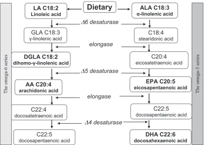
(Fig. 1). Depending on the number of attached ac-ids, mono- (1 acid), di- and triacylglycerols are formed. Simple triacylglycerols are character-ized by the presence of one type of acid; howev-er, there are two or three types of acids in mixed triacylglycerols. Fatty acids are the main compo-nents of membrane lipids and most frequently contain 12 to 24 carbon atoms forming hydrocar-bon chains. They may be represented by saturated (without double bonds), monounsaturated (one double bond) and polyunsaturated (with two or more double bonds) fatty acids – Fig. 2 presents polyunsaturated fatty acids omega-3 and omega-6 series [3–5].
Fatty acids commonly occurring in nature possess usual names (e.g. palmitic acid, linoleic ac-id, arachidonic acid), however due to a variety of forms and great possibilities of conversions, defi-nite rules regarding nomenclature of such struc-tures have been implemented. The order of num-bering carbon atoms in aliphatic fatty acids starts with the carboxyl group; and carbon in this group (COOH) is defined as C1, and further numbering is as follows: C2, C3, etc. According to another classi-fication, carbon attached to the carboxyl group, i.e. C2 is defined by the Greek letter alpha – α, C3 – β, C4 – γ etc., and the carbon atom furthest from the COOH group is defined by the letter omega – ω. When we start counting from carbon ω to the first
Fig. 1. Glycerol – stereospecific numbering
O CH2 O C R1
O CH2 O C R2
O CH2 O C R3
sn-1
sn-2
sn-3
Fig. 2. Pathways of biosynthesis of unsaturated fatty acids omega-6 and -3 series
Dietary
LA C18:2
Linoleic acid α-linolenic acid
ALA C18:3
GLA C18:3
γ-linolenic acid
DGLA C18:2
dihomo-γ-linolenic acid
AA C20:4
arachidonic acid
C22:5
docosapentaenoic acid
C22:4
docosatetraenoic acid
C18:4
stearidonic acid
C20:4
eicosatetraenoic acid
EPA C20:5
eicosapentaenoic acid
DHA C22:6
docosahexaenoic acid
C22:5
docosapentaenoic acid
∆
6 desaturase
elongase
∆
5 desaturase
elongase
∆
4 desaturase
The omega-3 series
double bond between carbon atoms in the hydro-carbon chain (-C = C-), we can determine the af-filiation of acid to series of omega-3, omega-6 or omega-9 fatty acids. The chemical structure of fat-ty acids is presented in the following way: the num-ber of carbon atoms (e.g. C22), the numnum-ber of dou-ble bonds and the group ω; e.g. docosahexaenoic acid is defined as C22 : 6ω-3, which means that it possesses 22 carbon atoms and six double bonds and it belongs to the group of omega-3 (possesses the first double bond when we count from the end at the third carbon atom) [3–5].
Metabolism of Fatty Acids
Saturated fatty acids such as palmitic (C16:0) or stearic (C18:0) acid, which mainly provide ener-gy, are produced in the human and other mammal organism. The formation of malonyl coenzyme A (malonyl-CoA) and acetyl-CoA is a fundamen-tal stage of fatty acid synthesis. The elongation pro-cess takes place with the involvement of fatty ac-id synthase. However, some mammals including
Homo sapiens do not possess enzymes (or possess them in slight amounts) capable of creating double bonds in fatty acid chains at a place further than at carbon C9. The human being is not able to pro-duce linoleic (LA; C18 : 2ω-6) and α-linolenic ac-id (ALA; C18 : 3ω-3) in the sufficient amounts to meet the requirements for these compounds, thus they are named exogenous acids. These two com-pounds give rise to the others, and all of them con-stitute a group of essential unsaturated fatty acids of high physiological significance (Fig. 2). Looking through literature reports the reader can find rich nomenclature connected with this group, such as polyunsaturated fatty acids (PUFAs), essential ty acids (EFAs) or long-chain polyunsaturated fat-ty acids (LCPUFAs). The human exhibits the abili-ty to elongate these two exogenous acids to a slight but de facto insufficient degree; however, the re-quirement for them is higher than ‘endogenous’ supply [3–5].
Linoleic and
α
-Linolenic Acid
Elongation
The omega-6 series derives from linoleic ac-id and includes arachac-idonic acac-id (AA or ARA; C20 : 4ω-6), the last one being docosapentae-noic acid (DPA; C22 : 5ω-6). Administration of α-linolenic acid into the body enables to form omega-3 fatty acid series such as eicosapentae-noic acid (EPA; C20 : 5ω-3) and docosahexaeno-ic acid (DHA; C22 : 6ω-3). Biosynthesis of these
acids requires the involvement of Δ-6, Δ-5 de-saturases (enzymes forming double bonds) and elongases (elongating hydrocarbon chain) occur-ring in the endoplasmic reticulum. The last stage of conversions, i.e. β-oxydase, requires translo-cation of substrates to peroxysomes. Omega-9 series of fatty acids also competes for the same enzymes and these reactions result in a final for-mation of eicosatrienoic acid (C20 : 3:ω-9) from oleinic acid (C18 : 1:ω-9), which is not so impor-tant as the remaining two series because it can be totally synthesized by humans from saturat-ed stearic acid. Moreover, a high concentration of eicosatrienoic acid, which normally occurs in trace amounts, indicates deficiency of substrates for the synthesis of omega-3 and omega-6 series of polyunsaturated fatty acids; this value might thus have a diagnostic importance. The same en-zymes participate in conversion of fatty acids of all three series, showing functional connections between metabolic paths of omega-3, -6 and -9 acids, which depend on competing for enzymes and regulating a given stage of transformation based on a negative feedback through a direct or indirect product [3, 4, 6].
Formation of Compounds
Capable of Inducing Extinction
of Inflammatory Processes
Prostanoids and leukotrienes are mobilized in response to damaging stimulus, i.e. inflammation. Acute inflammation that lasts for a relatively short time is a beneficial process in which threatening factors are removed and functions as well as tissue structures are restored. Inflammatory resolution/ /extinction is an important active stage, which is mediated by small molecules that are the products of the omega-6 and omega-3 acid metabolism. Un-der the influence of lipoxygenases (LOX: LOX-5, LOX-15 and LOX-12) ARA, EPA and DHA acids undergo conversion into lipid mediators actively extinguishing the inflammatory process such as li-poxin A4 and B4 (LXA4 and LXB4 – arising from ARA), E-series resolvins (RvE1 and RvE2) – gen-erated from eicosapentaenoic acid and D-series re-solvins (RvD1, RvD2, RvD3 and RvD4) generated from docosahexaenoic acid [9, 10]. Furthermore, at least two oxylipins are formed from DPA-ω6
acid, which also have properties extinguishing inflammation. At the same time, a particular in-volvement of acetylsalicylic acid (ASA), common-ly named aspirin/polopirin, has been observed,
which acetylates cyclooxygenase-2 (COX-2), and ASA-COX2 in turn metabolises ARA, EPA and DHA acids into intermediate products, which next form lipoxins and E- and D-series resolvins with participation of lipoxygenases. COX-2 acetylation inhibits formation of prostanoids produced by this enzyme; however, it maintains the ability to syn-thesize 15R-hydroxyeicosatetraenoic acid, which is next converted into resolvins through activated inflammatory cells. To emphasize the role of ASA in the initiation of these conversions, the achieved products are preceded by the symbol ‘AT’ derived from aspirin triggered: aspirin-triggered lipoxin – ATL, aspirin-triggered resolvin E –ATRvE or as-pirin-triggered resolvin D –ATRvD [11–13]. The described above ASA functions indicate an im-portant role of this drug as an anti-inflammatory agent, which not only inhibits the initiation of the inflammatory process but participates in the ex-tinction of ongoing inflammation as well.
DHA, influenced by lipoxygenase (LOX), is al-so converted into other compounds with protec-tive potentials, i.e. protectins PD1 (D1 indicates derivation from DHA and no. 1 defines the first compound in this series). The protectin produced in the central nervous system is named neuropro-tectin, NPD1, which has neuroprotective proper-ties. NPD1 occurs in photoreceptors and retinal
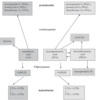
Fig. 3. Metabolic pathways of a polyunsaturated fatty acids omega-6 and omega-3 series arachidonic
acid (AA)
eicosapentaenoic acid (EPA)
cyclooxygenase
prostanoids prostaglandin E3 (PGE3)
prostacyclin I3(PGI3)
thromboxane A3(TXA3)
5-lipoxygenase
5-HPETE 5-HPEPE
leukotrienes
LTA4→LTB4
↓
LTC4→LTD4
LTA5→LTB5
↓
LTC5→LTD5
lipoxins
docosahexaenoic acid (DHA)
neuroprotektin D1 resolvins prostaglandin E2(PGE2),
prostacyclin I2(PGI2,)
pigment epithelium (RPE); it is responsible for the inhibition of expression and activity of proinflam-matory factors and proapoptotic caspase-3, as well as for the stimulation of antiapoptotic factors (i.e. proteins of Bcl-2 family) [14–19].
There is still another path of the DHA conver-sion affected by lipoxygenases, which leads to the formation of the next group of compounds, ma-resins, having antiinflammatory activity. Until now, only one compound from this group, MaR1, has been determined. The term “maresin” derives from the initial letters of the words macrophage, resolution, inflammation – which describe the site of formation of this compound and its biological function. Biological activity of MaR1 includes mul-tidirectional interactions leading to the limitation of polynuclear leukocyte aggregation in the area of inflammation resulting from the stimulation of phagocytic activity of macrophages [13, 20, 21].
Oxidation of
Poly-unsaturated Fatty Acids
Due to double bonds (–C = C–), PUFAs are especially susceptible to oxidation by radicals pro-duced in excessive amounts during the oxidative stress (homeostasis disorders resulting in intensive production of radical oxygen species that are not sufficiently deactivated by antioxidants). Lipid per-oxidation without the enzyme involvement com-prises the following processes: initiation, propa-gation and termination. The process of initiation depends on the OH• reaction with PUFA, as a sult of which a lipid radical is produced, which in re-action with oxygen provides LOO• (a radical of lip-id peroxlip-ide), having the ability to detach hydrogen from other molecules and to generate subsequent radicals L•. Such radicals undergo conversion in-to alcoxylic radicals LO•, in the presence of iron Fe2+, and next into peroxylic radicals that are de-composed into reactive aldehydes: 4-hydroxynon-enal, 4-hydroxyhexanal, malonic dialdehyde and acroleins, defined as secondary toxic transmitters. Omega 6-series fatty acids, such as linoleic or ar-achidonic acid, are mainly converted into 4-hy-droxy-2-nonenal (HNE), and omega-3 acids (EPA, DHA) into 4-hydroxy-2-hexanal (HHE) [22–24]. Monoperoxides are always primary products of PUFAs, which are defined as lipid peroxides with an additional group of LOOH. The number of monoperoxides that can be generated from unsat-urated fatty acids depends on the number of double bonds (n) and can be represented by the following formula: 2n-2, which means that 2 monoperoxides with –OOH groups will generate from linoleic ac-id (18 : 2) at the 9th and 13th carbon atom (9-OOH,
13-OOH). On the basis of the above formula, 6 dif-ferent monoperoxides are produced from ara-chidonic acid (20 : 4) – 8 from EPA (20 : 5) and 10 from DHA (22 : 6). As a consequence of PUFA oxidation, changes in the physical properties of the cell membrane (a decrease in electric potential dif-ferences on both sides of the membrane) occur, which results in the loss of functioning and struc-tural integrity of the cellular membrane [21–23].
Lipids are attacked by free radicals in a special way. The conversions described above, which occur under the influence of radical oxygen species (ROS), are not the only ones because Morrow et al. [25] in 1990 discovered isoprostanes, the compounds re-sembling prostaglandins, which are generated from arachidonic acid due to ROS oxidation, irrespec-tively of cyclooxygenase (COX). Further studies re-vealed that due to peroxylic transformations, iso-prostanes of different types can arise both in vitro
as well as in vivo conditions from omega-3 series of PUFAs, such as eicosapentaenoic (EPA) and doco-sahexaenoic (DHA) acids [26, 27]. Isoprostanes de-rived from DHA within the tissues of the central nervous system are named neuroprostanes; what is interesting, there are more of them as compared to other isoprostanes [28]. In in vivo conditions and in the presence of increased partial pressure of oxygen from arachidonic acid, additional compounds of the isofuran structure and DHA-structurally simi-lar compounds called neurofurans are formed [29]. Their higher concentration has been found in the cerebral cortex of animals used as a model of Al-zheimer disease [30]. Even the first observations re-garding free-radical formation of isoprostanes from arachidonic acid suggested that these compounds might be mediators of oxidative stress. Such a sug-gestion has been confirmed by further research on free-radical formation of isofurans and neurofurans in biological fluids such as urine, blood or cerebro-spinal fluid. Thus, the measurement of iso/neuro-furan concentration may be a reliable biomarker of intensity of oxidative stress and lipid peroxida-tion in the cell/tissue/organism, and a biomarker of an advanced process of neurodegeneration in the nervous tissue [27, 29]. There are some suggestions that supplementation of fish oil containing a high amount of EPA provides antiinflammatory prop-erties also as a result of considerable reduction in forming isoprostanes (F2-IsoPs) from arachidonic acid with strong proinflammatory activity [30].
Fatty Acid Functions
unsaturated fatty acid conversions in the human body, their role in forming prostanoids and leu-kotrienes have been observed. When they arise from ARA, like PGE2, PGD2, or 4-series leukot-rienes, they exhibit proinflammatory activity that is commonly known and described in the reports which discuss the mechanisms of nonsteroidal an-ti-inflammatory drug action.
Basic studies have indicated that DHA and EPA acids are beneficial for the human body ex-erting the following biological action [31]:
– maintaining cell-membrane fluidity, – inhibiting inflammatory processes,
– decreasing secretion of proinflammatory cytokines by monocytes/macrophages,
– decreasing susceptibility to ventricular rhythm disorders of the heart,
– improving functions of vascular endotheli-al cells,
– inhibiting blood platelet aggregation, – decreasing triglyceride synthesis in the liver.
Omega-3 Fatty Acid Effects
on Cell-Membrane Fluidity
The membrane of the cell as well as of mi-tochondria and other cellular elements is built up of proteins and lipids which contain saturat-ed or unsaturatsaturat-ed fatty acids. Saturatsaturat-ed fatty ac-ids have simple ‘tails’ because they do not possess double bonds. They are densely packed, so there is no space between the chains, resulting in a rig-id membrane. The presence of unsaturated fatty acids with numerous double bonds (occurring in nature in the cis conformation) causes ‘tail’/hy-drocarbon chain bending, which in turn results in forming free spaces and affects membrane fluidity and elasticity.
Polyunsaturated docosahexaenoic acid (DHA) commonly occurs in cellular and plasma mem-branes of the organism. Its high amount has been particularly found in the brain tissue and the retina (up to 50% and 60–80% membrane phospholipids, respectively). DHA can occur in the free state or combined with phosphatidylethanolamine (PEA) and phosphatidylocholine (PC), as well as with phosphatidylserine (PS) [5, 32].
DHA in the cell membranes (membrane ‘rafts’ are especially rich in DHA) exerts influence on their physical properties – ensures their proper fluidity, and also affects the proper functioning of membrane receptors, ion channels and transport-ing proteins, i.e. elements involved in adequate cell reactivity, its ability to react to stimuli and in inter-cellular communication [33].
Omega-3 Fatty Acid Effects
on Anti-Inflammatory Activity
Omega-3 and omega-6 PUFAs are incorpo-rated into cell membranes. They are released from membrane phospholipids and constitute substrates for eicosanoid synthesis, i.e. prostaglandins, pros-tacyclins, thromboxanes and leukotrienes.Eicosanoids arising from arachidonic acid (omega-6) induce an inflammatory response by 2-series prostanoids synthesize. PGE2 may also in-duce anti-inflammatory effect by increasing lipox-in production by lipox-induclipox-ing 15-LOX (lipooxygen-ase) [19]. Arachidonic acid-derived eicosanoids are responsible for proaggregation and vasocon-striction effect (TXA2 and TXB2) and proliferation of cancer cells (especially of breast, colorectal and prostate cancers) [7].
3-series prostanoids and 5-series leukotrienes arising from fatty acids of omega-3 series (mainly from EPA) possess weaker inflammation inducing properties or even anti-inflammatory properties, which means that the body response to factors in-ducing infection, impairment or inflammation de-pends on the composition of the cell membrane. When the proportions are favourable for ome-ga-3 PUFAs, the response to inflammatory factors is weaker. Production of lipoxins and resolvins, as well as oxylipins from both groups of polyun-saturated fatty acids allows to extinct the ongoing inflammatory process; lack of such reaction may contribute to the development of autoimmune diseases, chronic inflammation or excessive tis-sue damage and development of various diseases whose pathogenesis is associated with inflammato-ry disease. Omega-3 acid derivatives may also have antithrombotic activity counteracting blood vessel narrowing and inhibiting carcinogenesis [34, 35].
Omega-3 Fatty Acid Effects
on Cardiovascular System
Omega-3 fatty acids beneficially affect lipid metabolism. EPA and DHA decrease the triglyc-eride level in the plasma by 30% and in the case of patients with hyperglyceridemia even by 80%. They also decrease the level of total and LDL frac-tion cholesterol, while increasing HDL fracfrac-tion level [37, 38].
DHA and EPA normalize blood pressure through the rise in the level of prostacyclins and endothelium-derived relaxing factor (EDRF) – ni-trogen oxide (NO), belonging to vasodilated fac-tors, as well as through the reduction in the level of tromboxane A2 (TXA2), a strong vasoconstric-tor, and PGE2 (stimulates renin production and reversed sodium resorption). Hypotensive activity can be also caused by beneficial changes in the lip-id composition of the cell membrane at the recep-tor sites for vasoactive hormones and by the weak-ened response to them. A correlation has been found between the acid composition in the fatty tissue and the blood pressure value; an increase in
α-linolenic acid in the fatty tissue by 1% was
asso-ciated with a drop in the systolic pressure by 5 mm Hg. A complete hypotensive activity develops after 3–4 weeks of omega-3 acid consumption [36–40].
Omega-3 acids have antithrombotic activity. They prolong the bleeding time by decreasing the platelets tendency towards adhesion and aggrega-tion. This activity results from inhibiting the syn-thesis of prothrombotic compounds such as TXA2 and PAF (platelet-activating factor), decreasing fi-brinogen concentration, increasing prostacyclin level and activity of the tissue plasminogen activa-tor as well as of angiotensin III [41–43]. Omega-3 PUFA not only potentiate platelet response to an-tiplatelet drugs, but also reduce thrombin forma-tion. In coronary artery disease, it was found that in patients who received omega-3 PUFA together with aspirin and clopidogrel fibrin clots had a less compact structure, which made it less resistant to lysis [44, 45].
Due to omega-3 fatty acids, stabilization of the atheromatous plague takes place. In subjects using omega-3 supplementation, thicker fibrous capsule of the plaque and less inflammation have been ob-served. Beneficial changes may occur even in old atheromatous plaques. Some reports can be found in the literature, which indicate that omega-3 sup-plementation may contribute to a decrease in the incidence of restenosis after coronary angioplasty and to a decrease in the incidence of vessel closure after coronary arterial bypass graft surgery [46, 47].
Moreover, omega-3 acids play an impor-tant role in prevention of sudden death caused by
arrhythmia in patients with ischemic heart disease. They function as modulators affecting calcium flow via type L channels and controlling calcium release from the endoplasmic reticulum. Thanks to the presence of omega-3 fatty acids, elongation of the refraction period (by 150%) and elevation in the threshold of cardiomyocyte excitability (the power of electrical stimulation required for induc-ing a functional potential increases by 50%) have been noted. Long-term administration of omega-3 fatty acids at the dose of 1 g per day leads to a de-crease in the rate of hospitalization and death risk due to heart rhythm disorders [48, 49].
Omega-3 Fatty Acids Effects
on the Nervous System
DHA plays an essential role in a proper func-tioning of the nervous system of adults, as well as in its development during fetal life and childhood. It is one of the main constituents of phospholipids in neuron cell membranes, especially in the synapse. Omega-3 fatty acids are also indirectly involved in the synthesis of serotonin and dopamine [50]. They seem to have a protective function in mood impairments. Moreover, some reports state that they may exert a beneficial effect on concentration and hyper-reactivity in children with developmen-tal coordination disorders (DCD) [51]. Some re-searchers think that the consumption of omega-3 fatty acids by subjects with psychic disorders may provide health benefits not only due to their pro-tective activity exerted on the nervous system but also due to alleviation of metabolic adverse effects of psychotropic medications and obesity frequent-ly occurring in this group of patients [52].
further converted into 2-(ω-carboxyethyl) pyrrole
(CEP) and conjugated with the protein molecule (adduct CEP-protein). Peroxidative fragmentation and formation of immunogenic adducts are likely to be also associated with EPA and other acids or even all PUFAs consumed with food or in a form of diet supplementation. However, free radicals are also generated as a result of DHA oxidation, whose unfavourable action depends on local possibilities of their neutralization through antioxidative pro-tection systems [23, 24]. The above situation dem-onstrates that PUFAs are very important constit-uents for adequate functioning of the nervous system or the vision organ, however due to their special susceptibility to oxidation they can give rise to molecules exerting adverse effect on the above mentioned structures and contribute to the de-velopment of various diseases. Therefore, supple-mentation with DHA and other long-chain PUFAs should be combined with antioxidant compounds, e.g. vitamins E and C as well as lutein and zeaxan-thin, the latter ones are especially recommended for subjects at risk of AMD development.
Fatty Acids in Diet
Omega-3 and omega-6 PUFAs are also de-fined as essential polyunsaturated fatty acids (EFA), which emphasizes their significant role in the functioning of the organism and the necessi-ty of supplying it with food. EFAs are absorbed in the digestive tract (diet, supplementation), reach the liver, where they are esterified into phospholip-ids and next they are released into the bloodstream in the form of lipoproteins. EFAs are necessary for proper growth, development and functioning of all tissues and organs, especially the retina, brain and heart. Taking into consideration the importance of EFAs, and particularly omega-3 series, for proper functioning of the human body, the internation-al heinternation-alth organizations highlight the need for con-stant and regular consumption of about 200 mg of DHA/day by adults in the form of various foods rich in DHA and EPA or pharmacological prepa-rations containing these acids [5, 53].
Marine fish predators are the richest food sources of DHA and EPA. Other types of fish like salmons, herrings, sardines, mackerels, tu-nas, halibuts, flounders and trout contain ω-3 se-ries of fatty acids in slightly lower amounts. They also occur in different seafoods and algae. Culti-vated microalgae, Cryptehecodinium cohnii, are one of ω-3 acid sources, whose oil contains 40% of DHA (i.e. DHASCO – DHA Single Cell Oil) – the product which has obtained a positive opinion of the United States Food and Drug Administration
(US FDA) and is recommended to be given to in-fants and small children. Another recommended source of fatty acids is the oil produced by micro-algae Schizochytrium sp., containing 40% of DHA, 2.5% of EPA and additionally 15% of docosapen-taenoic acid (DPA), belonging to ω-6 (DPA-ω6) acids. It has been stated that bioequivalence and effectiveness of supplementation are similar to those achieved by taking capsules containing oils from both types of algae and do not differ from an equivalent portion of ready to eat salmon. In dif-ferent countries, food enriched with small amounts of fatty acids, i.e. bread, milk products, margarine or juice, has been produced. These products are regarded as functional food, which beneficially af-fects the human body owing to the presence of bio-active components (natural or added), regardless of its nutritional properties [54, 55].
Numerous national and international health organizations dealing with health protection rec-ommend regular consumption at least 500 mg/ /day of EPA and DHA [56]. The National Group of Experts in their recommendations regarding consumption and supplementation of diet with omega-3 fatty acids advise adults to take fatty ac-ids, DHA and EPA, in the amount from 0.5 do 1.5 g (mean 1 g) per day. In order to achieve ben-eficial health effects, the ratio of omega-6 to ome-ga-3 fatty acids in the diet should be 4 : 1 [4, 31]. Be-cause omega-3 acids decrease platelet aggregation in the blood, which prolongs the time of bleeding, their simultaneous application with anticoagulation and antiplatelet drugs may potentiate the response to drugs [45, 57, 58]. Therefore, in patients taking these drugs, an additional dose of omega-3 fatty ac-ids should not exceed 1 g per day. This dose, which may be covered by food, exerts cardioprotective ac-tion. In patients with high risk of cancer, cardiac, rheumatoid and neurodegenerative diseases, the EPA and DHA dose can be increased up to 1.5 g per day. In the treatment of hypertriglicerydemia, ome-ga-3 fatty acids can be used as supplements un-der the physician’s control at a dose of 2–4 g/day (capsules containing 465 mg of EPA and 375 mg of DHA in the form of ethylene esters) [59]. It is par-ticularly beneficial to consume omega-3 fatty ac-ids in everyday diet, combined with statin or fibrate treatment – this combination is recommended es-pecially in mixed dyslipidemias [60]. According to the FDA data, consumption up to 3 g of omega-3 fatty acids per day should not induce side effects; however, higher doses should be reserved for spe-cial situations (specified therapeutic indications) and used under the physician’s supervision [4, 31].
natural products, e.g. a meal containing fatty sea fish twice a week, what approximately corresponds to 500 mg/day of EPA + DHA + DPA. The Europe-an Food Safety Authority (EFSA) draws attention to the fact that due to the growing environmental pol-lution unlimited consumption of meat of large fish predators (especially tuna, shark, marlin and pike) may lead to the excessive exposure to mercury [61]. Since toxic activity of mercury compounds is most dangerous for fetuses, infants and small children, women planning to be pregnant, pregnant wom-en, breastfeeding mothers and younger children are advised to choose smaller species of fish that do not cumulate such a high amount of pollutants. In this group of people possibilities of supplementa-tion with PUFA preparasupplementa-tions should be considered.
Conclusions
Unsaturated fatty acids of omega-6 and ome-ga-3-series are not synthesized by humans in the sufficient amounts and must be provided with
food. That is why they are called essentials. Sea fish oil is a source of DHA and EPA, which con-siderably limits their availability in the diet as com-pared to omega-6 fatty acids present in plants. However, omega-3 series fatty acids exhibit par-ticularly beneficial effect on proper functioning of the brain, cardiovascular system or the eye retina, owing to the presence and the number of double bonds in the molecule. Double bonds, so signifi-cant for molecular properties, easily enter into re-actions with radicals, which promotes their oxida-tion and changes their characteristics. At the same time, it is worth emphasizing that no significant adverse reactions of this group of compounds have been reported. At the same time, it should be re-membered that they cannot replace a pharmaco-logical therapy. However, due to their involvement in maintaining heath they should be supplied in proper amounts, if possible in an adequately bal-anced diet or as pharmacological preparations, i.e. diet supplements that should also contain com-pounds possessing the properties of lipophilic an-tioxidants, e.g. vitamin E.
References
[1] Bang HO, Dyerberg DJ, Sinlair HM: The composition of the Eskimo food in north western Greenland. Am J Clin Nutr 1980, 33, 2657–2661.
[2] Sinclair HM: Prevention of coronary heart disease: the role of essential fatty acids. Postgrad Med J 1980, 56, 579–584.
[3] Das UN: Essentials Fatty Acids – a review. Curr Pharm Biotechnol 2006, 7, 467–482.
[4] Nowak JZ: Wielonienasycone kwasy tłuszczowe omega-3 w siatkówce i praktyce medycznej – blaski i cienie. Mag Lek Okul 2009, 3, 208–220.
[5] San Giovanni JP, Chew EY: The role of omega-3 long-chain polyunsaturated fatty acids in health and disease of the retina. Prog Retin Eye Res 2005, 24, 87–138.
[6] Le HD, Meisel JA, de Meijer VD, Gura KM, Puder M: The essentiality of arachidonic acid and docosahexaenoic acid. Prostaglandins Leukot Essent Fatty Acids 2009, 81, 165–170.
[7] Calder PC: n-3 Polyunsaturated fatty acids, inflammation, and inflammatory diseases. Am J Clin Nutr 2006, 83, 1505–1519.
[8] Simopoulos AP: Omega-3 fatty acids in inflammation and autoimmune diseases. J Am Coll Nutr 2002, 21, 495–505.
[9] Arita M, Bianchini F, Aliberti J, Sher A, Chiang N, Hong S: Stereochemical assignment, anti-inflammatory properties, and receptor for the omega-3 lipid mediator resolving E1. J Exp Med 2005, 201, 713–722.
[10] Arita M, Ohira T, Sun YP, Elangovan S, Chiang N, Serhan CN: Resolvin E1 selectively interacts with leukotriene B4 receptor BLT1 and ChemR23 to regulate inflammation. J Immunol 2007, 178, 3912–3917.
[11] Serhan CN, Chiang N: Endogenous pro-resolving and anti-inflammatory lipid mediators: a new pharmacologic genus. Br J Pharmacol 2008, 153, 200–215.
[12] Schwab JM, Serhan CN: Lipoxins and new lipid mediators in the resolution of inflammation. Curr Opin Pharmacol 2006, 6, 414–420.
[13] Nowak JZ: Biosynthesis and characteristics of anti-inflammatory proresolving derivatives of omega-3 and omega-6 polyunsaturated fatty acids. Mil Pharm Med 2011, 3, 20–41.
[14] Bazan NG: Neurotrphins induce neuroprotective signaling in the retinal pigment epithelial cell by activating the synthesis of the anti-inflammatory and anti-apoptotic neuroprotectin D1. Adv Exp Med Biol 2008, 613, 39–44.
[15] Bazan NG: Neuroprotectin D1-mediated anti-inflammatory and survival signalling in stroke, retinal degenera-tions, and Alzheimer disease. J Lipid Res 2009, 50, 400–405.
[16] Bazan NG, Molina MF, Gordon WC: Docosahexaenoic acid signalopidomics in nutrition: significance in aging, neuroinflammation, macular degeneration, Alzheimer’s, and other neurodegenerative diseases. Annu Rev Nutr 2011, 31, 321–351.
[17] Mukherjee PK, Marcheselli VL, Barreiro S, Hu J, Bok D, Bazan NG: Neurotrophins enhance retinal pigment epithelial cell survival through neuroprotectin D1 signaling. Proc Natl Acad Sci USA 2007, 104, 13152–13157.
[19] Nowak JZ: Inflammation: course and role of PUFA-derived lipid mediators in the resolution of inflammatory reaction. Mil Pharm Med 2011, 1, 20–30.
[20] Serhan CN, Yang R, Martinod K, Kasuga K, Pillai PS, Porter TF, Oh SF, Spite Maresins M: Novel macrophage mediators with potent antiinflammatory and proresolving actions. J Exp Med 2009, 16, 206, 15–23.
[21] Catala A: Lipid peroxidation of membrane phospholipids generates hydroxyl-alkenals and oxidized phospholipids active in physiological and/or pathological conditions. Chem Phys Lipids 2009, 157, 1–11.
[22] Esterbauer H: Cytotoxicity and genotoxicity of lipid-oxidation products. Am J Clin Nutr 1993, 57, 779–786.
[23] Nowak JZ: W poszukiwaniu biomarkerów dla zwyrodnienia plamki związanego z wiekiem (AMD). Mag Lek Okul 2009, 3, 132–143.
[24] Nowak JZ: Oxidative stress, polyunsaturated fatty acids-derived oxidation products and bisretinoids as potential inducers of CNS diseases: focus on age-related macular degeneration. Pharmacol Rep 2013, 65, 288–304.
[25] Morrow JD, Hill KE, Burk RF, Nammour TM, Badr KF, Roberts 2nd LJ: A series of prostaglandin f2-like
com-pounds are produced in vivo in humans by a non-cyclooxygenase, free radical-catalyzed mechanism. Proc Natl Acad Sci USA 1990, 87, 9383–9387.
[26] Lawson JA, Kim S, Powell WS, FitzGerald GA, Rokach J: Oxidized derivatives of ω-3 fatty acids: identification of IPF3α-VI in human urine. J Lipid Res 2006, 47, 2515–2524.
[27] Song WL, Paschos G, Fries S, Reilly MP, Yu Y, Rokach J, Chang CT, Patel P, Lawson JA, FitzGerald GA:
Novel eicosapentaenoic acid-derived F3-isoprostanes as biomarkers of lipid peroxidation. J Biol Chem 2009, 284, 23636–23643.
[28] Roberts LJ 2nd, Montine TJ, Markesbery WR, Tappert AR, Hardy P, Chemtob S, Dettbarn WD, Morrow JD:
Formation of isoprostane-like compounds (neuroprostanes) in vivo from docosahexaenoic acid. J Biol Chem 1998, 273, 13605–13612.
[29] Song WL, Lawson JA, Reilly D, Rokach J, Chang CT, Giasson B, Fitzgerald GA: Neurofurans, novel indices of oxidant stress derived from docosahexaenoic acid. J Biol Chem 2008, 283, 6–16.
[30] Roberts LJ 2nd, Milne GL: Isoprostanes. J Lipid Res 2009, 50, 219–223.
[31] Nowak JZ: Wielonienasycone kwasy tłuszczowe omega-3: aspekty biochemiczne, funkcjonalne i praktyczne. Farmakoter Psychiatr Neurol 2009, 3–4, 127–146.
[32] Fliesler SJ, Anderson RE: Chemistry and metabolism of lipids in the vertebrate retina. Prog Lipid Res 1983, 22, 79–131.
[33] Feller SE, Gawrisch K: Properties of docosahexaenoic-acid-containing lipids and their influence on the function of rhodopsin. Curr Opin Struct Biol 2005, 15, 416–422.
[34] Gajewska-Meszaros S, Meszaros J: Ryby morskie i owoce morza: luksus czy konieczność. Ter Leki 2001, 2, 26, 31.
[35] Committee on Diet and Health, Food and Nutrition Borad, National Research Council. Diet and Health: Implications for Reducing Chronic Disease Risk. National Academy Press, Washington 1989.
[36] Drevon CA: Marine oils and their effects. Nutr Rev 1992, 50 (4 (Pt 2)), 38–45.
[37] Chan JM, Gann PH, Giovannucci EL: Role of diet in prostate cancer development and progression. Clin Oncol 2005, 23, 8152–8160.
[38] Strauss MH, Dorian P, Verma S: Fish oil supplementation and arrhythmias. JAMA 2005, 294, 2165–2166.
[39] Banning M: The role of omega-3-fatty acids in the prevention of cardiac events. Br J Nurs 2005, 14, 503–508.
[40] Mori TA: Omega-3 fatty acids and hypertension in humans. Clin Exp Pharmacol Physiol 2006, 33, 842–846.
[41] McEwen BJ, Morel-Kopp MC, Chen W, Tofler GH, Ward CM: Effects of omega-3 polyunsaturated fatty acids on platelet function in healthy subjects and subjects with cardiovascular disease. Semin Thromb Hemost 2013, 39, 25–32.
[42] Kristensen SD, Iversen AM, Schmidt EB: N-3 polyunsaturated fatty acids and coronary thrombosis. Lipids 2001, Suppl 36, 79–82.
[43] Lee KW, Blann KD, Lip GY: Effects of omega-3 polyunsaturated fatty acids on plasma indices of thrombogenesis and inflammation in patients post-myocardial infarction. Thromb Res 2006, 118, 305–312.
[44] Gajos G, Rostoff P, Undas A, Piwowarska W: Effects of polyunsaturated omega-3 fatty acids on responsive-ness to dual antiplatelet therapy in patients undergoing percutaneous coronary intervention: the OMEGA-PCI (OMEGA-3 fatty acids after pci to modify responsiveness to dual antiplatelet therapy) study. J Am Coll Cardiol 2010, 55, 1671–1678.
[45] Gajos G, Zalewski J, Rostoff P, Nessler J, Piwowarska W, Undas A: Reduced thrombin formation and altered fibrin clot properties induced by polyunsaturated omega-3 fatty acids on top of dual antiplatelet therapy in patients undergoing percutaneous coronary intervention (OMEGA-PCI clot). Arterioscler Thromb Vasc Biol 2011, 31, 1696–1702.
[46] Thies F, Garry JM, Yaqoob P, Rerkasem K, Williams J, Shearman CP, Gallagher PJ, Calder PC, Grimble RF:
Association of n-3 polyunsaturated fatty acids with stability of atherosclerotic plaques: a randomised controlled trial. Lancet 2003, 361(9356), 477–485.
[47] Eritsland J, Arnesen H, Grønseth K, Fjeld NB, Abdelnoor M: Effect of dietary supplementation with n-3 fatty acids on coronary artery bypass graft patency. Am J Cardiol 1996, 77, 31–36.
[48] Leaf A: The electrophysiologic basis for the antiarrhythmic and anticonvulsant effects of n-3 polyunsaturated fatty acids: heart and brain. Lipids 2001, Suppl 36, 107–110.
[50] Salem N Jr, Litman B, Kim HY, Gawrisch K: Mechanisms of action of docosahexaenoic acid in the nervous sys-tem. Lipids 2001, 36, 945–959.
[51] Ross BM, Seguin J, Sieswerda LE: Omega-3 fatty acids as treatments for mental illness: which disorder and which fatty acid? Lipids Health Dis 2007, 18, 6–21.
[52] Freeman MP, Hibbeln JR, Wisner KL, Davis JM, Mischoulon D, Peet M, Keck PE Jr, Marangell LB, Richardson AJ, Lake J, Stoll AL: Omega-3 fatty acids: evidence basis for treatment and future research in psychia-try. J Clin Psychiatry 2006, 67, 1954–1967.
[53] Kris-Etherton PM, Hill AM: N-3 fatty acids: food or supplements? J Am Diet Assoc 2008, 108, 1125–1130.
[54] Arterburn LM, Hall EB, Oken H: Distribution, interconversion, and dose response of n-3 fatty acids in humans. Am J Clin Nutr 2006, 83, Suppl 6, 1467–1476.
[55] Arterburn LM, Oken HA, Bailey Hall E, Hamersley J, Kuratko CN, Hoffman JP: Algal-oil capsules and cooked salmon: nutritionally equivalent sources of docosahexaenoic acid. J Am Diet Assoc 2008, 108, 1204–1209.
[56] National Heart Foundation of Australia. Position statement on Fish, fish oils, n-3 polyunsaturated fatty acids and cardiovascular health. Presented at AIFST conference July 2008. (online) Available at: www.heartfoundation.org. au/SiteCollectionDocuments/Fish-position-statement.pdf (Accessed September 17, 2014).
[57] Gajos G, Zalewski J, Nessler J, Zmudka K, Undas A, Piwowarska W: Polyunsaturated omega-3 fatty acids improve responsiveness to clopidogrel after percutaneous coronary intervention in patients with cytochrome P450 2C19 loss-of-function polymorphism. Kardiol Pol 2012, 70, 439–445.
[58] Larson MK, Ashmore JH, Harris KA, Vogelaar JL, Pottala JV, Sprehe M, Harris WS: Effects of omega-3 acid ethyl esters and aspirin, alone and in combination, on platelet function in healthy subjects. Thromb Haemost 2008, 100, 634–641.
[59] Kris-Etherton PM, Harris WS, Appel LJ: Fish consumption, fish oil, omega-3fatty acids, and cardiovascular dis-ease. Circulation 2002, 106, 2747–2757.
[60] Krajowa Grupa Ekspertów. Rekomendacje Grupy Ekspertów dotyczące spożycia i suplementacji diety kwasami omega-3 w populacji ludzi dorosłych. Family Med Prim Care Rev 2007, 9, 175–177.
[61] EFSA Panel on Dietetic Products, Nutrition and Allergies: Scientific opinion on the tolerable upper intake level of eicosapentaenoic acid (EPA), docosahexaenoic acid (DHA) and docosapentaenoic acid (DPA). EFSA J 2012, 10, 2815.
Address for correspondence:
Anna Wiktorowska-Owczarek
Department of Pharmacology and Toxicology Medical University of Lodz
Żeligowskiego 7/9 90-752 Łódź Poland
E-mail: [email protected]
Conflict of interest: None declared