J.micropalaeontol., 3 (2): 73-94, September 1984
Re-study
of
some dinoflagellate cysts from the Oligocene and
Miocene of Germany
WILLIAM A. S. SARJEANT
Department of Geological Sciences, University of Saskatchewan, Saskatoon, Canada
ABSTRACT-The type material of six dinoflagellate cyst species from the Late Oligocene to Middle Miocene of northwest Germany, described originally by Gerlach (1 961), is reillustrated and redescribed. It is shown to include representatives of nine species. Areosphaeridium (ex: Baltisphaeridium) pectiniforme is found to be a senior synonym of Areosphaeridium multi- cornutum Eaton. Systematophora placacantha is considered to be a senior synonym of Cleistosphaeridium (ex: Baltisphaeridium) panniforme (Gerlach). The new combination Rhynchodiniopsis tenuitabulata (Gerlarch) is proposed. Revised diagnoses for these three species and for Leptodinium membranigerum (Gerlach), Achomosphaera triangulata (Gerlach) and Lejeunecysta hyalina (Gerlach) are proposed. The morphology of a form described here for the first time, and tentatively attributed to Phrhanoperidinium, is considered perhaps to imply a separate origin for the Phthanoperidiniaceae: for that reason familial, rather than tribal, rank is preferred for that group. The stratigraphical ranges of the nine species here recognised and of two others of Gerlach’s species redescribed in earlier papers are detailed; elimination of misattributed forms means that these ranges are shorter than the published literature suggests.
INTRODUCTION
The history of early studies of Oligocene and Miocene dinoflagellate cysts has been summarised in an earlier paper (Sarjeant, 1983). Whilst Dorothea Maier was completing her studies of cysts from borehole cores and outcrops in the Niederrheingebiet, Nordrhein- Westfalen, Schleswig-Holstein and Jutland (Denmark) at the Uni- versity of Kiel, Ellen Gerlach was examining assem- blages from two boreholes through sediments of similar age in the Emsland region of northwest Germany at the University of Tubingen. Maier found extreme difficulty in dealing with the diverse morphologies with which she found herself confronted and her research supervisor, Walter Wetzel, had too poor a comprehension of these microfossils (as his own papers make evident; see Wetzel, 1952, 1955) to give her the guidance she needed. In contrast, though Ellen Gerlach’s research supervisor, Otto Schindewolf, was not himself a micro- palaeontologist, she had the great advantage of being able to consult Alfred Eisenack for advice when in difficulties. In consequence, whilst Maier’s work (1959) was to occasion confusion among other palynologists and to be the focus for much criticism, Gerlach’s single paper (1961) was of higher quality and has proved of greater utility.
Four new genera were proposed by Gerlach. One of these, Pentudinium, has come to be regarded as of great importance in Tertiary stratigraphy (see Benedek, Gocht & Sarjeant, 1982 and Edwards, 1982, for recent interpretations). Another, Lejeunia, proved to be the
junior homonym of a modern liverwort (see Artzner & Dorhofer, 1978). However, under the substitute name Lejeunecysta Gerlach’s genus remains one that is widely recognised; its type species is reillustrated, and its distri- bution discussed, herein. Her third genus, Membrano- phoridiurn, fell into disuse but has been effectively reinstated by Stover & Evitt (1978), under a revised definition. A misunderstanding concerning the morpho- logy of her fourth genus, Emsfundiu, caused it to be formally abandoned for a while; however, recently it has been likewise reinstated under a revised definition (Benedek & Sarjeant, 1981).
In contrast, the species assigned by Gerlach to genera already proposed before 196 1 have falled largely into disuse; only one of them, Spiniferires (ex: Hystricho- sphueru) cornutus (Gerlach, 1961) Sarjeant, 1970, has been regularly reported. There are three reasons for this: first, because the advances in knowledge of dino- flagellate cysts since 1961 mean that her diagnoses can be no longer considered adequate for confident identifi- cation of those taxa; second, because her illustrations were produced at too small a magnification; and third, because her holotypes and paratypes are, in some instances, markedly dissimilar in morphology.
My work on Gerlach’s holotypes was undertaken during visits to the University of Tubingen in 1979 and 1981. During those visits, I received continuous and courteous co-operation from Dr. Hans Gocht, many of whose percipient comments are reflected in the pages that follow. Six of Gerlach’s species are reillustrated,
and five of them redescribed, herein; nine taxa are recognised among them. I hope to complete work on her other types during a future visit to Tiibingen.
The seventeen samples from which Gerlach’s assem- blages were obtained stemmed from two shallow borings,
Emsbiiren 7 (mapsheet Lohne 3509, R = 2586100/H = 54812830) and Emsbiiren 9 (mapsheet Lohne 3509, R = 2588090/H = 5808343), put down through the Oligocene and Miocene sediments of Hannover in the triangle of land bounded by Nordhorn, Lingen and
A
B
C
D
Oligocene and Miocene dinoflagellates from Germany
Rheine. Full details of the petrography of the samples and of the method of preparation are given by Gerlach
All described specimens are lodged in the Gerlach collection, Institut fur Palaontologie, University of Tubingen. Their present condition is highly variable. Some remain in as good order as when Gerlach described them, but others have split open or suffered in varying degree from bacterial or fungal attack, and two could not be located on the slide supposed to contain them. Details are given in the ensuing pages and lectotypes designated when necessary.
(1961, pp. 146-149).
SYSTEMATIC DESCRIPTIONS Kingdom Plantae
Division Pyrrhophyta Pascher Class Dinophyceae Fritsch
Suborder Gonyaulacystineae Norris, 1978 Family Gonyaulacystaceae Sarjeant & Downie, 1966,
emend. Sarjeant, 1982
Genus Leptodiniurn Klement, 1960, emend. Sarjeant, 1982
Leptodiniurn rnembranigerurn Gerlach, 1961, emend. nov.
(Pl. 1, figs. 1 , 3 ; P1. 2, figs. 1,2; P1. 3, fig. 3; P1.4, fig. 6;
1961 1964 1964 1967 1973 1975 1977 1978 1981 Fig. 1)
Leptodinium rnernbranigerurn Gerlach, 162- 164, pl. 26, figs. 1-4, 7, text-figs. 4-5.
Leptodinium membranigerurn Gerlach; Eisenack & Klement, 495-596.
Leptodiniurn rnembranigerum Gerlach; Downie & Sarjeant, 126.
Leptodiniurn rnernbranigerurn Gerlach; Sarjeant, tab. I1 (p. 328).
Leptodiniurn rnernbranigerum Gerlach; Lentin & Williams, 87.
L.eptodiniurn rnembranigerurn Gerlach; Harker & Sarjeant, chart 17 (p. 250).
Leptodinium rnembranigerurn Gerlach; Lentin & Williams, 98.
Zmpagidinium? rnernbranigerurn (Gerlach); Stover & Evitt, 166.
Impagidiniurn? membranigerum (Gerlach); Lentin & Williams, 154.
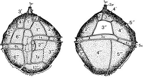
Original Diagnosis. "A species of the genus Leptodiniurn with oval theca. Tabulation formula as given in the generic diagnosis; 4', 6 , 5'", lp and 1"". Epitheca larger and more elongate than the rounded hypotheca. Girdle furrow spiral, formed of six plates. Offset of its ends somewhat more than a furrow breadth. Longitudinal furrow wholly undifferentiated. Sutures of plates marked by low hyaline crests, shell membrane appearing to bear a thornlike point at the position of convergence of two crests. Plates finely granulate, membrane rela- tively thin" (Gerlach, 1961, p. 162-163, new transl.).
Emended Diagnosis. Cyst proximate, holotabulate, and acavate; relatively thin but composed of two closely appressed wall layers. Ambitus broadly ovoidal, with epitract markedly larger than hypotract. Low crests, undulate to irregularly scalloped or echinate distally, define the paratabulation 4', 6", 6c, 6'", 2p, l"", ?6s. Paraplate 4' is quite large and asymmetrically penta- gonal, having a fairly long boundary with a quadrate 6"; this boundary intersects that of 1' in a position anterior to that of 1' with the sulcus. All precingular paraplates are larger than their postcingular equivalents. The right boundary of the small, elongate paraplate 1"' is poorly marked; for this reason the sulcus, which broadens posteriorly, may appear to have the form of an inverted, broad-hafted axe with its blade to the right. Faint lines divide the sulcus into one anterior, at least four median and one (or two?) posterior paraplates. Two posterior intercalary paraplates of similar size separate the sulcus from the rather small antapical paraplate. The cingulum is of moderate breadth, forming a feeble laevorotatory spiral such that its two ends differ in anteroposterior position only by the cingulum's breadth. Surface of phragma uniformly granulate.
Archaeopyle single-plate precingular (type P) formed by the opening or loss of paraplate 3"'.
Holotype. Preparation 1170/14(676), illustrated by Gerlach, 1961, pl. 26, figs. 1-3, lodged in the Gerlach collection, University of Tubingen [Not found: believed to have disintegrated]. Lectotype. Preparation 1 170/23, illustrated by Gerlach, 1961, pl. 26, fig. 7; text-fig. 5 and herein, pl. 1, figs. 1, 3; Figs. 1A-B; same depository. Paratype. Preparation 11 70/16 (670; illustrated herein, pl. 2, figs. 1-2; Figs. 1C-D).
Type Horizon and Locality. Upper Oligocene, depth 151 m, Emsbiiren boring no. 7, northwest Germany.
Dimensions. Holotype (in lateral view): length 55 pm, breadth 47pm. Lectotype (in slightly oblique dorso- ventral view and slightly flattened): length 74pm, breadth 66pm. Paratype (in lateral view): length 63pm, breadth 53pm. Range of dimensions: length 55-74pm), breadth 47-63pm (near 51 pm). Material: 10 speci- mens. [Note: Gerlach's original, lower measurement of the lectotype width is used here
I.
collapsed and is in lateral view (PI. 2, figs. 1-2, Fig. 1C- D). Moreover, it contains the detached operculum of another dinoflagellate cyst with dissimilar ornament (see PI. 3, fig. 3). For these reasons, it was considered unsuitable to serve as lectotype. Instead, another specimen figured, but not named as a paratype, by Gerlach (1961, pl. 26, fig. 7) was chosen. It is in dorso- ventral view and, though split open at the left side, remains as well preserved as when she illustrated it.
This species was allocated tentatively to Impagidinium by Stover & Evitt (1978); but the relative size and the shapes of paraplates 4‘ and 6”, and their relation to para- plate l’, do not accord with the diagnosis of that genus. Yet the occurrence of a species of Leptodinium in sedi- ments as young as the Late Oligocene is surprising. Since L . membranigerum has not yet been reported from Tertiary sediments elsewhere, the possibility that these specimens were reworked from more ancient strata cannot be ruled out. However, since this species has not been reported from earlier stratigraphic levels and since no other reworked species are present, there is as yet no good reason to doubt that it is indigenous.
Genus Rhynchodiniopsis Deflandre, 1935, emend. Sarjeant, 1982
Rhynchodiniopsis tenuitabulata (Gerlach, 1961) comb. nov., emend.
(PI. 2, fig. 3; P1. 4, fig. 3; Fig. 2)
(pars) 196 1 Gonyaulux tenuitabulata Gerlach, 159- 161, pl. 25, figs. 10, 11, text-figs. 1-3. ? 1963 Gonyaulax cf. G . tenuitabulatum (sic)
Gerlach; Brosius, 37, pl. 1, fig. 5 . 1964 Gonyaulux tenuitabulata Gerlach;
Eisenack & Klement, 407-408.
1964 Gonyaulax tenuitabulata Gerlach; Downie & Sarjeant, 115.
1967 Gonyaulacysta tenuitabulata (Gerlach); Sarjeant, tab. I1 (p. 328), nomen nudum. 1968 Gonyaulacysta tenuitabulata (Gerlach) ;
De Coninck, 23 (taxonomic change only).
1969 Gonyaulacysta tenuitabulata (Gerlach); Sarjeant in Davey et al., 11.
1973 Gonyaulacysta tenuitabulata (Gerlach); Lentin & Williams, 64.
?
non
non
1975 Gonyaulacysta tenuitabulata (Gerlach); Harker & Sarjeant, chart 17 (p. 250). 1977 Gonyaulacysta tenuitabulata (Gerlach);
Lentin & Williams, 10.
1978 M i l l i o u d o d i n i u m tenuitabulatum (Gerlach); Stover & Evitt, 174.
979 Gonyaulacysta tenuitabulata (Gerlach) ; Barss, Bujak & Williams, 42.
981 M i l l i o u d o d i n i u m tenuitabulatum (Gerlach); Lentin & Williams, 191. 968 Gonyaulacystu tenuitabulata (Gerlach);
De Coninck, 23, pl. 5 figs. 9,10, 13-16. 975 Gonyaulacysta tenuitabuluta (Gerlach);
De Coninck, 21, 25.
non 1975 ?Gonyaulacysta tenuitabuluta (Gerlach); De Coninck, 18, 72.
non 1976 Gonyuulacystu tenuitabulata (Gerlach); Eaton, 226, pl. 8, fig. 9, text-figs. 14a, b. non 1 983 Mill i o u d o d in i u m te n u i ta b u la t u m (Gerlach); Matsuoka, 106, pl. 1, figs. 6a, b.
Original Diagnosis. “Shell thin-walled, broadly spheroi- dal, with short, blunt apical horn. Tabulation pattern: 4’, 6”, 6’”, Ip, Ipv, 1””. Plates enclosed by low, narrow crests. Girdle furrow spiral, with an offset of about 1 1/2 furrow widths. Membrane finely granulate.” (Gerlach, 1961, p. 160, new transl.).
Emended Diagnosis. Cyst proximate, holotabulate, apically cornucavate (monocornucavate). Ambitus spheroidal to broadly rounded-subpolygonal, with epitract and hypotract of almost equal shape and relative size. Apical horn short, tapering and blunt. Low, narrow crests, entire to undulate distally, delimit the paraplates: the crests on the horn may impart to it a trifid appear- ance. Paratabulation 2pr, 4’, 6”, 6c, 6“’, Ip, l””, ?8s. Paraplate 4’ is quite large and asymmetrically quadrate, having a long boundary with an almost rectangular 6”; their mutual boundary intersects that of paraplate 1’ just anterior to the junction of the latter paraplate with the sulcus. Paraplate 1’ broadens considerably in its pos- terior portion. Paraplate 3“ is unusually small, whereas paraplates 2‘ and 3‘ are somewhat larger than usual. Paraplate 1”’ is reduced and elongate and 2“‘ reduced
Explanation of Plate 1 All figures are x 1000.
Figs. 1,3. Leptodinium membranigerum Gerlach, 1961, emend. nov. The lectotype: fig. 1, oblique ventral view; fig. 3,
Fig. 2. Areosphaeridiumpectiniforme (Gerlach, 1961) Stover & Evitt, 1978, emend. nov. The holotype, in polar view. Figs. 4, 5. Achomosphaera triangulata (Gerlach, 1961) Davey & Williams Daveyetal., 1979, emend. nov. The holo-
oblique dorsal view, by transparency.
Oligocene and Miocene dinoflagellates from Germany
and quadrate, both paraplates being separated from the antapex by paraplate lp. Cingulum of moderate breadth and degree of spirality, its two ends differing in antero- posterior position by 1 1/2 times its width. The sulcus is moderately broad, extending from mid-point on the epitract to the antapex; it is subdivided by faint lines into at least six, perhaps eight or more, small paraplates. Surface of phragma granulate.
Archaeopyle single-plate precingular (type P), formed by loss of paraplate 3”: operculum free. Holotype. Preparation 1170/10 (269), illustrated by Gerlach, 1961, pl. 25, figs. 1 0 , l l ; lodged in the Gerlach collection, University of Tubingen. [Not found; see later remarks]. Lectotype. Preparation 11704 1 (283), illustrated herein, pl. 2 fig. 3, pl. 4 fig. 3, Fig. 2; same depository. Paratype. Specimen 1170/12 (280); same depository [not found].
Type Horizon and Locality. Middle Oligocene, depth 179m. Emsburen boring no. 7, northwest Germany. Dimensions. Holotype: overall length 84pm, breadth 74.5pm. Lectotype: overall length 88pm, breadth 79.7pm. Paratype: overall length 84pm, breadth 77.8pm. Range of dimensions: overall length 84-93 pm (mean 88pm), breadth 74.5-95pm (mean 82pm). Material: 10 specimens. (These figures for size range are quoted from Gerlach, but almost certainly include the forms here distinguished as Phthanoperidinium sp.).
Remarks. The holotype was not found; however, the slide supposed to contain it included a specimen suf- ficiently similar to it in general form, to have been probably considered referable to this species by Gerlach. It is of closely similar size and shape, but lacks any trace of the archaeopyle so prominently seen in Gerlach’s figure (1961, pl 25, fig. 11). It is here redescribed as Phthanoperidinium sp. In consequence, there are two possibilities First, that the holotype is contained in that slide, but has either disintegrated to unrecognisability or was simply missed in traversing. Second, that the slide was mislabelled, the specimen here illustrated (PI. 3 figs. 4-5) being mistaken for the holotype at some stage of Gerlach’s work. Whatever the reason, the holotype must be considered lost and a lectotype selected.
Gerlach designated two paratypes (ibid., p. 159), though neither of them was illustrated. Only one of these was found; it is here designated as lectotype. Nevertheless, it is far from being ideal, containing dark organic material and showing some evidences of fungal attack, as well as being slightly folded at left. In conse- quence, the paratabulation could not be determined with complete confidence, the form and relative size of paraplates 2’” and Ip and the exact number and shape of the midventral sulcal paraplatelets remaining uncertain, as is indicated by the broken lines in my diagram (Fig. 2). Fortunately, the anterior ventral and dorsal para- tabulation could be confidently elucidated. Unusual features are the relatively small size of 3“ (lost in archaeo- formation) and, as a result, the proportionate enlarge- ment of 2’ and 3‘ (see PI. 4, fig. 3). An unusually broad paraplate 1’ and a long, quite broad sulcus furnish additional distinguishing characters. The diagnosis is emended to stress these characteristics and to include mention of the presence of preapical paraplates. The anterior ventral paratabulation demonstrates that this species belongs, not in the genus Millioudodinium as presently defined, but in Rhynchodiniopsis.
None of the specimens attributed to this species since Gerlach’s time accords beyond doubt with the revised diagnosis. The German Upper Oligocene specimen compared with this species by Brosius (1 963) is the most similar, but the ambitus is rather more angular and the apical horn broader. The record from the early Miocene of the Grand Banks, offshore eastern Canada (Barss et al., 1979) is unsubstantiated by an illustration. Other records must be rejected. The Belgian Eocene specimen illustrated by De Coninck (1968) has an indistinct para- tabulation, an ambitus and apical horn of dissimilar character and a much larger archaeopyle; it cannot be referable to this species. De Coninck’s later record (1975) must, in consequence, also be rejected. Eaton’s English Eocene specimen (1 976), illustrated in oblique ventral view, has a shorter apical horn, a small posterior ventral paraplate, a much larger paraplate 3” (lost in archaeopyle formation) and, in consequence, smaller dorsal apical paraplates. The specimens from the Early to Middle Miocene of Japan described by Matsuoka (1983) are too elongate, and have too dissimilar a ventral paratabulation, for retention in this species.
Explanation of Plate 2 All figures are x 1000.
Figs. 1,2. Leptodinium membrunigerum Gerlach, 1961, emend. nov. The paratype: fig. 1, left lateral view; fig. 2, right Fig. 3. Rhynchodiniopsis tenuitabufata (Gerlach, 1961) comb. nov., emend. The lectotype, in dorsal view. Fig. 4. Chiropteridium galea (Maier, 1959) emend. Sarjeant, 1983. Specimen in oblique ventral view. [ A formerpara-
lateral view, by transparency.
Oligocene and Miocene dinoflagellates from Germany
1'
5 c
Fig. 2. Rhynchodiniopsis tenuitabulata (Gerlach, 1961) comb. nov., emend. Left: in ventral view. Right: in dorsal view. Broken lines indicate uncertainty concerning exact parasuture position: dotted lines indicate a tentative interpretation ( x 1,000).
Explanation of Plate 3
Fig. 1. Systernatophora placacantha (Deflandre & Cookson, 1955) Davey 1969, emend. May, 1980. A specimen in
Fig. 2. Lejeunecysta hyalina (Gerlach, 1961) Artzner & Dorhofer, 1978, emend. nov. The holotype, in dorsal view.
Fig. 3. Leptodinium membranigerum Gerlach, 1961, emend. nov. The lectotype in median focus, showing the
Figs. 4, 5. ?Phthanoperidinium sp. Specimen in preparation 1170/10: fig. 4, dorsal view; fig. 5, ventral view, by ventral view. [The lectotype of Baltisphaeridium panniforme Gerlach, 1961
3
( x 1,000).(The archaeopyle can be seen by transparency, as a lighter area) ( x 750).
extraneous operculum that has become lodged within the split-open cyst ( x 1,000).
Oligocene and Miocene dinoflagellates from Germany
For the moment, Rhynchodiniopsis tenuitabulata can be considered, in view of the fact that two morphological types were assigned to it by Gerlach, to be known confi- dently only from the Middle Oligocene (with a question- able extension of range up to the Middle Miocene) and only from Germany.
Family Spiniferitaceae Sarjeant, 1970, emend. Sarjeant & Downie, 1974
Genus Achomosphaera Evitt, 1963
Achomosphaera triangulata (Gerlach, 1961) Davey & Williams in Davey et al., 1969, emend. nov.
(Pl. 1, figs. 4, 5)
1 96 1 Baltisphaeridium triangulatum Gerlach, 1 94- 195, pl. 29, fig. 1.
1963 Baltisphaeridium triangulatum Gerlach; Downie & Sarjeant, 92.
1964 Baltisphaeridium triangulatum Gerlach; Downie & Sarjeant, 97.
1966 Achomosphaera triangulata (Gerlach); Davey & Williams, 52, nomen nudum.
1967 Achomosphaera triangulata (Gerlach); Sarjeant, tab. 8 (p. 334).
1969 Achomosphaera cf. A. sagena Davey & Williams; Gocht, 36, pl. 7, figs. 1-2.
1969 Achomosphaera triangulata (Gerlach); Davey & Williams in Davey et al., 4.
1971 Achomosphaera triangulata (Gerlach); Eisenack & Kjellstrom, 83.
1972 Achomosphaera triangulata (Gerlach); Benedek, tab. 3.
? 1972 Achomosphaera aff. A . triangulata (Gerlach); p. 22, pl. 5, figs. 3a, b.
1973 Achomosphaera triangulata (Gerlach); Lentin & Williams, 10.
1974 Achomosphaera cf. A. ramulifera (Deflandre); Cookson & Eiseanck, 54 (pars), pl. 23, fig. 14. 1975 Achomosphaera sp. Williams & Brideaux,
pl. 17, fig. 9.
1975 Achomosphaera triangulata (Gerlach); Harker
& Sarjeant, chart 18 (p. 251).
1977 Achomosphaera triangulata (Gerlach); Lentin & Williams, 2.
1978 Achomosphaera triangulata (Gerlach); Stover & Evitt, 139.
198 1 Achomosphaera triangulata (Gerlach); Lentin & Williams, 4.
Original Diagnosis. “Theca in outline oval to circular. Processes numerous, massive, divided into three branches, which bear at their ends two hooklets. Pro- cesses two-and three-rooted [lit. “footed” ]. Pylome trapezoidal. Membrane granulate.” (Gerlach, 1961, p. 194, new trans].).
Emended Diagnosis. Proximochorate, spiniferate cysts, hercotabulate. Ambitus ovoidal, with epitract somewhat larger than hypotract. Epitract in the form of a broad hemiellipsoid, hypotract almost hemispheroidal. Pro- cesses gonal and intergonal in situation, varying in length according to position on the cyst from about 25% to 28% of the cyst breadth, sometimes appearing longer because of distortions in the orientation of their branches. Gonal processes slender, arising from broad bases but of very constant thickness between the base and the position of branching; trifurcate, their branches long and directed almost parallel (or at only a low angle) to the phragma surface, with bifid terminations, the branchlets typically recurved. Intergonal processes present in situations corresponding to the sutures between precingular and between postcingular plates; slimmer than the gonal processes and bifurcate, with extremely slender branches that may be simple or bifid. Paratabulation not directly indicated. Surface of phragma coarsely granulate to shagreenate or verrucate. Archaeopyle single-plate precingular (type P), formed by loss of a portion of the cyst wall corresponding to plate 3“. Operculum shield-shaped, free or attached.
Holotype. Preparation 11 70/50(409), illustrated by Gerlach, 1961, pl. 29, fig. 1, and herein, pl. 1, figs. 3-4 lodged in the Gerlach collection, University of Tubingen. Paratypes. A. Preparation 1170/51(437). B. Preparation 1170/52(397); same depository.
Type Horizon and Locality. Middle Miocene, depth 85 m, Emsburen borehole no. 9, northwest Germany. Dimensions. Holotype: length of central body 58pm, breadth 53pm, length of processes c. 12-15pm. Para- type A: length of central body 60pm, breadth 48pm, length of processes c. 12-14pm. Paratype B: length of central body 53 pm, breadth 42pm, length of processes c. 9-1 1 pm. Range of dimensions: length of central body 36-60pm (mean 53wm), breadth 33-48pm (mean 44pm). 12 specimens measured (material 36 speci- mens). [Note: Gerlach quotes unrealistically high lengths for the processes, up to 22pm. No specimens seen had processes of such high proportionate length].
Remarks. The Spiniferites/AchomosphaeralNemato- sphaeropsis group of dinoflagellate cysts is a particularly
Oligocene and Miocene dinoflagellates from Germany
studies of variation in Achomosphaera ramulifera (Deflandre, 1937) Evitt, 1963 may show A . triangulata to merit merely subspecific or varietal status. For the moment, however, it is retained as a separate species and distinguished from A . ramulifera by the slimness of its spines and the length of their branches.
Although not reported as such from other localities since its first description, A . triangulata may be recog- nised under different names in illustrations by some other authors. Gocht’s form from the Late Eocene of Germany (1 969) is illustrated in polar view, but seems to correspond with A . triangulata in all particulars. Benedek’s single specimen from the Middle Oligocene of Germany, described asA. aff. triangulata, is stated to have only two process branches (‘hooklets’); however, in all other respects it is closely comparable and may well prove, on restudy, to have trifurcate gonal processes. One of the forms from the Late Eocene of Victoria, Australia, illustrated by Cookson & Eisenack (1974) seems, though crushed, to accord with this taxon, as does that from the Cenozoic of the Grand Banks, offshore eastern Canada, illustrated by Williams & Brideaux (1 975). On these bases, A . triangulata is seen to have a wide geographic range and a stratigraphic range from Late Eocene to Middle Miocene. (Gerlach’s records include Middle Oligocene representatives).
Suborder Hystrichosphaeridiineae Norris, 1978 Family Hystrichosphaeridiaceae Evitt, 1963, emend.
Sarjeant & Downie, 1974 Genus Areosphaeridium Eaton, 1971
Areosphaeridium pectiniforme (Gerlach, 1961) Stover & Evitt, 1978, emend. nov.
(pars) 1961
1963
1963
1963
(pars) 1965
1966
1966
1967
(Pl. 2, fig. 2; P1. 4, fig. 2)
Baltisphaeridium pectiniforme Gerlach, 195-196, pl. 28, fig. 14; text-fig. 18. Baltisphaeridium pectiniforme Gerlach;
Brosius, 43-44, pl. 1, fig. 7; text-fig. 2, nos. 9a-b.
Baltisphaeridium pectiniforme Gerlach; Downie & Sarjeant, 92.
Baltisphaeridium pectiniforme Gerlach; Downie & Sarjeant, 94.
C o r d o s p h a e r i d i u m capricornum Cookson & Eisenack, pl. 15, fig. 3
Baltisphaeridium pectiniforme Gerlach; Davey, Downie, Sarjeant & Williams, 17. Cleistosphaeridium pectiniforme (Gerlach); Davey, Downie, Sarjeant & Williams, 170.
Cleistosphaeridium pectiniforme (Gerlach); Sarjeant, tab. 5 (p. 331), nomen nudum.
(only) *
1969 Cleistosphaeridium pectiniforme (Gerlach); Davey, Downie, Sarjeant & Williams, 16.
1971 Areosphaeridium multicornutum Eaton, 363-364, pl. 4, figs. 1-7; text-fig. 6. 1973 Cleistosphaeridium pectiniforme
(Gerlach); Lentin & Williams, 29. 1973 Areosphaeridium multicornutum Eaton;
Lentin & Williams, 16.
1975 Areosphaeridium multicornutum Eaton; Auffret & Gruas-Cavagnetto, 647,650. 1975 Areosphaeridium multicornutum Eaton; Eisenack & Kjellstrom, 31-32 (96c-d). 1975 Cleistosphaeridium pectiniforme (Gerlach); Harker & Sarjeant, chart 20
1975 Areosphaeridium multicornutum Eaton; Harker & Sarjeant, chart 22 (p. 255). 1976 Areosphaeridium multicornutum Eaton;
Costa, Downie & Eaton, tab. I. 1976 Areosphaeridium multicornutum Eaton;
Bujak, 107, pl. 2, figs. 1-8; text-figs. A. 1976 Areosphaeridium multicornutum Eaton;
Eaton, 250, pl. 6, fig. 3; text-fig. 30. 1977 Cleistosphaeridium pectiniforme
(Gerlach); Lentin & Williams, 29. 1977 Areosphaeridium multicornutum Eaton;
Lentin & Williams, 12.
1978 A reosp h a erid i u m ? pectin i f o rme (Gerlach); Stover & Evitt, 20.
1 978 Areosphaeridium multicornutum Eaton ; Stover & Evitt, 20.
1979 Areosphaeridium multicornutum Eaton; Barss, Bujak & Williams, 49,51,52,57, 58, 81, 86, 94.
? 1979 Areosphaeridium cf. A. multicornutum Eaton; Barss, Bujak & Williams, 90. 1980 Areosphaeridium multicornutum Eaton;
Liengjarern, Costa & Downie, tab. 1. 1980 ? A re o s p h a e r i d i u m p ec tin i f o r m e
(Gerlach); Weiler in Doebl et al., 58, tab. 4.
1980 Areosphaeridium multicornutum Eaton; Bujak, Downie, Eaton & Williams, 23, tab. 7c (p. 20).
198 1 Cleistosp h aerid iu m pectin i fo r m e (Gerlach); Eisenack & Kjellstrom, 105 (214a).
1981 A r e o s p h a e r i d i u m ? p e c t i n i f o r m e (Gerlach); Lentin & Williams, 2 1. 198 1 Areosphaeridium multicornutum Eaton;
Lentin & Williams, 20. (P. 25).
Original Diagnosis. “A species of the genus Balti- sphaeridium with slender solid processes, which are widely extended on both sides [of the tip] and set with
numerous small hooklets. Form of capsule circular to oval. Membrane granulate.” (Gerlach, 1961, p. 195, new transl.).
Emended Diagnosis. Proximate, skolochorate cysts, intratabulate and acavate. Central body subspherical to subovoidal or subpolygonal; phragma thin, two layered. Processes ranging in length between about 35% to 45% of the cyst breadth. Each process is distally expanded and bifurcate (licrate); the attitude of the bifurcations ranges from patulate to recurved. The two branches are of variable relative and absolute length; they vary in breadth from slender, with a denticulate distal margin, to broad, with a denticulate or irregular distal margin and with some development of fenestration. Para- tabulation 4’, 6”, 7c, 6”’, Ip, 5pa, 1””. The paraplates, however, are not always fully represented; the processes equivalent to 6“ and Ip, and up to three of the cingular processes, may be lacking and the preantapical pro- cesses are often very incompletely developed (as few as one may be present in some specimens). The antapex, with its single process, is typically offset to the right of the midventral line. Surface of phragma laevigate to finely or more coarsely granulate.
Archaeopyle apical (type tA): operculum (or oper- cular pieces?) free.
Holotype. Preparation 1170/53(290), illustrated by Gerlach, 1961, pl. 28, fig. 14, text-fig. 18, and herein, pl. 1, fig. 2; pl. 4, fig. 2; lodged in the Gerlach collection, University of Tubingen.
Type Horizon and Locality. Middle Oligocene, depth 179m, Emsburen borehole no. 7, northwest Germany.
Dimensions. Holotype (in apical view): diameter of central body 32pm, length of processes c. 1 3 p m .
Remarks. Even when Eaton first proposed his species Areosphaeridium multicornutum, he was aware that it might prove to be a junior synonym of Gerlach’s Balti- sphaeridium pectiniforme, for he commented: “B. pectiniforme has been recorded by two writers,
GERLACH (1 961) from the Middle Oliogocene of Germany, and BROSIUS (1963) from the Upper Oligocene of Germany. The specimens figured by these two writers have processes which are identical to those of A . [ reosphaeridium ] arcuatum [now A . dictyostilum (MenCndez, 1965) emend. Sarjeant, 19811 and A . multicornutum. It is possible that one of the species
described from the Bracklesham Beds may be identical to B. pectiniforme, but the precise relationship between the English and the German forms cannot be deter- mined until the number and distribution of the processes in B. pectiniforme is known” (Eaton, 1971, p. 364).
In the ensuing thirteen years, though the two species recognised by Eaton have been widely reported, Gerlach’s material has remained unstudied. My examin- ation of it shows Eaton’s hesitancy to have been fully justified, since the two specimens reported by Gerlach represent, not one, but both species! The holotype, a thin-walled specimen in apical view and having numerous, very slender processes, corresponds in all particulars with Eaton’s Areosphaeridium multi- cornutum. In consequence, the latter name must be regarded as a subjective junior synonym of Areo- sphaeridium pectiniforme and its use abandoned.
The paratype, in contrast, is thicker-walled, with more massive processes that are broader at base and tip than those of the holotype and are much less numerous. It is reattributed below to A . dictyostilum.
Areaosphaeridium pectiniforme has a known range from middle Eocene to late Oligocene; specimens found in Miocene sediments by Barss et al. (1979) were con- sidered to be reworked. It has been reported from
Explanation of Plate 4 All figures are x 1000.
Figs. 1, 5. Chiropteridium galea (Maier, 1959) emend. Sarjeant, 1983: fig. 1, oblique ventral view; fig. 2, oblique dorsal view, by transparency. ( A former paratype of Baltisphaeridium panniforme Gerlach, 1961
1.
Phase-contrast microphotographs.Fig. 2. Areosphaeridium pectiniforme Gerlach, 1961, emend. nov. The holotype, in polar view. Phase-contrast micro- photograph.
Fig. 3. Rhynchodiniopsis tenuitabulata (Gerlach, 1961) comb. nov., emend. The holotype: detail of apex, showing the dorsal apical paraplates.
Fig. 4. Areosphaeridium dictyostilum (MenCndez, 1965) emend. Sarjeant, 1981. Damaged specimen in (?) polar view. [The former paratype of Baltisphaeridium pectiniforme Gerlach, 1961
I.
Oligocene and Miocene dinoflagellates from Germany
Germany, England, marine sediments of the English Channel (Auffret & Gruas-Cavagnetto, 1975) and the Grand Banks, offshore eastern Canada (Barss et al., 1979). It has also been tentatively identified from the Labrador Shelf, offshore eastern Canada (ibid., p. 90).
Areosphaeridium dictyostilum (MenCndez, 1965) emend. Sarjeant, 1981.
(Pl. 4, fig. 4)
(pars) 1961 Baltisphaeridium pectiniforme Gerlach, 195-196. [Not illustrated 1.
[A full synonymy for this species to 1980 is given by Sarjeant, 1981, p. 1151.
1981 Areosphaeridium arcuatum Eaton; Lentin & Williams, 20.
198 1 Areosphaeridium dictyostilum (MenCndez); emend. Sarjeant, 1981, 115-116.
Remarks. As noted above, the “paratype” of Gerlach’s Baltisphaeridium pectiniforme proved to be a specimen of Areosphaeridium dictyostilum. It is in oblique polar view and is somewhat crushed and obscured (see PI. 4 fig. 4).
This species has been reported hitherto only from the late Eocene (see Sarjeant, 1981, p. 116). Its occurrence in the Middle Oligocene may represent an extension of its range or (since only a single, poor specimen is present) may be a consequence of reworking. Certainly the record should be distrusted until further middle Oligo- cene specimens are reported.
Family Systematophoraceae Sarjeant & Downie, 1974 Genus Systematophora Klement, 1960
Systematophora placacantha (Deflandre & Cookson, 1955) Davey, Downie, Sarjeant & Williams, 1969,
emend. May, 1980. (Pl. 3, fig. 1)
1955 Hystrichosphaeridium placacanthum Deflandre & Cookson, 276-277, pl. 9, figs. 1-3.
(pars) 1961 Baltisphaeridium panniforme Gerlach, 196-198, pl. 28, fig. 13.
1963 Bal tisp haerid iu m p l a c aca nth u m (Deflandre & Cookson); Downie & Sarjeant, 92.
1963 Baltisphaeridium panniforme Gerlach; Downie & Sarjeant, 92.
1964 Baltisphaeridium placacanthum (Deflandre & Cookson); Downie and Sarjeant, 94.
1964 Baltisphaeridium panniforme Gerlach; Downie & Sarjeant, 94.
1966 Systematophora placacantha (Deflandre & Cookson); Davey, Downie, Sarjeant & Williams, 173, nomen nudum.
1966 1966 1966 ? 1967 1969 1970 1970 1971 1972 1973 1973 1975 1975 1975 1975 1975 1976 1977 1977 1977 1977
Baltisphaeridium panniforme Gerlach; Davey, Downie, Sarjeant & Williams, 174.
Impletosphaeridium placacanthum (Deflandre & Cookson; Morgenroth, 35-36, pl. 9, figs. 10-11.
Impletosphaeridium panniforme (Gerlach); Morgenroth, 35 [taxonomic change only].
Baltisphaeridium cf. B. placacanthum (Deflandre & Cookson); De Coninck, 217, 226.
Systematophora placacantha (Deflandre & Cookson); Davey, Downie, Sarjeant & Williams, 17.
Adnatosphaeridium caulleryi (Deflandre) ; Heisecke, 250,252, pl. 9, fig. 4; pl. 10 figs. 1-2.
Systematophora placacantha (Deflandre & Cookson); Verdier, 13, pl. 9, figs. 1-2. Systematophora placacantha (Deflandre & Cookson); Eisenack & Kjellstrom, 1007- 1008.
Impletosphaeridium placacanthum (Deflandre & Cookson); Gruas- Cavagnetto, 72, pl. 3, fig. 1.
Systematophora placacantha (Deflandre & Cookson); Lentin & Williams, 133. Impletosphaeridium panniforme (Gerlach); Lentin & Williams, 82. Systematophora placacantha (Deflandre
& Cookson); Haskell & Wilson, pl. 3,
fig. 7.
Systematophora placacantha (Deflandre & Cookson); Benedek, 45.
Systematophora placacantha (Deflandre & Cookson); Williams & Brideaux, pl. 27, fig. 1; text-figs. 7, 8, 12, 14. Systematophora placacantha (Deflandre & Cookson); Harker & Sarjeant, chart 23 (p. 256); chart 54 (p. 287).
Impletosphaeridium panniforme (Gerlach); Harker & Sarjeant, chart 21 (p. 254).
Systematophora placacantha (Deflandre & Cookson); Benson, 228, pl. 14, figs.
Systematophora placacantha (Deflandre & Cookson); Ioannides and Colin, fig. 2. Systematophora placacantha (Deflandre
& Cookson); Stover, text-fig. 2. Systematophora placacantha (Deflandre
& Cookson); Lentin & Williams, 159.
Oligocene and Miocene dinoflagellates from Germany 1978 1978 1979 1979 1980 1980 1981 1981 1983
non 1966
Systematophora placacantha (Deflandre & Cookson); Stover & Evitt, 84. C l e i s t o s p h a e r i d i u m p a n n i f o r m e (Gerlach); Stover & Evitt, 31.
Systematophora placacantha (Deflandre
& Cookson); Barss, Bujak & Williams,
15,44,52,55,56,58,62,67,76,83,86. Systematophora placacantha (Deflandre & Cookson); Piasecki, pl. 3, fig. 8; text- figs. 4, 5.
Systematophora placacantha (Deflandre & Cookson); Bujak, 88, pl. 21, figs. 11, 12.
Systematophora placacantha (Deflandre
& Cookson); emend. May, 68, pl. 7,
figs. 8-1 1.
Systematophora placacantha (Deflandre & Cookson); Lentin & Williams, 273. C l e i s t o s p h a e r i d i u m p a n n i f o r m e (Gerlach); Lentin & Williams, 50. Systematophora placacantha (Deflandre & Cookson); Goodman & Ford, 866, pl. 8, figs. 6-7; text-fig. 3.
I m p l e t o s p h a e r i d i u m p a n n i f o r m e (Gerlach); Morgenroth, 35, pl. 9, figs. 8-9.
Original Diagnosis of Baltisphaeridium panniforme Gerlach: “Central bodies ellipsoidal to spheroidal, almost exclusively without apical caps. Upper and lower sides and margin of the theca set with numerous long, slender, solid processes, which may be simple or divided, commonly with adjacent processes grown together in fanshaped pattern and distally branched into several points. Shell membrane delicate, finely reticulate”. (Gerlach, 1961, p. 197, new transl.).
Remarks. The holotype of Baltisphaeridium panniforme, as originally illustrated (preparation 11 70/55(315); Gerlach, 1961, pl. 28, fig. 13) was far from being an ideal specimen; it was badly obscured by adherent material and contained black pyritic inclusions. In the intervening years, it has deteriorated into unrecognisability and its exact character can no longer be determined. Two para- types were named, though neither was illustrated. One of these, preparation 1170/56(585), though in the morphological range included in Gerlach’s diagnosis, differs too markedly from the holotype to serve as a suitable lectotype (see P1. 2., fig. 4; PI. 4, figs. 1, 5, herein); it is discussed later. The other paratype, prep- aration 1170/56(624), though much obscured internally and externally by pyrite crystals, corresponds much more closely to the holotype. This is reillustrated here (PI. 3 fig. 1) and selected as lectotype.
The morphology of this specimen cannot be fully determined. Nevertheless, the nature of its processes and their arrangement into groups shows it to be a Systematophora; moreover, it is sufficiently similar in morphology to S. placacantha (Deflandre & Cookson) to be regarded as referable to that species. Accordingly, Cleistosphaeridium (ex: Baltisphaeridium) panniforme is here regarded as a subjective junior synonym of Systematophora placacantha and abandoned.
May (1980) has proposed a revised diagnosis for S.
placacantha which has tightened its definition consider- ably. This revision was based, not on the Australian Miocene type material, but o n latest Cretaceous (Maastrichtian) specimens from New Jersey. Whether all the specimens attributed to this species (the type material included!) accord with the emended diagnosis, remains to be determined.
The German Lower Eocene specimens allocated to Impletosphaeridium panniforme by Morgenroth (1 966) have a circular opening and processes of a very different nature; they cannot be included into S. placacantha. If all other records listed in the synonymy prove acceptable on rescrutiny, then this species has a stratigraphic range from latest Cretaceous to late Miocene. Its geographic range is wide; it has been reported from Germany (Gerlach, 1961 ; Benedek, 1975), doubtfully from Belgium (De Coninck, 1967), more positively from France (Gruas-Cavagnetto, 1972), England (Bujak, 1980), Denmark (Piasecki, 1980), Maryland and New Jersey, U.S.A. (Benson, 1976; May, 1980), Argentina (Heisecke, 1970) and Australia (Deflandre & Cookson, 1953; Verdier, 1970). In addition, it has been recorded from submarine sediments in the North Atlantic Ocean (Ioannides & Colin, 1977), the Grand Banks and Scotian Shelf, offshore eastern Canada (Williams & Brideaux, 1975; Barss et al., 1979), offshore from the southeastern United States (Stover, 1977), in the south- western Atlantic Ocean (Goodman & Ford, 1983) and from the Tasman Sea off Tasmania (Haskell & Wilson, 1975). All records appear to come from sediments laid down in cool to warm temperate marine waters; it has yet to be reported from polar or tropical marine sediments.
Family Areoligeraceae Evitt, 1963, emend. Sarjeant & Downie, 1966
Genus Chiropteridium Gocht, 1960
Chiropteridium galea (Maier, 1959) emend. Sarjeant, 1983
(Pl. 2, fig. 4; PI. 5, figs. 1, 5)
(pars) 196 1 Baltisphaeridium panniforme Gerlach,
[ A full synonymy for this species is given in Sarjeant, 1983, pp. 108-1091.
196-1 98.
Remarks. One of the two paratypes, preparation 11701 56(585), proposed by Gerlach though not illustrated by her, falls within the wide morphological range exhibited by this very variable species. Since it is known to range from Early Oligocene to Middle Miocene (all Eocene records are, in varying degree, dubious) its presence in Gerlach's Middle Oligocene assemblage occasions no surprise.
Sub-order Peridiniineae Fott, 1959, emend. Bujak & Davies, 1983
Family Phthanoperidiniaceae Drugg & Loeblich, 1967, emend. Bujak & Davies, 1983
Remarks. Certain features in the morphology, not only of the specimen described below, but also of existing species of this genus, are considered by me to cast doubt on Bujak & Davies' (1983) belief that Phthanoperidinium should be considered an infrafamilial grouping within the Family Deflandreaceae. Thus, though their emen- dation is adopted, I prefer to continue to rank the Phthanoperidinaceae as a separate family rather than reducing it to the status of a tribe.
Genus Phthanoperidinium Drugg & Loeblich, 1967, emend. Islam, 1982
?Phthanoperidiniurn sp. (Pl. 3, figs. 4-5; Fig. 3)
(pars) 1961 Gonyaulax tenuitabulata Gerlach, 159- 161.
?(pars) 1975 Gonyaulacysta giuseppei m a j o r (Morgenroth, 1966); De Coninck, 70, pl. 10, fig. 15 (only).
Description. Proximate, holotabulate cyst, monocornu- cavate. Ambitus ovoidal to rounded-subpolygonal, with an apical horn of moderate length. Phragma two-layered, but relatively thin and delicate. Epitract and hypotract of about equal size. Epitract almost exactly conical, with only a slight inbulge about the base of the apical horn; horn broad-based and slightly rounded at the tip, formed from periphragm only. Crests delimiting paraplates low and entire. Paratabulation ?lpr,
4',
3a, 7", 6c, 6'", lp, 2"". Paraplate 1' is relatively narrow, its boundary with the sulcus not clear in the specimen but certainly anterior to the junction of paraplates 4' and 7"- a junction of moderate dimension. All dorsal precingulars are reduced to accommodate the three dorsal intercalaries, paraplate 4" especially so and much broader than long. Paraplate 2a is the largest of these inteicalaries, linteloid and with side HI less than half the width of H,, whilst H, andI&
are relatively elongate (see Bujak & Davies, 1983, text-fig. 3 for explantion). Cingulum broad and strongly laevorotatory, its two ends differing in antero- posterior position by more than twice its width.Oligocene and Miocene dinoflagellates from Germany Paraplate 1”’ is elongate and narrow, having an oblique boundary with elongate and relatively narrow lp, which also separates the quadrate paraplate 2”‘ from the antapex. Paraplate 6”’ is relatively small, whereas 4”’ is the largest of all the paraplates. The two antapical paraplates are both comparatively small, their mutual boundary poorly seen in this specimen. Sulcus broad, extending from apex to antapex and undivided. Archaeo- pyle not developed.
Figured Specimen. Preparaton 11 70110, illustrated herein, PI. 3, figs. 4-5; text-fig. 3. Lodged in the Gerlach collection, University of Tubingen.
Horizon and Locality. Middle Oligocene, depth 179 m, Emsburen borehole no. 7, northwest Germany.
Dimensions. Figured specimen: overall length 83 pm, length of apical horn 10pm, breadth 70.5pm. Unique.
Remarks. This specimen is present in the preparation labelled by Gerlach as containing the holotype of Rhynchodiniopsis (ex: Gonyaulax) tenuitabulata. Though it is similar in size and ambitus to the holotype (as illustrated by Gerlach, 1961, pl. 25, figs. 1 0 , l l ) it is certainly not that specimen, since it has no archaeopyle and is conspicuously different in paratabulation. However, it appears certain that Gerlach attributed this specimen to R . tenuitabulata and quite probable that, at some stage, the preparations containing it and the holo- type came to be inadvertently transposed.
The typical paratabulation pattern of Phthanoperidi- nium, both according to the emendations of Edwards & Bebout (1981) and the more recent emendation of Islam (1982), contains only five postcingulars and no posterior intercalary paraplate. However, this difference is not so great as may appear at first sight. Some species of Phthanoperidinium
,
for example P. brooksii Edwards &Bebout 1981 (see especially their text-fig. 3) show an inbend to the left of the sulcal margin of paraplate 1”’ equivalent of paraplate 1”’ of this specimen); moreover, that paraplate does not impinge upon the antapex, being separated from it by a space comparable to that occupied by paraplate l p in the German specimen. It seems, therefore, that the two left ventral paraplates seen in this specimen have been lost in later species of Phthano- peridinium.
If this interpretation is correct, then it is possible that Phthanoperidinium was not a product of the Deflan- dreoid lineage, but was instead a “parallel evolution”, derived directly from the Gonyaulacoids. This might account for the fact, remarked on by Bujak & Davies (1983, p. 134), that its paratabulation is so unusually clear; and it calls into question the intrafamilial hierarchy proposed by those authors to include this genus. For that
reason, I prefer to continue to treat the Phthanoperi- diniaceae as a separate family, rather than reducing them to the rank of a tribe within the Subfamily Palaeo- peridinioideae, as advocated by Bujak & Davies.
Since only a single specimen was available for study, however (and in particular because its style of archaeo- pyle is unknown), no new generic or specific name is proposed for it.
Family Protoperidiniaceae Bujak & Davies, 1983 Subfamily Protoperidinioideae Bujak & Davies, 1983
Genus Lejeunecysta Artzner & Dorhofer, 1978 Lejeunecysta hyalina (Gerlach, 1961) Artzner &
1961 1964 1964 1967 1967 1968 1972 1973 1975 1975 1975 1976 1977 1977 1978 1978 1979 1980 1980 1981
Dorhofer 1978, emend. nov. (PI. 3, fig. 2)
Lejeunia hyalina Gerlach, 1961,169-1 71, pl. 26, figs. 10-11.
Lejeunia hyalina Gerlach; Eisenack & Klement, 485-486.
Lejeunia hyalina Gerlach; Downie & Sarjeant, 125.
Lejeunia hyalina Gerlach; Vozzhennikova, 105-106 [English transl., 1971,161-1621. Lejeunia hyalina Gerlach; Sarjeant, tab.
IV (p. 330).
Lejeunia hyafina Gerlach; De Coninck, 19, pl. 1, figs. 28-29; pl. 2, figs. 6-7.
Lejeunia hyalina Gerlach; emend. Kjellstrom, 469 [taxonomic change only]. Lejeunia hyafina Gerlach; Lentin &
Williams, 85.
Lejeunia hyalina Gerlach; De Coninck, 15, 21, 59.
Lejeunia hyalina Gerlach; Auffret & Gruas-Cavagnetto, 650.
Lejeunia hyalina Gerlach; Harker & Sarjeant, chart 18 (p. 251).
Lejeunia hyalina Gerlach; Lentin & Williams, 68, 69, 71, pl. 10, fig. 145. Lejeunia hyalina Gerlach; Lentin & Williams, 1976.
Lejeunia hyalina Gerlach; De Coninck, encl. 1.
Lejeunecysta hyalina (Gerlach); Artzner & Dorhofer, 1381.
Lejeunia hyalina Gerlach; Stover & Evitt, 112.
Lejeunecysta hyalina (Gerlach); Artzner et al., fig. 244 (p. 109).
Lejeunia hyalina Gerlach; Liengjarern, Costa & Downie, tab. 1.
Lejeunia hyalina Gerlach; Bujak, Downie, Eaton & Williams, tab. 7B (p. 19). Lejeunecysta hyalina (Gerlach); Lentin &
Williams, 170.
1983 Lejeunia hyalina Gerlach; De Coninck, Geets & Willems, 89.
non 1972 Lejeunia hyalina Gerlach; Kjellstrom, figs. 1-2.
non 1976 Lejeunia hyalina Gerlach; Lentin & Williams, pl. 10, fig. 153 [Kjellstrom’s specimen I.
non 1978 Lejeunia hyalina Gerlach; Wilson, 152- 153, pl. 12, fig. 8.
non 1983 Lejeunecysta hyalina (Gerlach); Matsuoka 106, pl. 15, figs. 5-6.
Original Diagnosis. “Typical species of the genus Lejeunia. Apical horn and antapical horns more or less widely splayed out. Membrane thin, striped by vertically directed foldlets. Girdle furrow narrow, only slightly sunken, with distance of offset of 1/2 to 1 furrow breadth. Longitudinal furrow indicated”. (Gerlach, 1961, p. 169, new transl.).
Emended Diagnosis. Proximate, cingulotabulate peri- dinioid cysts, acavate, apparently formed of autophragm only. Ambitus broadly rounded-subpentagonal, pro- longed into a single, very short apical horn and two larger antapical horns, set widely apart. Overall length and overall breadth typically equal. Epitract almost hemispheroidal, surmounted by a broad-based, nipple- like apical horn; hypotract in the form of a slightly obliquely truncated cone, the horns arising from the angles. Left antapical horn slightly longer and somewhat more acute than right. Cingulum extremely narrow and bordered by raised ridges that form acute angles on the left and right flanks, imparting pentagonality to the ambitus; only slightly laevorotatory, its two ends scarcely differing in anteroposterior position. Sulcus marked by the gap between the ends of the cingulum and by an inbulge of the ventral surface that is more marked posteriorly than anteriorly. Surface of phragma laevigate or very finely granulate, marked by irregular longitudinal lines running from anterior to posterior across both surfaces of the cyst. Adjacent lines may be parallel or may converge or diverge.
Archaeopyle single-plate intercalary, type I, sym- metrically positioned on the dorsal surface and formed by loss of a portion
of
the cyst wall equivalent to a linte- loid anterior intercalary paraplate (2a) in which sides H, and H, are of similar size and the whole outline closely comparable to text-fig. 3A of Bujak & Davies (1983).Holotype. Preparation 1 170/23(677), figured by Gerlach, 1961, pl. 26, figs. 10-11, and herein, pl. 3, fig. 2. Lodged in the Gerlach collections, University of Tubingen. Paratypes A. Preparation 1170124 (670). B. Preparation 11 70/25(679). Same depository.
Type Horizon and Locality. Upper Oligocene, depth 151 m, Emsburen borehole no. 7, northwest Germany.
Dimensions. Holotype: length 93 pm, breadth 93pm. Paratype A: length 84pm, breadth 74pm (slightly obliquely positioned). Paratype B: length 92pm, breadth 92pm. Range of dimensions: length 61-93pm (mean 85pm), breadth 61-93pm (mean 81pm). Measured specimens: 5.
Remarks. The longitudinal surficial lines that characterise this species are difficult to interpret. They are too narrow, and too uniform, to seem a likely product of post-mortem shrinkage. These lines are absent from the hollow between the two antapical horns, nor do they seem to traverse the cingulum. It is likely that they include, or mask, traces of a paratabulation and it is possible that they reflect growth stages of the motile theca.
The style of archaeopyle formation in L . hyalina remained long a source for controversy.. Had Gerlach’s illustrations been reproduced at larger scale, this might have been avoided, for an intercalary archaeopyle is present in the lower (dorsal) surface of the holotype itself (see P1. 3 fig. 2).
The Swedish Upper Cretaceous specimen attributed to Lejeunecysta (then Lejeunia) hyalina by Kjellstrom (1972) has a very much larger intercalary archaeopyle and appears to be tricornucavate. I consider it referable to Phelodinium rnagnificurn (Stanley, 1965) Stover & Evitt, 1978. The Late Cretaceous specimen figured by Wilson (1978) also corresponds better withPhelodinium magnificum
.
The Japanese Miocene forms described by Matsuoka (1983) are too elongate, and have antapical horns that are too short, for retention in L . hyalina; in ambitus they are perhaps closest to L . beninensis Biffi & Grignani, 1983, but they lack the tiny antapical hornlets which characterise that species. It is likely that Matsuoka’s specimens should be placed into a new species of Lejeunecysta. Vozzhennikova’s record (1 967) is unaccompanied by illustrations and must be distrusted accordingly. The NorwegiadGreenland Sea forms of Middle Oligocene to Early Miocene date reported by Manum (1976) are, from his illustration, too dissimilar in morphology to be retained in this species.Oligocene and Miocene dinoflagellates from Germany
STRATIGRAPHICAL RESULTS
In Table 1 is given a list of species described originally by Gerlach and redescribed in this paper or earlier ones by Benedek & Sarjeant (1981) and Benedek, Gocht & Sarjeant (1982). For simplicity of reference, the arrange- ment adopted is alphabetical. (The justifications for the ranges quoted are set forth above or in the earlier papers). Species in Gerlach’s assemblage that have not yet been re-examined are omitted from this Table.
Two species, Leptodinium membranigerum and Rhynchodiniopsis tenuitabulata, and the form here styled ?Phthanoperidinium sp. are known as yet only from Gerlach’s assemblage. One species, Systematophora placacantha, is long-ranging and another, Achomo- sphaera triangulata, potentially so. All other species appear to be of stratigraphical utility in the identification of middle to late Palaeogene strata at outcrop or in sub- surface.
ACKNOWLEDGEMENTS
I am deeply indebted to the University of Tiibingen for making Gerlach’s type material available for restudy and to Dr. Hans Gocht for his co-operation and valuable comments, both when we were working together and subsequently. I travelled to Europe and undertook my subsequent research with the financial support of National Research Council Operating Grant A8393 and a Staff Travel Grant from the University of Nottingham. The photographs were taken in Tiibingen with Dr. Gocht’s assistance and were processed at the University of Saskatchewan by Miss Robin Currie; Mrs. Linda Dietz helped in the preparation of the manuscript.
Table 1. Present and original names and stratigraphical distribution of re-examined species in the Gerlach Collection, University of Tubingen.
PRESENT SPECIES ORIGINAL NAME KNOWN STRATIGRAPHIC
NAME (GERLACH, 1961) RANGE
Achomosphaera triangulata
A reosphaeridium . dicty ostilum
Areosphaeridium pectiniforme
Chiropteridium galea (Maier) (Gerlach)
(MenCndez)
(Gerlach)
Emslandia emslandensis Gerlach
Lejeunecysta hyalina (Gerlach) Leptodinium membranigerum
Pentadinium laticinctum Gerlach Gerlach
?Phthanoperidinium sp. Rhynchodiniopsis tenuitabulata
(Gerlach)
Systematophora placacantha (Deflandre & Cookson)
Baltisphaeridium triangulatum Late Eocene-Middle Miocene
Baltisphaeridium pectiniforme Late Eocene-?Middle Oligocene
Baltisphaeridium pectiniforme Middle Eocene-Late Oligocene
Baltisphaeridium panniforme Early Oligocene-Middle Miocene (pars)
(pars)
(pars)
Emslandia emslandensis Middle Oligocene- Middle Miocene Lejeunia hyalina Early Eocene-Late Leptodinium membranigerum Late Oligocene
Oligocene
Pentadinium laticinctum Late Eocene-Middle Miocene Pentadinium taeniagerum
Gonyaulax tenuitabulata (pars) Middle Oligocene
Gonyaulax tenuitabulata (pars) Late Oligocene-?Middle Miocene
Baltisphaeridium panniforme Latest Cretaceous-Late Miocene (pars)
REFERENCES
Artzner, D., Davies, E. H., Dorhofer, G. Fasola, A. Norris, G.
& Poplawski, S. 1979. A Systematic Illustrated Guide to
Fossil Organic-walled Dinoflagellate Genera. 11 9 pp., 276 figs., 1 text-fig. Life Sciences Miscellaneous Publications, Royal Ontario Museum.
Artzner, D. & Dorhofer, G. 1978. Taxonomic note: Lejeune- cysta nom. nov. pro. Lejeunia Gerlach 1961 emend. Lentin and Williams 1976 -dinoflagellate cyst genus. Can. J . Bot., 56, 1381-1382.
Auffret, J. P. & Gruas-Cavagnetto, C. 1975. Les Formations Paleogenes sousmarines de la Manche orientale; donnees palynologiques. Bull. SOC. ge‘ol. Fr. 7e ser., 17(5), 641-655, pls. 1-4, text-fig. 1 .
Barss, M. S., Bujak, J. P. & Williams, G. L. 1979. Palynological zonation and correlation of sixty-seven wells, Eastern Canada. Geol. Surv. Can., Paper 78-24, 118 pp., figs. 1-9, tabs. 1-2.
Benedek, P. N. v. 1972. Phytoplanktonten aus dem Mittel-und Oberoligozan von Tonisberg (Niederrheingebiet). Palaeon- tographica ser. B, 137, 1-71, pls. 1-16, text-figs. 1-28, tabs. 1-2.
Benedek, P. N. v. 1975. Phytoplankton from the type Chattian (Upper Oligocene). VIth Congress Regional Committee on
Mediterranean Neogene Stratigraphy, 43-47, tab. 1 .
Benedek, P. N. v., Gocht, H. & Sarjeant, W. A. S. 1982. The dinoflagellate cyst genus Pentadinium Gerlach: a re- examination. Neues Jb. Geol. Palaont. Mh.. 162(3), 265- 285, figs.. 1-8.
Benedek, P. N. v. & Sarjeant, W. A. S., 1981. Dinoflagellate cysts from the Middle and Upper Oligocene of Tonisberg (Niederrheingebiet): a morphological and taxonomic restudy. Nova Hedwigia, 35, 313-356, figs. 1-11.
Benson, D. G., 1976. Dinoflagellate taxonomy and biostrati- graphy at the Cretaceous-Tertiary boundary, Round Bay, Maryland. TulaneStud. Geol. Paleont., 12(4), 169-234, pls. 1-15, text-figs. 1-4, tab. 1.
Biffi, U. & Grignani, D. 1983. Peridinioid dinoflagellate cysts from the Oligocene of the Niger Delta, Nigeria. Micro- paleontology, 29(2), 126-145, pls. 1-7, text-figs. 1-2, tab. 1. Brosius, M. 1963. Plankton aus dem nordhessischen Kasseler
Meeressand (Oberoligozan). Z. dt. geol. Ges. 114(1), 32- Bujak, J. P. 1976. An evolutionary series of late Eocene dino- flagellate cysts from Southern England. Mar. Micropaleont.,
1, 101-117, pls. 1-4, text-figs. 1-4.
Bujak, J. P. & Davies, E. H. 1983. Modern and Fossil Peridini- ineae.Am.Ass. Stratigr. Palyn. Contr. Ser. no. 13, ix
+
202 pp., 12 pls., 49 text-figs., 10 tabs.Bujak, J. P., Downie, C., Eaton, G. L. & Williams, G. L. 1980. Dinoflagellate cysts and acritarchs from the Eocene of southern England. Palaeont. Assoc. Spec. Paps., no. 24, 15- 26, text-figs. 7a-7d.
Cookson, I. C. & Eisenack, A. 1965. Microplankton from the Browns Creek clays, S.W. Victoria. Proc. Roy. SOC. Victoria, 79(1), 119-131, pls. 11-15, text-fig. 1.
Cookson, I. C. & Eisenack, A. 1974. Microplankton aus Australischen Mesozoischen und Tertiaren Sedimenten. Palaeontographica, ser. B, 148, 44-93, pls. 1-29, text-figs.
1-3.
Costa, L. I., Downie, C. & Eaton, G. L. 1976. Palynostrati- graphy of some Middle Eocene sections from the Hampshire Basin (England). Proc. Geol.Ass., 87(3), 273-284, text-figs. 56, pls. 1-8.
1-3.
Davey, R. J., Downie, C., Sarjeant, W. A. S. &Williams, G. L. 1966. Fossil dinoflagellate cysts attributed to Baltisphaeri- dium. In Davey, R. J., Downie, C., Sarjeant, W. A. S. &
Williams, G . L., Studies on Mesozoic and Cainozoic dino- flagellate cysts. Bull. Br. Mus. nut. Hist., London, (Geol.), Suppl. 3, 157-173, pls. 2, 3, 8, 9, 11, text-figs. 41-45. Davey, R. J., Downie, C., Sarjeant, W. A. S. &Williams, G. L.
1969. Appendix to “Studies on Mesozoic and Cainozoic Dinoflagellate Cysts”. Bull. Br. Mus. nut. Hist., London, (Geol.), Appendix to Supp. no. 3, 24 pp.
Davey, R. J. & Williams, G. L. 1966. The genera Hystricho- sphaera andAchomosphaeru. In Davey, R. J., Downie, C., Sarjeant, W. A. S. & Williams, G. L., Studies on Mesozoic and Cainozoic dinoflagellate cysts. Bull. Br. Mus. nut. Hist., London, (Geol.), Suppl. 3,28-52, pl. 1-5,9, text-figs. 8-12, tab. 2.
De Coninck, J. 1967. Hystrichospheren en Dinoflagellaten. In Moorkens, T. Het Fossielhoudend Ieperiaan van Merelbeke. Natuurw. Tijdschr., 48, 215-218 & 225-227.
De Coninck, J. 1968. Dinophyceae et Acritarcha de I’YprCsien du Sondage de Kallo. M t m . lnst. r. Sci. nut. Belg., 161, 67 pp, pls. 1-17, figs. 1-2.
De Coninck, J. 1975. Microfossiles a paroi organique de I’Yprtsien du bassin Belge. Prof. Pap. Serv. geol. Belg., no.
12, vi
+
151 pp., pls. 1-22, figs. 1-3, maps 1-3.De Coninck, J. 1977. Organic walled microfossils from the Eocene of the Woensdrecht borehole, southern Netherlands. Meded. Rijks. geol. Dienst., new Ser. 28(3), 33-64, pls. 1-8, figs. 1-3, enclosure 1.
De Coninck, J., Geets, S. & Willems, W. 1983. The Mont- Heribu Member: Base of the Ieper Formation in the Belgian Basin. Tert. Res. 5(2), 83-104,pls. 1-3, text-figs. 1-3, tabs. Deflandre, G. 1935. Considerations biologiques sur les organismes d’origine planctonique conserves dans les silex de la craie. Bull. biol. Fr. Belg., 69,213-244, pls. 5-9, text- figs. 1-11.
Deflandre, G . 1937. Microfossiles des silex cretaces. Pt. 11.
Flagelles incertae sedis, Hystrichosphaerides, Sarcodines, Organismes divers. Annls. Paleont., 26, 51-103, pls. 8-18. Deflandre, G. & Cookson, I. C. 1955. Fossil microplankton
from Australian late Mesozoic and Tertiary sediments.Aust. J . mar. Freshwat. Res., 6(2), 242-313, pls. 1-9, text-figs.
Doebl, F., Martini, E., Sonne, V. & Weiler, H. 1980. Mikro- fauna und -flora des Unteren Meeressandes (Rupel) 2. Sand- grube am “Zeilstuck” bei Alzey-Weinheim (Mainzer Becken). Mainzer geowzss. Mitt., 8, 31-71, figs. 1-14, tabs. Downie, C. & Sarjeant, W. A. S. 1963. On the interpretation and status of some Hystrichosphere genera. Palaeontology ,
Downie, C. & Sarjeant, W. A. S. 1964. Bibliography and index of fossil Dinoflagellate and Acritarchs. Mem. geol. SOC. A m .,
no. 94, 180 pp., graphs 1-3.
Drugg, W. S. & Loeblich, A. R. jr. 1967. Some Eocene and Oligocene phytoplankton from the Gulf Coast, U.S.A. Tulane Stud. Geol., 5(4), 181-194, pls. 1-3, text-fig. 1-8. Eaton, G. L. 1971. A morphogenetic series of dinoflagellate
cysts from the Bracklesham Beds of the Isle of Wight, Hampshire, England. Proceedings 11 Plankton Conference, Roma 1970. 355-379, pls. 1-4, text-figs. 1-7.
1-5.
1-59.
1-6.