Current Research in Microbiology and Biotechnology
Vol. 7, No. 2 (2019): 1681-1685Research Article Open Access
I
ISSSSNN::22332200--22224466
Seropositivity of
Helicobacter pylori
among Iraqi
patients with Atherosclerosis and coronary artery
disease
May K. Ismael
1*, Mahmood R. Al-Haleem
2and Rawaa S. Salman
11 Department of Biology, College of Science, University of Baghdad, Baghdad, Iraq. 2 Ibn Al-Bitar Specialist Center for Cardiac Surgery, Baghdad, Iraq.
* Corresponding author:May K. Ismael; e-mail: may_bio2007@yahoo.com
ABSTRACT
The present study evaluated the anti- Helicobacter pylori IgG, IgA and the role of virulence factor of H. pylori
Vacuolating associated cytotoxin gene (Vac A) as a risk factors for CAD.The levels of serum IgG and IgA was done by indirect immunofluorescent (IIF) whereas Vac A measured by enzyme linked immunosorbent assay (ELISA). Ibn Al-Bitar specialist center for cardiac surgery laboratory and Ministry of Health/ Baghdad/ Iraq, between May and October 2018.Seventy Iraqi patients with CAD were enrolled in this study, their ages ranged between 40-84 years ; and 20 individuals as a control group which was divided into 2 subgroups: 10 apparently healthy volunteers (negative control) and the other subgroup contained 10 with normal coronary artery but had other heart disease except CAD (positive control).There were high significant differences (P< 0.01) of anti- H. pylori
IgG between CAD patients and both positive and negative controls. 78.57% (55/70), 100% (10/10) and 0% (0/10) respectively. Also, a highly significant difference was observed in the serum level of anti H. pylori IgA between CAD patients and both positive and negative controls37.14 % (26/70), 30% (3/10) and 0% (0/10) respectively. There is significant difference in the mean value of Vac A antigen of CAD patients when compared to control group (1.299± 0.04), and (1.41±0.13) respectively. As well as a significant difference found between another CAD patient group as compared to control group (1.79±0.17), (1.41±0.13) respectively (P˂ 0.05). The results suggest a relationship between Helicobacter pylori infection and coronary artery disease.
Keywords:
Helicobacter pylori, Antibodies, Atherosclerosis, Coronary artery disease, Vac A1. INTRODUCTION
Coronary artery disease (CAD) is multifactorial disease that results from the accumulation of atheromatous plaque within the walls of the coronary arteries which ends with the decrease of oxygen and ischemic heart disease (IHD). Chest pain (angina) happened when the artery is partially blocked, if the artery becomes completely blocked, cells in the heart began to die and heart attack may occur [1, 2].
Coronary artery disease is the leading cause of morbidity and mortality in the world. Every year, more than 19 million people worldwide die of an acute coronary event [3]. Altogether, atherosclerosis is considered to be a chronic inflammatory disease and
inflammation play important role in the pathophysiology; Interleukin-6 (IL-6), Interleukin-8 (IL-8) or Monocyte Chemoattractant Protein-1(MCP-1) are elevated in acute myocardial infarction (AMI) and are predictive for recurrent plaque instability [4]. C-reactive protein (CRP) can be increased more than 10000 times as a response to an infection and represent direct and quantitative measure of acute and chronic inflammatory reaction[5]. Infectious agents may cause a spectrum of systemic effects and induce coronary atherosclerosis in several different ways. For instance, by increasing the production of circulating cytokines, through the generation of acute phase reactants (white blood cells and C- reactive protein)
and the stimulation of immune-mediated responses, such as the production of antibodies targeted to the invading pathogens, etc [6]. Several authors have also reported that infections might stimulate smooth muscle cell proliferation, migration, and lipid accumulation; apoptosis of endothelial cells can be inhibited and many procoagulant effects could be produced H. pylori infection is one of the most widely spread infectious diseases in human. This microorganism infects half the world population and causes chronic gastritis [7, 8]. Some authors have suggested a potential role of HP infection in several extra-intestinal pathologies including haematological, cardiovascular, neurological, metabolic, autoimmune, and skin diseases [9], whereas many reports found a positive relationship between the risk of cardiovascular disease and,H. pylori infection whereas the others have not confirmed these finding [10]. They revealed the presence of H. pylori, especially infective CagA-seropositive strains, in atherosclerotic plaques and showed H. pylori’s association with atherosclerotic diseases [11]. There is now clear evidence that the cytotoxic strains of H. pylori bearing
the CagA have a greater potential for eliciting a
systemic immune response and are associated with increased inflammation in the development of atherothrombosis [12].
This study aimed to evaluate the seroprevalence of anti- Helicobacter pylori antibodies IgG, IgA and the seroprevalence of the virulence factor Vac A in the sera of atherosclerotic patients.
2. MATERIALS AND METHODS
Seventy patients with coronary artery disease (CAD) aged 40-84 years, Mean 58.80 ± 5.13 years, were enrolled in this study. They were admitted to Ibn Al-Bitar specialist center for cardiac surgery in Baghdad All patient and control groups were smoker, non-diabetic, non-hypertension patients and none of them had a history of any underlying autoimmune or chronic infectious disease. Control group was divided into 2 subgroups: 10 apparently healthy volunteers (with normal coronary angiography and without other heart
diseases) which called negative control, the other subgroup contained 10 positive control with normal coronary artery but had other heart disease. Negative and positive control included in this study as a control group, with ages mean 54.85 ± 5.12 years ranged between 37-70 years. Tubes containing CAD and control sera were labeled and carried out to measure anti-Helicobacter pylori IgG , IgA by using indirect immunofluorescent (IIF) where as measured and H.
pylori Vacuolating associated cytotoxin gene (VacA) by
using enzyme linked immunosorbent assay (ELISA). Ministry of Health- Baghdad.
2.1 STATISTICAL ANALYSIS:
The Statistical Analysis System used included T-test (or Least significant difference–LSD) and Chi square test. P value <0.05 was considered statistically significant, and P value < 0.01 was considered statistically highly significant [13].
3. RESULTS AND DISCUSSION
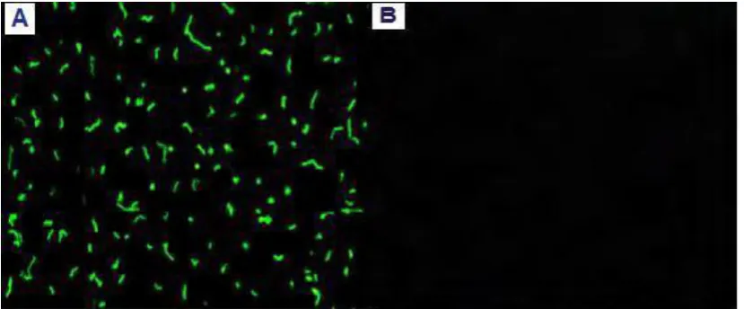
A total of 70 cases with coronary artery disease and 20 (negative and positive control) subjects were enrolled in this study. Their ages mean were 58.80 ± 5.13 years ranged between 40-84 years and ages mean 54.85 ± 5.12 years ranged between 37-70 years for patients and healthy control respectively. The male: female ratio was 9:1(M:F= 63:7). The subjects were selected from non-smoker, non-diabetic, non-hypertension patients (without any risk factors). Many of biochemical tests were done on patient´s blood such as glucose test, urea test, creatinine test and lipid profile test (cholesterol, triglyceride, LDL and HDL), all results showed normal value. Figure 1 and 2 showed the seroprevalence of anti-H. pylori IgG. It showed seroprevalence of anti - H.
pylori IgG. The overall positive of anti- H. pylori IgG in
Figure 2: Seroprevalence of anti- H. pylori IgG among patients and control groups.
Whereas fig.3 showed Anti H. pylori IgA seropositivity was 37.14 % (26/70) among patients while 30% (3/10) among another CAD patient group and 0% (0/10) among control group. A highly significant
difference was observed when the serum levels of anti
H. pylori IgA antibodies of both CAD patients and
another CAD patient as compared to control group were analyzed based on the Chi-square value (P˂ 0.01).
Figure 3: Seroprevalence of anti- H. pylori IgA among patients and control groups.
On the other hand a significant difference in the mean value of Vac A antigen was noticed in CAD patients when compared to control group (1.299± 0.04), and (1.41±0.13) respectively (P= 0.05). As well as a
Figure 4: Ratio of Vac A among studied group
Chronic infection by H. pylori occurs in approximately half of the world’s population causing gastrointestinal and extra-gastrointestinal disorders such as heart diseases [9, 14, 15]. Its pathogenesis and disease outcomes are mediated by a complex interplay between bacterial virulence factors, host and environmental factors [16].
Results of current study agree with other recent studies which revealed that 80% of CAD patients showed seropositive anti-H. Pylori IgGs [17]. Altogether, others showed that the prevalence of seropositivity for H.
pylori was higher in patients with CAD compared to
controls [18, 19]. One of the possible mechanisms contribute in the effects on the cardiovascular system induced by H. pylori is the platelet aggregation has been suggested as certain strains of H. pylori interact with the main platelet receptor glycoprotein in addition to plasma factors such as von Willebrand factor (vWf ) and IgG to provoke platelet aggregation [20]. H. pylori infection has been suggested to influence the development of atherosclerotic changes in coronary arteries indicating a damaging effect of this bacterium or its products (e.g. cytokines, endotoxins, cytotoxins, and other virulence factors) on the coronary endothelium, while no relation between lipid profile and CAD [10, 21].
Virulent H. pylori strains have cytotoxin-associated gene-A (Cag A) toxin [2]. It was documented that local gastric inflammatory and immune responses against H. pylori can induce systematic immune response [22]. Moreover, an auto immune reaction and local inflammation of the artery may be caused by this bacterium due to the presence of a protein on their cell surface which is similar to the heat shock protein-60 found in endothelial cells. This may induce immune cross-reaction between human and bacterial heat shock
H. pylori strains produce VacA. However, there is
significant variation among strains in their ability to stimulate cell vacuolization. This variation is responsible for the genetic structural diversity of the VacA gene which can produce different polymorphic rearrangements [24]. The former studies on VacA determined two major polymorphic regions; the sign (s)- and the middle (m)- regions. The s- region suppose two forms s1 or s2 allele and the m- region encoded the VacA m1 or m2 allele. The combination of s- and m-region allelic types determines the production of the cytotoxin and is associated with pathogenicity of H.
pylori strains. VacA s1/m1 strains produce a large
amount of toxin, s1/ m2 strains produce moderate amounts and s2/m2 strains produce very little or no toxin [25, 26]. Besides that, some studies concluded that seropositivity to Cag A was widely used as a surrogate of infections with toxic H. pylori strains; individual seronegative to Cag A may still be infected with virulent H. pylori expressing Vac A in culture [27]. Interestinglly, some recent reports evaluated the role of
H. pylori removal on coronary artery lumen in patients
who underwent percutaneous intervention for CAD. A higher loss of coronary lumen was observed in those patients who were seropositive for H. pylori [15, 28].
All in all, the main leading role in the cause of cardiovascular anomalies is the improper functioning of the immune system, at the cellular and humoral levels together and characterization of H. pylori virulent markers may help in determination of high-risk patients, as the prevalence rate of H. pylori infection among Iraqi CAD patients is high.
© 2019; AIZEON Publishers; All Rights Reserved This is an Open Access article distributed under the terms of the Creative Commons Attribution License which permits unrestricted use, distribution, and reproduction in any medium, provided the original work is properly cited.
4. REFERENCES
1. Hussein, S. A. Levels of Interferon-8 and von wellibrand factor among Iraqi patients with angina pectoris. M. Sc. Thesis. 2009.College of health and medical technology, Iraq.
2. Ismael, M. K.; Al-Haleem M.R.and Salman, R.S. Evaluation of Anti-Helicobacter pylori Antibodies in A group of Iraqi Patients with Atherosclerosis and Coronary Artery Diseas. Iraqi Journal of Science. 2015. 56(1A): 81-88.
3. Li, T.; Li, X.; Zhao, X.; Cai, W. Z. Z.; Yang, L.; Guo, A.and Zhao, S. Classification of human coronary atherosclerotic plaques using ex vivo high-resolution multicontrast-weighted MRI compared with histopathology. A.J.R. 2012. 198:1069– 1075.
4. Tousoulis, D.; Oikonomou, E.; Economou, E. K.; Crea, F. and Kaski, J. C. Inflammatory cytokines in atherosclerosis: current therapeutic approaches. European Heart Journal. 2016. 37, 1723–1735.
5. Ismael, M. K. Estimation of anti-intrinsic factor IgG and high sensitive C- reactive protein levels in patients with multiple sclerosis. Journal of Babylon university-pure and applied science. 2013. 21(2):398-404.
6. Filep JG. Platelets affect the structure and function of C-reactive protein. Circ Res. 2009.105:109-111.
7. En-Zhi, J.; Zhao, F.; Hao, B.; Zhu, T.; Wang, L.; Chen, B.; Cao1, K.; Huang, J.; Ma, W.; Yang, Z. and Zhang, G. Helicobacter pylori infection is associated with decreased serum levels of high density lipoprotein, but not with the severity of coronary atherosclerosis. Lipids in Health and Disease. 2009. 8:59.
8. Dinakaran, V. Microbial Translocation in the Pathogenesis of Cardiovascular Diseases: A Microbiome Perspective. J Cardiol Curr Res, 2017. 8(6): 00305. DOI: 10.15406/jccr.2017.08.00305.
9. Abd-Alrahman, T. Z.; Aboud, R. S.; Al-Ahmer, Saife D. and Muhammad, T. Y. The role of Helicobacter pylori infection in skin disorders. Iraqi Journal of Science, 2016. 57(4A): 2406-2411.
10. Vafaeimanesh, J.; Hejazi, S.F.; Damanpak, V.; Vahedian, M.; Sattari, M. and Seyyedmajidi, M. Association of Helicobacter pylori Infection with coronary artery disease: Is Helicobacter pylori a Risk Factor?.The Scientific World Journal 2014. Article ID 516354, 6 pages.
11. Al-Qurashi, A. M. and Hodhod,T. E. The association of CagA-positive Helicobacter pylori serotype and atherosclerosis in Najran area, Saudi Arabia. J Am Sci. 2013. 9(4):355-361. 12. Zhang, S.; Yang, G. and Yue, T. Cytotoxin-associated
gene-A-seropositive virulent strains of Helicobacter pylori and atherosclerotic diseases: a systematic review. Chin Med J; 2008. 121(10):946-951.
13. SAS. Statistical Analysis System, User's Guide. Statistical, Version 9.1th (ed). SAS. Inst. Inc. Cary. N.C. USA. 2012. 14. Fagoonee, S.; Angelis, C.D.; Elia, C.; Silvano, S.; Oliaro, E.;
Rizzetto, M. and Pellicano, R. Potential link between
Helicobacter pylori and ischemic heart disease: does the bacterium elicit thrombosis?. Minerva Med. 2010. 101:121–125.
15. Sharma, V. and Aggarwal, A. Helicobacter pylori: Does it add to risk of coronary artery disease. World J Cardiol. 2015. 7(1): 19-25.
16. Kao C, Sheu B, Wu J. Helicobacter pylori infection: an overview of bacterial virulence factors and pathogenesis. Bioomed. J. 2016. 39:14–23.
17. AL-Obeidy, E. S. and Saeed, B. N. H. Pylori Infection in Iraqi patients with ischemic heart diseases. J. Fac Med Baghdad. 2011.53(1):29-31.
18. Vcev, A.; Nakič, D.; Mrden, A.; Mirat, J.; Balen, S. ; Ružic, A. et al. Helicobacter pylori infection and coronary artery disease. Coll. Antropol. 2007. 31 (3): 757–760.
19. Izadi, M.; Fazel, M.; Sharubandi, S. H.; Saadat, S.H.; Farahani, M.M.; Nasseri, M.H.; et al. Helicobacter species in the atherosclerotic plaques of patients with coronary artery disease, Cardiovascular Pathology. 2012. 21(4): 307–311. 20. Byrne, M.F.; Murphy, J.F.; Corcoran, P.A.; Atherton, J.C.;
Sheehan, K.M. Helicobacter pylori induce cyclooxygenase-1 and cyclooxygenase-2 expression in vascular endothelial cells. Scand J Gastroenterol. 2003. 38: 1023-1030. 21. Pérez-Méndez, Ó; Pacheco, H. G.; Martínez-Sánchez, C. and
Franco, M. HDL- cholesterol in coronary artery disease risk: Function or structure? Clinica Chimica Acta, 2014. 429(15) 111–122.
22. Riccioni, G. and Sblendorio, V. Atherosclerosis: from biology to pharmacological treatment: J. Geriatr. Cardiol. 2012. 9: 305−317.
23. Jamkhande, P.G.; Gattani, S. G. and Farhat, Sh. A.
Helicobacter pylori and cardiovascular complications: a mechanism based review on role of Helicobacter pylori in cardiovascular diseases. integr med res. 2016. 5: 244–249. 24. Ferreira RM, Machado JC, Figueiredo C. Clinical relevance
of Helicobacter pylori vacA and cagA genotypes in gastric carcinoma. Best Pract Res Clin Haematol. 2014. 28:1003– 15.
25. Van Doorn L.J.; Figueiredo, C.; Sanna, R.; Pena, A.S. and Midolo, P. N. Expanding allelic diversity of H. pylori vacA. J Clin Microbiol. 1998. 36:2597–603.
26. El-Shenawy,A.; Diab, M.; Shemis, M.; El-Ghannam, M.; Salem, D.; Abdelnasser et al. Detection of Helicobacter pylori vacA, cagA and iceA1 virulence genes associated with gastric diseases in Egyptian patients.The Egyptian Journal of Medical Human Genetics. 2017. 18: 365–371. 27. Mayr, M.; Kiechl, S.; Mendall, M. A.; Willeit, J.; Wick, G. and
Xu, Q. Increased Risk of Atherosclerosis Is Confined to CagA-Positive Helicobacter pylori Strains. Stroke. 2003. 34:610-615.
28. Wang, J-W.; Tseng, K-L.; Hsu, Ch-N.; Liang, Ch-M.; Tai, W Ch. et al. Association between Helicobacter pylori eradication and the risk of coronary heart diseases. PLOS ONE.2018. 13(1): e0190219.


