Azari Mehrdad Mohammad Ali. Periodontal condition in rats with experimental diabetes mellitus after orthodontic surgery. Journal of Education, Health and Sport. 2019;9(10):246-252. eISSN 2391-8306. DOI http://dx.doi.org/10.5281/zenodo.3522313
http://ojs.ukw.edu.pl/index.php/johs/article/view/7610
The journal has had 7 points in Ministry of Science and Higher Education parametric evaluation. Part B item 1223 (26/01/2017). 1223 Journal of Education, Health and Sport eISSN 2391-8306 7
© The Authors 2019;
This article is published with open access at Licensee Open Journal Systems of Kazimierz Wielki University in Bydgoszcz, Pola nd
Open Access. This article is distributed under the terms of the Creative Commons Attribution Noncommercial License which permits any noncommercial use, distribution, and reproduction in any medium, provided the original author (s) and source are credited. This is an open access article licensed under the terms of the Creative Commons Attribution Non commercial license Share a like.
(http://creativecommons.org/licenses/by-nc-sa/4.0/) which permits unrestricted, non commercial use, distribution and reproduction in any medium, provided the work is properly cited. The authors declare that there is no conflict of interests regarding the publication of this paper.
Received: 03.10.2019. Revised: 08.10.2019. Accepted: 29.10.2019.
UDK 616.31:615.37:616.179:379:008
PERIODONTAL CONDITION IN RATS WITH EXPERIMENTAL DIABETES
MELLITUS AFTER ORTHODONTIC SURGERY
Azari Mehrdad Mohammad Ali
Odessa National Medical University
Abstract
Background. Determine periodontal condition in rats with experimental diabetes mellitus
after orthodontic surgery.
Methods. In rats, type 1 diabetes mellitus (DM1) was reproduced with aloxane (100 mg /
kg, intraperitoneally) once. Orthodontic surgery was performed by fixing the spring, starting
from the 12th day. Animal euthanasia was performed on the 35th day of the experiment. The
activity of urease, lysozyme, catalase, elastase, as well as the content of malondialdehyde (MDA)
and hyaluronic acid were determined in the gum homogenate. In the alveolar bone homogenate,
the activity of alkaline phosphatase (AlP) and acid phosphatase (AcP) and elastase was
determined, as well as the content of calcium and protein. The antioxidant-prooxidant index
(API) was calculated by the ratio of catalase activity and MDA content, and the degree of
dysbiosis according to A. P. Levitsky was calculated by the ratio of the relative activities of
of alkaline phosphatase (AlP) and AcP in the bone tissue and the mineralization (MD) degree was
calculated by the ratio of the concentration of calcium, and protein.
Results. In rats with DM1, the level of elastase, urease, MDA and the degree of dysbiosis
increase in the gum, however, the level of lysozyme, hyaluronic acid, and the API index
decrease. In the bone tissue of the periodontium of rats with type 1 diabetes, the level of alkaline
phosphatase and MA decreases, but the level of AC increases. Orthodontic surgery significantly
reduces the degree of dysbiosis in the gums and shows a tendency to increase API and decrease
elastase activity. After orthodontic surgery, rats significantly increase the level of alkaline
phosphatase and MA.
Conclusion. With type 1 diabetes, periodontitis, dysbiosis develops and the mineralizing
activity of periodontal bone tissue decreases. Orthodontic surgery tends to improve periodontal
conditions.
Keywords: periodontium; diabetes mellitus; orthodontics; dysbiosis; inflammation;
mineralizing activity.
INTRODUCTION
Diabetes mellitus is characterized by the development of pathological processes in tissues
of mouth [1-5]. Established development with diabetes oral dysbiosis [6, 7], inflammatory
dystrophic processes in the mucosa oral and periodonticus. Of course, any operational
intervention in the mouth on the background of diabetes must have a special character of the flow
and require special of approach to prevent possible complications [8-10]. Purpose of this research
was the definition of the state of the gums and bone periodontal rats after orthodontic operation
on the background of diabetes.
MATERIAL AND RESEARCH METHODS
Experiments were carried out by 20 White rats line Wistar (males, 3 months, the average
live weight 125 grams), distributed in 3 groups: 1st (7 rats) – intact, 2nd (8 rats), and 3-I (5 of
rats) – with diabetes type 1, which caused by alloxane (100 mg/kg, in /abdominal, once). Rats 3rd
group, since the 12th day of experience, fixed the spring [11]. Euthanasia animals was carried out
on the 35th day of experience under thiopental anesthesia (20 mg/kg) by total loos blood from of
elastase (biochemical marker of inflammation) [13], catalase (antioxidant enzyme) [13] and the
content of malonic acid dialdehyde (MDA), indicator of lipid peroxidation [13], as well as
content hyaluronic acid [14]. In homogenate of periodontal bone was determined activity alkaline
(AlP), and acid (AcP) phosphatases [15], the content of calcium [15], protein [15] and activity
elastase [13]. Ratio of relative activities urease and lysozyme calculated the degree of dysbiosis
by A. P. Levitsky [12]. Ratio activity catalase and the content of MDA was calculated
antioxidant-prooxidant index API [13]. Ratio AlP and AcP calculated mineralizing index (MI)
[16], and on the ratio of the content of calcium and protein was determined the mineralization
degree (MD) of bone [16]. The resiults of experiments were subjected to a standard statistics
processing [17].
RESULTS AND DISCUSSION
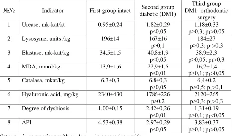
In table 1 presents the results of determine the number of biochemical parameters of rats
[image:3.612.73.540.402.676.2]gums with diabetes type 1, who carried out orthodontic operations.
Table 1. Biochemical parameters of the gums of rats with type 1 diabetes mellitus
and orthodontic treatment
№№ Indicator First group intact Second group diabetic (DM1)
Third group DM1+orthodontic
surgery 1 Urease, mk-kat/kt 0,95±0,24 1,82±0,29
р<0,05
1,18±0,33 р>0,3; р1>0,05
2 Lysosyme, units /kg 196±14 167±16 р>0,1
184±27 р>0,3; р1>0,3
3 Elastase, mk-kat/kg 34,5±1,5 40,8±1,9 р<0,05
38,9±2,3 р>0,05; р1>0,3
4 MDA, mmol/kg 13,9±1,6 22,9±1,5 р<0,01
16,7±1,4 р>0,1; р1>0,05
5 Catalasa, mkat/kg 6,3±0,3 6,8±0,3 р>0,05
6,4±0,2 р>0,5; р1>0,1
6 Hyaluronic acid, mg/kg 2340±430 1786±226 р>0,2
2120±265 р>0,3; р1>0,3
7 Degree of dysbiosis 1,00±0,15 2,42±0,26 р<0,01
1,31±0,19 р>0,1; р1<0,05
8 API 4,53±0,38 2,97±0,29
р<0,05
3,83±0,37 р>0,1; р1>0,05
As seen from these data activity urease rats with diabetes significantly increases (2 times),
indicating the increase in microbial seedling periodontal tissues. After orthodontic operations
activity urease normal, indicating a favorable the influence of this operation on oral
microbiocenosis. Activity elastase in gums rats with diabetes significantly increases, indicating
the development of inflammation (gingivitis). The second marker of inflammation – MDA also
increased 65 % in rats with diabetes. Orthodontic operation almost normalizes both a marker of
inflammation that again shows favorable the influence of orthodontic operations on parodontis.
Activity lysozyme, catalase and the content of hyaluronic acid in gums of rats with diabetes
change is insignificant, but also little change after surgery.
Degree of dysbiosis gums of rats with diabetes is increased 2.4 times, and after
orthodontic operations was normal. Index IPA, on the contrary, decreases with diabetes and
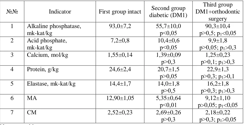
normal after orthodontic operations. In table 2 results of determine the number of biochemical
parameters bone periodontal. It is clear that the activity of alkaline phosphatase significantly
reduced in rats with diabetes and fully recovered after orthodontic operations. Activity acid
phosphatase significantly increases with diabetes and little changed after surgery. Calcium
content, protein and activity elastase in bone not undergo significant changes in diabetes or in
combination of diabetes with orthodontic operation. Does not change also exponent
mineralization periodontal bone, but meniralizing index MA determined by the relation AlP/AcP,
reduced rats with diabetes more than 2 times, but orthodontic operation it restores almost to the
norm.
Thus, conducted by us study have confirmed data literature [6, 7] on the development of
dysbiosis and inflammation in parodont at diabetes and reducing mineralizing activity of bone.
New is that we managed to show beneficial effect orthodontic operations on the rats with
diabetes: they have decreased microbial seedling, the degree of dysbiosis, inflammation and
normalized mineralizing activity of bone.
Obtained us data indicate of the possibility of the implementation of orthodontic
operations in patients with diabetes, without fear of the development of complications. On the
Table 2. Biochemical parameters of periodontal bone tissue of rats with type 1 diabetes mellitus
and orthodontic treatment.
№№ Indicator First group intact Second group diabetic (DM1)
Third group DM1+orthodontic
surgery 1 Alkaline phosphatase,
mk-kat/kg
93,0±7,2 55,7±10,0 р<0,05
90,3±10,4 р>0,5; р1<0,05
2 Acid phosphate, mk-kat/kg
7,2±0,8 10,4±0,6 р<0,05
9,9±1,8 р>0,05; р1>0,3
3 Calcium, mol/kg 1,55±0,14 1,39±0,09 р>0,3
1,25±0,23 р>0,1; р1>0,3
4 Protein, g/kg 24,6±2,4 20,7±1,5
р>0,05 р>0,3; р22,9±1,3 1>0,1
5 Elastase, mk-kat/kg 14,4±1,7 14,0±1,8 р>0,5
16,2±1,8 р>0,3; р1>0,3
6 МA 12,90±1,05 5,35±0,64
р<0,01
9,12±1,10 р>0,05; р1<0,05
7 СM 2,52±0,23 2,69±0,26
р>0,3
2,18±0,22 р>0,3; р1>0,05
Note: see tables
CONCLUSIONS
1. Diabetes is the development of the parodont dysbiosis, inflammation and reduction
mineralizing activity bone.
2. Orthodontic operation has a benefit with diabetes, which gives a reason to implement
such operations in persons with diabetes.
REFERENCES
1. Fesenko UA, Malygina DA. Influence of diabetes mellitus on clinical and laboratory
indices of inflammatory diseases of maxillofacial area in children. Dentist. 2010; 1(139): 32-33.
(in Russian)
2. Skiba AV, Tereshina TP, Dmitrieva NB. Diabetes and periodontal disease Visceral
dentistry. Spec. release. 2012; 6: 82-86. (in Russian)
3. Moroz BT, Zhavoronkova NV, Khromova EA. Condition of periodontal tissues and hard
tissues of teeth in patients with type 2 diabetes mellitus. Institute of Dentistry. 2013; 3(60):
4. Ota M, Seshima F, Okubo N [and others]. A collaborative approach to care for patients
with periodontitis and diabetes. Bull. Tokyo Dent. Coll. 2013; 54(1): 51-57.
5. Basov AA, Bykov IM, Melnonyan KI. Changes in immunological reactivity and
processes of free radical oxidation in the oral fluid in patients with type 2 diabetes mellitus.
International Journal of Applied and Fundamental Research. 2014; 2: 31-34.
6. Levitsky AP, Tsiselsky Yu. V. Dysbiosis, diabetic retinopathy and prebiotics. Odessa,
KP ОГТ, 2012: 197. (in Russian)
7. Mutoh T., Honda E., Matsumoto K [and others]. Study of oral microflora in diabetes
mellitus patients. J. Dent. res. 2000; 79(Spec. is.): 2013.
8. Sykes LM, Sukha A. Potential risk of serious oral infections in the diabetic patient: a
clinical report. J. Proshet. Dent. 2001; 86: 569-573. (in Russian)
9. Badanov RM. Development of a differentiated approach to the prevention and treatment
of prosthetic stomatitis in patients with diabetes mellitus. Odessky Journal of Medicine. 2011; 1
(123): 36-40. (in Ukrainian)
10. Demyanenko SA, Morozov AL, Denga AE. Influence of the experimental orthodontic
operation on the periodontal condition in rats with metabolic syndrome. Parodontology. 2017;
XXIIIb(4(85): 4-7. (in Russian)
11. Gorokhovskiy VN, Denga OV, Dentga AE, Mirchuk BN. Modeling of the orthodontic
movement of teeth in rats. In book. Schneider SA, Levitsky AP "Experimental stomatology". Part
1. Experimental models of dental diseases. Odessa, 2017: 128-132. (in Russian)
12. Levitsky AP, Makarenko OA, Selivanskaya IA [and others]. Enzymatic method for
determining oral dysbiosis for screening pro- and prebiotics: guidelines. Kiev, State
Pharmacological Center, 2007: 22. (in Russian)
13. Levitsky AP, Denga OV, Makarenko OA [and others]. Biochemical markers of
inflammation of the tissues of the oral cavity: methodical recommendations. Odessa, KP of the
OSG, 2010: 16. (in Russian)
14. Asatiani VS. New methods of biochemical photometry. Moscow, Science, 1965: 298.
(in Russian)
15. Levitsky AP, Makarenko OA, Denga OV [and others]. Experimental methods for
16. Levitsky AP, Makarenko OA, Khodakov ІV [and others]. Enzymatic method otsinki to
become kistkovoi fabrics. Odessky meditsyny magazine. 2006; 3: 17-21. (in Ukrainian)
17. Lapach SN, Chubenko AV, Babich PN Statistical methods in biomedical research using