R E S E A R C H
Open Access
Histamine modulates microglia function
Raquel Ferreira
1, Tiago Santos
1, Joana Gonçalves
2,3, Graça Baltazar
4, Lino Ferreira
1,5, Fabienne Agasse
1and Liliana Bernardino
1,4,6*Abstract
Background:Histamine is commonly acknowledged as an inflammatory mediator in peripheral tissues, leaving its role in brain immune responses scarcely studied. Therefore, our aim was to uncover the cellular and molecular mechanisms elicited by this molecule and its receptors in microglia-induced inflammation by evaluating cell migration and inflammatory mediator release.
Methods:Firstly, we detected the expression of all known histamine receptor subtypes (H1R, H2R, H3R and H4R), using a murine microglial cell line and primary microglia cell cultures from rat cortex, by real-time PCR analysis, immunocytochemistry and Western blotting. Then, we evaluated the role of histamine in microglial cell motility by performing scratch wound assays. Results were further confirmed using murine cortex explants. Finally, interleukin-1beta (IL-1β) and tumor necrosis factor-alpha (TNF-α) levels were evaluated by ELISA measurements to determine the role of histamine on the release of these inflammatory mediators.
Results:After 12 h of treatment, 100μM histamine and 10μg/ml histamine-loaded poly (lactic-co-glycolic acid) microparticles significantly stimulated microglia motility via H4R activation. In addition, migration involvesα5β1 integrins, and p38 and Akt signaling pathways. Migration of microglial cells was also enhanced in the presence of lipopolysaccharide (LPS, 100 ng/ml), used as a positive control. Importantly, histamine inhibited LPS-stimulated migrationviaH4R activation. Histamine or H4R agonist also inhibited LPS-induced IL-1βrelease in both N9 microglia cell line and hippocampal organotypic slice cultures.
Conclusions:To our knowledge, we are the first to show a dual role of histamine in the modulation of microglial inflammatory responses. Altogether, our data suggest that histamine per se triggers microglia motility, whereas histamine impedes LPS-induced microglia migration and IL-1βrelease. This last datum assigns a new putative anti-inflammatory role for histamine, acting via H4R to restrain exacerbated microglial responses under inflammatory challenge, which could have strong repercussions in the treatment of CNS disorders accompanied by microglia-derived inflammation.
Keywords:Histamine, Microglia, Inflammation, Histamine 4 receptor, Migration
Background
Microglial cells play a pivotal role in the immune surveil-lance of the central nervous system (CNS) by avidly sur-veying the brain parenchyma in search of infection, injury, or other sources of pathology [1,2]. In this sense, micro-glial cells become activated and migrate to the injury site in order to fully develop a concerted immune response, in-volving the release of both trophic and pro-inflammatory
factors [1]. The study of the classical components that constitute the microglia response in the inflammatory process has been copiously supported by the use of lipo-polysaccharide (LPS), a gram-negative cell wall compo-nent. LPS binds to the CD14/TLR4/MD2 receptor complex, located on the cell membrane, triggering clas-sical microglial responses such as proliferation, migration, phagocytosis and release of inflammatory mediators [3,4]. Histamine is an endogenous biogenic amine mostly stored in the granules of mast cells and basophils that readily re-lease their content upon stimulation [5]. Other sources of histamine include histaminergic neurons, gastric entero-chromaffin-like cells, leukocytes and platelets, to name a * Correspondence:[email protected]
1
CNC - Center for Neuroscience and Cell Biology, University of Coimbra, Coimbra, Portugal
4
CICS-UBI - Health Sciences Research Center, University of Beira Interior, Covilhã, Portugal
Full list of author information is available at the end of the article
few. Moreover, in the CNS, histamine is released by microglial cells [6]. Histamine exerts its various functions through the activation of four distinct subtypes of
G-pro-tein coupled receptors: H1 receptor (H1R), H2 receptor
(H2R), H3receptor (H3R) and H4receptor (H4R) [7]. Their actions range mainly from the modulation of the allergic reaction (H1R), regulation of heart and gastric acid secre-tion (H2R) to neurotransmitter release (H3R). H4R is mainly expressed by cells of the immune system (e.g., B-and T-lymphocytes, dendritic cells, eosinophils, fibro-blasts, mast cells, monocytes, natural killer cells and neu-trophils), and its expression is modulated by an inflammatory context [8]. At the moment, H4R is primar-ily known for its chemotactic effect on mast cells and eosi-nophils (reviewed by [9]).
In our study, we showed for the first time that all known histamine receptors are expressed in microglial cells. We also showed that histamineper sestimulates microglia mo-tility. However, and most interestingly, in an LPS-induced inflammatory context, histamine has an inhibitory action in microglia migration and in the release of interleukin-1beta (IL-1β). In summary, we uncovered a novel dual role for histamine in the regulation of neuroinflammation mediated by microglia activity by modulating cell recruitment and the release of pro-inflammatory cytokines, such as IL-1β and tumor necrosis factor-alpha (TNF)-α.
Methods
All experiments were performed in accordance with Euro-pean Union (2010/63/EU) guidelines for the care and use of laboratory animals. All efforts were made to minimize animal suffering and the number of animals used.
Cell line culture
The murine N9 microglia cell line (a kind gift from Prof. Claudia Verderio, CNR Institute of Neuroscience, Cellular and Molecular Pharmacology, Milan, Italy) was grown as previously described [10]. Cells were plated at a density of 2 × 10⁴cells per well in 24-well trays (immunocytochem-istry), 5 × 10⁴cells per well in 12-well trays (scratch wound assays) or plated at a density of 5 × 10⁵cells per well in 6-well trays (for the remaining experiments). Cell treatments included the following incubation setup: histamine dihy-drochloride (1–100μM, Sigma), LPS (100 ng/ml, Sigma),
α5β1 blocking antibody (10 μg/ml, Millipore Corp., Bed-ford, MA, USA), H1receptor antagonist,
2-((2-(dimethyla-mino)ethyl)(p-methoxybenzyl)amino)-pyridine maleate
(mepyramine maleate, 1μM), H2receptor antagonist,
N-cyano-N’-methyl-N”-[2-[(5-methyl-1 H-imidazol-4-yl)me-thyl]thio]ethyl]guanidine (cimetidine, 5 μM), H3 receptor antagonist 3-amino-N-[2-(1 H-imidazol-4-yl)ethyl]propa-namide ditrifluoroacetate (carcinine ditrifluoroacetate, 5
μM), H4receptor antagonist, 1-[(5-chloro-1 H-indol-2-yl)
carbonyl]-4-methylpiperazine (JNJ7777120, 5μM) and H4
receptor agonist, 5-(2-aminoethyl)-4-methylimidazole
dihydrochloride (4-methylhistamine dihydrochloride, 20
μM) (all from Tocris, Ballwin, MO, USA) for 3 h (receptor expression evaluation), 6 h (cytokine release) or 12 h (mi-gration studies). Wortmanin (50 nM, Alomone Labs Ltd., Jerusalem, Israel), p38 inhibitor SB239063 (20μM, Sigma) and all histamine receptor antagonists/agonists were added 40 min prior to cell treatment.
Primary microglia cell cultures from cortex
Mixed glial cultures from the cortex were prepared as pre-viously described by Saura and colleagues (2003) [11]. Briefly, neonatal Wistar rats (P2-4) were killed, and the brains were placed in ice-cold 0.15 M sterile PBS. After re-moval of the meninges, cortex explants were digested in cysteine solution (1.9 mM CaCl2, 1.3 mM cysteine) and H&B solution (116 mM NaCl, 5.4 mM KCl, 26 mM NaHCO3, 12 mM NaH2PO4.H2O, 1 mM MgSO4.7H2O, 0.5 mM EDTA, 25 mM glicose, pH 7.3) supplemented with 20 U/ml papain and 0.001% phenol red at 37°C for 4 min, under constant agitation. Then, the tissue was rinsed with high glucose Dulbecco’s modified Eagle’s medium (DMEM, Invitrogen, Paisley, UK) supplemented with 10% fetal
bo-vine serum (FBS), 100 U/ml penicillin and 100 μg/ml
streptomycin. After mechanical dissociation, cells were pel-leted by centrifugation (3 min, 405× g; 3K18C Bioblock Sci-entific; Sigma Laboratory Centrifuges) and suspended in DMEM. The cells were then plated into 12-well cell culture plates at a density of 0.087 × 10⁶cells per well. The cultures
were kept at 37°C in a 5% CO2and 95% air atmosphere,
and the cell medium was changed every 7 days. On day 20–21, a mild trypsinization (trypsin dilution 1:3 in DMEM without FBS) was done for 40 min at 37°C to remove astro-cytes. The resultant adherent microglial cells were washed
twice with DMEM and kept at 37°C in a 5% CO2and 95%
air atmosphere for a further 5 days before RT-PCR and Western blot experiments. More than 98% of the cells in culture were immunopositive for the microglia marker CD11b.
Brain cortex explants
Adult wild-type (WT) C57BL6 mice were used for the study of cell motility in cortex explants. Briefly, mice
were killed, and the brains were placed in Hank’s
balanced salt solution (HBSS), supplemented with 100
U/ml penicillin and 100 μg/ml streptomycin (all from
serum (25%) and HBSS (25%), supplemented with D-glucose to a final concentration of 25 mM and 100 U/ml penicillin and 100μg/ml streptomycin (all from Invitro-gen). The motility assay occurred for 24 h, after medium
replacement, at 5% CO2and 95% atmospheric air at 37°
C, before fixation.
Results are expressed as the number of CD11b-posi-tive cells, denoting microglial and/or CNS macrophages, that migrated from the explants within a 300-μm radius from the explant edge and normalized per explant area
(number of CD11b-positive cells/mm2). Cell migration
was only evaluated in explants with an area ranging
from 1 to 1.5 mm2. Explant images were acquired using
MetaFluor Software (Universal Imaging, Downingtown, PA, USA), and the explant area and radius were analyzed with NIH ImageJ Software.
Organotypic hippocampal slice cultures
Briefly, 7-day-old C57BL6 WT mice were killed by de-capitation, their brains removed under sterile conditions,
and the hippocampi isolated and cut in 350-μm coronal
sections using a McIlwain tissue chopper. Individual slices were placed in ice-cold Gey’s balanced salt solu-tion (Biological Industries, Israel) supplemented with 25 mM D-glucose (Merck, Darmstadt, Germany), 100 U/ml
penicillin and 100 μg/ml streptomycin, before being
placed on porous insert membranes (Millipore Corp.). Six slices were put onto each membrane, and the inserts were transferred to a six-well culture tray (Corning Costar, Corning, NY, USA). Each well contained 1 ml culture medium, composed of 50% Opti-minimal essen-tial medium, 25% heat-inactivated horse serum, and 25% HBSS supplemented with 25 mM D-glucose, 50 U/ml penicillin and 50 μg/ml streptomycin (all from Invitro-gen). Slices were allowed to grow for 2 weeks before the ELISA experiments.
Real-time polymerase chain reaction analysis of histamine receptors expression
Total RNA was isolated from the N9 murine microglial cell line, from primary microglia cell cultures from the cortex and from hippocampal extracts (positive control) according to the illustra RNAspin Mini RNA Isolation
Kit manufacturer’s instructions (GE Healthcare Life
Sciences, Buckinghamshire, UK). Precipitated RNA was
resuspended in 20 μl of diethylpyrocarbonate
(DEPC)-treated water and quantified by optical density (Nano-Drop 2000, ThermoScientific, Waltham, MA, USA). For
cDNA synthesis, 2 μg of total RNA was mixed with 2.5
μM anchored-oligo-p(dT)18 primers, 1× PCR reaction
buffer, 20 U RNase inhibitor, dNTPs (1 mM each) and 10 U AMV Reverse Transcriptase in a 20-μl final volume (Roche Molecular Biochemicals). The reaction was per-formed at 55°C for 30 min and stopped by 85°C for a
5-min step. For gene expression analysis, 1 μl of sample
cDNA was added to 10 μl EvaGreen Supermix (BioRad,
Hercules, CA, USA), and the final concentration of each
primer was 10 μM in 20μl total reaction volume. The
thermocycling reaction was initiated with activation of Taq DNA polymerase by heating at 94°C for 3 min, fol-lowed by 30 cycles of a 15-s denaturation step at 94°C, and a 30 s annealing and elongation step at 57°C for all sets of primers. For housekeeping gene hypoxanthine phosphoribosyltransferase 1 (HPRT-1), primer sets were obtained from selected QuantiTect Primer Assays (Qia-gen, Austin, Texas). Fluorescence was measured after the extension step by the iQ5 Multicolor Real-Time PCR Detection System (BioRad). After the thermocycling re-action, the melting step was performed with slow heat-ing, starting at 55°C and with a rate of 0.5°C per 10 s, up to 95°C, with continuous measurement of fluorescence, allowing detection of possible nonspecific products. The assay included a non-template control (sample was sub-stituted by sterile water), a negative transcriptase reac-tion sample and a standard curve (in 10-fold steps) of DNA for assessing the efficiency of each set of primers. All reactions were run in duplicates. Ct values were measured in the exponential phase and were set at the
same fluorescence value in each run performed.ΔCt was
calculated by subtracting each Ct sample from in-plate Ct of the housekeeping reference gene HPRT-1. A
smal-ler ΔCt value represents higher gene expression. Data
analysis was performed with Biorad iQ5 software (BioRad). The PCR products were subjected to electro-phoresis in a 1% agarose gel stained with ethidium bromide. Digital photographs were taken in a Versa-Doc Imaging System (Model 3000, BioRad).
Primer sequences: H1R, forward primer 5’-GGG CTC
AAA GGC CAA TGA C-3’and reverse primer 5’-TCC
GCC GGC AAG TAC TCA-3’ (74 bp); H2R, forward
primer 5’-CTG GCT GTC AGC TTG AAT CG-3’and
reverse primer 5’-GCT GCC AGG GAC ACA ATG A-3’
(65 bp); H3R, forward primer 5’-CTT CTC TCT CCC
AAG ACG ATC TG-3’and reverse primer 5’-GGC TCC
GGG ATT AAG GAA GA-3’(65 bp); H4R, forward
pri-mer 5’-GCT ACG ATC GAT ACC AGT CA-3’and
re-verse primer 5’-AAG AAA GCC AGT ATC CAA ACA
G-3’ (109 bp) (synthesized by Eurofins MWG Operon,
Ebersberg, Germany).
Preparation and characterization of PLGA microparticles The single emulsion technique was used to prepare
microparticles of approximately 2μm in diameter. PLGA
v)]. The resulting suspension was stirred for 3 h, washed with distilled water and finally freeze-dried. The morph-ology and diameter of PLGA particles were evaluated by scanning electron microscopy according to our previous reports [12]. Release experiments in 0.15 M PBS at 37°C were performed in order to evaluate the release profile of histamine over 30 days and to assess the loading effi-ciency of the microparticles. The loading capacity of
PLGA microparticles was approximately 5.3μg of
hista-mine per mg of microparticles. Approximately 1 μg of
histamine was released per mg of microparticles over 4 days [13]. Blank microparticles, i.e., without histamine, were also prepared to test the effect of the microparticle formulation per se [13].
Motility assay
Before N9 microglial cell seeding, two parallel lines were carved on the underside of each well with a scalpel. These lines served as a guidance axis together with the line provided by the scratch wound. The cell monolayer was approximately 95% confluent before the migration assay took place. One hour before performing the wound, medium was replaced by serum-free medium to ensure no proliferation occurred during experiments. The wound was made by a perpendicular scratch made with a P10 pipette tip (Gilson S.A.S., Villiers-le-Bel, France). After N9 microglial cell treatment, images were taken with an inverted Axiovert 200 microscope (Carl Zeiss, Göttingen, Germany), with a 5× objective and a CoolSNAP digital camera (Roper Scientific, Tucson, AZ, USA). Differential interference contrast (DIC) images were acquired using MetaFluor Software (Universal Im-aging, Downingtown, PA, USA) and analyzed with NIH ImageJ Software. For the N9 microglia cell line, cell mo-tility was determined by counting the number of cells that migrated towards the middle of the wound within a 12-h period of treatment. The protocol was adapted from Valster and colleagues (2005) [14].
Enzyme-linked immunosorbent assay for IL-1β
Cells were plated and treated as described above (see section Cell line culture). Cells were left at room temperature (RT) for 5 min in lysis buffer [137 mM
NaCl, 20 mM Tris–HCl, 1% Triton X-100, 10% glycerol,
1 mM phenylmethylsulfonyl fluoride (PMSF), 10 μg/ml
aprotinin, 1μg/ml leupeptin, 0.5 mM sodium
orthovana-date (all from Sigma), pH 8.0]. Total protein concentra-tion was determined by the bicinchoninic acid method
(BCA), and samples were stored at−80°C. For the
quan-tification of IL-1β protein levels, a mouse IL-1βELISA kit was used following the manufacturer’s instructions (eBioscience, San Diego, CA). For that purpose, Microti-ter plates (MaxiSorp, Nunc A/S, Roskilde, Denmark) were used. Optical density was recorded at 450 and 570
nm (values later subtracted from those obtained with 450 nm) in an ELISA plate (SPECTRA max 384 Plus, Molecular Devices).
Enzyme-linked immunosorbent assay for TNF-α
Cells were plated and treated as described above (see section Cell line culture). After cell lysis and protein quantification, as described above (see section
Enzyme-linked immunosorbent assay for IL-1β), a mouse TNF-α
ELISA kit was used according to the manufacturer’s
instructions (eBioscience). Optical density was recorded at 450 and 620 nm (values later subtracted from those obtained with 450 nm) in an ELISA plate (SPECTRA max 384 Plus, Molecular Devices).
Western blotting
For total extracts, cells were incubated with lysis cocktail
solution [137 mM NaCl, 20 mM Tris–HCl, 1% Triton
X-100, 10% glycerol, 1 mM phenylmethylsulfonyl fluoride,
10 μg/ml aprotinin, 1 μg/ml leupeptin, 0.5 mM sodium
vanadate (all from Sigma), pH 8.0]. Samples were centri-fuged for 20 min at 4,300 g at 4°C, and the supernatant collected.
The total amount of protein was quantified using the
BCA assay. Afterwards, 50-μg samples were loaded onto
12% acrylamide/bisacrilamide gels (BioRad). Proteins were separated by SDS-PAGE using a bicine/SDS (Sigma) elec-trophoresis buffer (pH 8.3) and then transferred to PVDF membranes (Millipore) on the following conditions: 300 mA, 90 min at 4°C in a solution containing 10 mM CAPS and 20% methanol, pH 11.0). Membranes were blocked in
Tris-buffer saline containing 5% BSA and 0.1% TweenW
20 (Sigma) for 1 h, at RT, and then incubated overnight at 4°C with the primary antibody solution diluted in 0.1% TBS-Tween, 5% BSA. The following primary antibodies were used: goat polyclonal anti-H4receptor (1:5,000, Santa Cruz Biotechnology, Inc., Santa Cruz, CA, USA) and mouse monoclonal anti-GAPDH (1:10,000, Millipore) (all from Cell Signaling). After rinsing with TBS-T, mem-branes were incubated for 1 h at RT with an alkaline phos-phatase-linked secondary antibody goat or
anti-mouse IgG 1:10,000, in 5% BSA, 0.1% TweenW20 (Sigma)
and 1% TBS-T (GE Healthcare UK Limited, Buckingham-shire, UK). Protein immunoreactive bands were visualized in a Versa-Doc Imaging System (Model 3000, BioRad), after incubation of the membrane with ECF reagent (GE Healthcare UK Limited) for 5 min.
Immunocytochemistry
for 2 h at RT with the corresponding secondary anti-body. Antibodies were used as listed: goat polyclonal
anti-H4 receptor (1:5,000, Santa Cruz Biotechnology,
Inc., CA, USA), rat monoclonal anti-CD11b (1:1,000, AbD Serotec, Oxfordshire, UK) and rat monoclonal
α5β1 antibody (10 μg/ml, Millipore) in 0.1% Triton X-100, 0.3% BSA solution; Alexa Fluor 594 donkey anti-goat and Alexa Fluor 488 anti-goat anti-rat (all 1:200 in PBS, from Molecular Probes, Eugene, OR, USA). For nuclear labeling, cell preparations were counterstained with
Hoechst 33342 (2 μg/ml) (Molecular Probes) in 0.15 M
PBS for 5 min at RT and mounted in Dakocytomation fluorescent medium (Dakocytomation Inc.). Fluorescent images were acquired using a confocal microscope with a Plan-ApoChromat 63×/1.40 oil objective (N9 micro-glial cells) or Plan-ApoChromat 40×/1.3 oil objective (cortex explants) (LSM 510 Meta, Carl Zeiss).
Data analysis
Statistical analysis was performed using GraphPad Prism 5.0 (GraphPad Software, San Diego, CA). Statistical signifi-cance was considered relevant for p values<0.05 using one-way analysis of variance followed by Bonferroni’s post hoc test for comparison among experimental settings and Dunnett’s post hoc test for comparison with control con-ditions. Data were presented as means ± standard error of mean (SEM). For the N9 microglia cell line motility assay, four images per experimental condition were acquired. For cortex explants, a series of images around the whole specimen were taken to count every cell within a 300-μm radius from the explants. Every experimental condition was tested in three sets of independent experiments, un-less stated otherwise, and performed in duplicates.
Results
Microglial cells express histamine H4receptor
In this work, we studied the role of histamine in micro-glial-induced inflammatory responses, and for that pur-pose, we used complementary approaches using a murine microglia cell line, primary microglia cultures from the cortex, hippocampal organotypic slice cultures and cortex explants. We previously validated the use of the N9 cell line in the study of microglial responses by showing that neuropeptide Y dampens both LPS- and IL-1β-stimulated cell migration, phagocytosis and the re-lease of potent pro-inflammatory mediators [10,15,16].
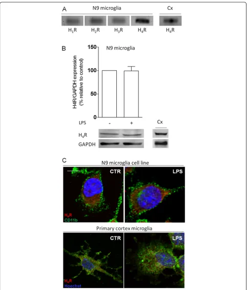
Firstly, we performed real-time polymerase chain reac-tion (qPCR) to identify the expression of all known his-tamine receptors. In that sense, we amplified cDNA coding for H1R, H2R, H3R and H4R, and normalized the relative expression of each gene against a previously selected housekeeping gene, hypoxanthine phosphoribo-syltransferase 1 (HPRT-1) [17]. We found that N9 micro-glial cells expressed low basal levels of H1R, H2R and H3R,
whereas H4R appeared to be more abundantly expressed (ΔCtH1R = 13.4 ± 0.7; ΔCtH2R = 15.0 ± 1.3; ΔCtH3R = 16.4 ± 2.3;ΔCtH4R = 13.0 ± 0.3; n = 3-4) (Figure 1A). A smaller
ΔCt value represents higher gene expression. Negative
controls showed no amplification (data not shown). H4R is mainly expressed by cells of the immune system. Since its role in the CNS has been poorly discussed, and to our knowledge, we are the first to report H4R expression by microglial cells, we then focused our study on this recep-tor [8,18]. Accordingly, we performed Western blotting to determine whether differences regarding the pattern of re-ceptor expression existed in an inflammatory context. Interestingly, LPS challenge (100 ng/ml, for 12 h) did not alter H4R protein expression by N9 microglial cells (Figure 1B). For both experimental approaches, primary microglia cultures obtained from the cortex (Cx) were used as positive controls. Furthermore, for qPCR analysis we also used whole hippocampus cDNA as a positive con-trol [19] (data not shown). Then, we confirmed by im-munocytochemistry that both N9 microglial cells (top panel) and cortex primary microglia cell cultures (bottom panel) expressed H4R (in red), in a similar fashion, in con-trol conditions and under LPS challenge (Figure 1C). As a positive control, coronal brain slices were used for H4R-positive immunodetection on the hippocampus [19] (data not shown). To visualize cell morphology we labeled the alpha chain of αMβ2-integrin, CD11b (in green), a well-known surface marker for microglia and leukocytes, whose over-expression is associated to microglial and/or CNS macrophage activation [20].
Histamine induces microglia motility through H4R
activation
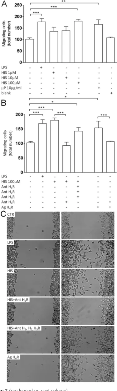
To evaluate the effect of histamine on microglia motility, we determined the number of N9 microglial cells that migrated in vitro across a scratch wound (Figures 2, 3, 4, 5). Accordingly, we performed a dose–response curve to assess a functionally relevant concentration of histamine (Fig-ure 2A). Microglial cells were treated for 12 h with
concen-trations of histamine ranging from 1 to 100μM, and we
observed that only 100 μM histamine significantly
induced cell motility (meanCTR= 101.9 ± 4.9 cells;
meanHIS100μM= 174.4 ± 5.9 cells; p<0.001, n= 13-20). At this concentration, histamine did not interfere with microglia cell death or proliferation (data not shown).
We also developed histamine-loaded poly (lactic-co
Figure 1Microglial cells express histamine receptors. (A)Histamine receptor expression analysis by real-time PCR showed that microglial cells constitutively express all known histamine receptors–H1R (74 bp), H2R (65 bp), H3R (65 bp) and H4R (109 bp).(B)LPS stimulation (100 ng/ml) did not alter
the expression of H4R, quantified by Western blotting. Data are expressed as percentage of control (n= 3-4). A representative blot is shown below the
graph (H4R, 44 kDa; GAPDH, 37 kDa). In both experimental paradigms, primary microglia cultures from the cortex (Cx) were used as a positive control. For
qPCR, whole hippocampal cDNA was used as an additional positive control (data not shown).(C)N9 microglial cells (top panel) and primary cortical microglia cultures (bottom panel) expressed H4R (inred) under control conditions and upon LPS challenge (100 ng/ml). Cells were stained for morphology
extracellular release of histamine [13]. In previous work by our group, histamine-loaded PLGA microparticles were shown to efficiently promote neurogenesis with-out any cytotoxic effect [13]. Accordingly, 10μg/ml his-tamine-loaded microparticles significantly promoted cell motility, whereas blank (void formulation) particles had no effect (meanuP10μg/ml= 166.4 ± 19.7 cells; mean-blank= 107.7 ± 3.2 cells;p<0.01, n= 3-4). LPS challenge
(100 ng/ml) was used as a positive control (meanLPS=
167.0 ± 11.2 cells;p<0.001,n= 13) (Figure 2A). To un-cover which histamine receptor was involved, we
trea-ted N9 microglial cells with histamine (100 μM)
together with an antagonist for each receptor individu-ally (data not shown), and only H4R antagonist
(JNJ7777120, 5 μM) significantly reduced
histamine-induced migration (meanHIS+H4R ant= 93.5 ± 11.1 cells;
p<0.001, n= 4). Moreover, the simultaneous blockade of other receptors [H1R antagonist, mepyramine
male-ate (1μM), H2R antagonist, cimetidine (5μM) and H3R
antagonist, carcinine ditrifluoroacetate (5 μM)] did not abolish histamine-induced migration (meanHIS+AntH1,2,3R= 142.6 ± 12.8 cells;p<0.05, n= 4). Noteworthily, applica-tion of an H4R agonist (4-methylhistamine dihydrochlor-ide, 20μM) mimicked the effect induced by histamine per se (meanH4R ag= 159.2 ± 9.3 cells; p<0.05, n= 8) (Fig-ure 2B). Representative digital images depict the migratory effect induced by histamine through the activation of H4R (Figure 2C). These data suggest that histamine per se induced microglia motility via H4R activation.
Histamine-induced migration requiresα5β1 integrin
signaling
An important feature for cell motility to occur is the presence of membrane integrins that promote the
Figure 2(See legend on next column)
(See figure on the previous column)
Figure 2Histamine induces microglia migration via H4receptor
activation. (A)Within the range of concentrations tested, only 100
μM histamine significantly induced microglia motility. Moreover, 10
μg/ml histamine-loaded PLGA microparticles (μP, 10μg/ml) mimicked the effect of 100μM histamine, whereas blank microparticles did not enhance motility.(B)Histamine-induced motility was inhibited in the presence of H4R antagonist (Ant H4R:
JNJ7777120, 5μM). Accordingly, H4R agonist application (Ag H4R:
4-methylhistamine dihydrochloride, 20μM) resembled histamine treatment. The involvement of other receptors was excluded since the application of their respective antagonists did not interfere with the migration-inducing effect of histamine [H1R antagonist
(mepyramine maleate), 1μM; H2R antagonist (cimetidine), 5μM; H3R
antagonist (carcinine ditrifluoroacetate), 5μM)].(C)Representative photomicrographs depict the migratory effect induced by histamine and H4R agonist in N9 microglia cell line, an effect abolished in the
presence of the H4R antagonist. Data are expressed as mean ± SEM
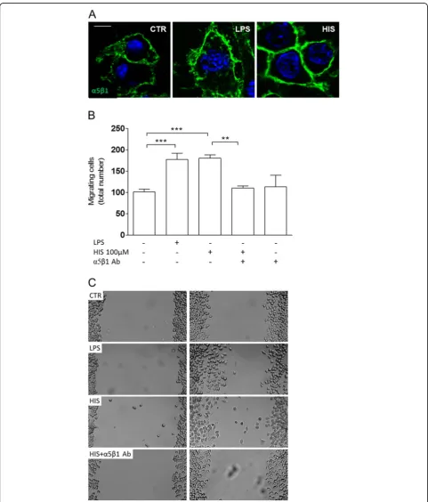
formation of adhesion sites to the substrate [21,22]. For this purpose, we immunolabeled alpha5/beta1 (α5β1) in-tegrin and observed that upon LPS challenge or hista-mine treatment, N9 microglial cells displayed a robust
α5β1 integrin expression (in green) on their cell surface (Figure 3). Accordingly, when we blocked this integrin by
adding 10 ug/ml of the anti-α5β1
integrin-neutralizing antibody, histamine-induced motility was compromised (meanHIS100μM= 174.4 ± 5.9 cells; meanHIS
+α5β1= 110.3 ± 5.3 cells; p<0.01, n= 3-13) (Figure 3B).
The neutralizing antibody against α5β1 alone did not
have any effect. Representative digital images depict the
histamine-induced migratory effect promoted by α5β1
integrin (Figure 3C). Other integrins, such as the integrin
α6 subunit, which forms a heterodimer with integrin β1
or β4, and recognizes laminin as a ligand, was not
involved in histamine-induced motility, as tested using a neutralizing antibody directed against α6 subunit (data not shown). Therefore, the integrin α5β1 is involved in the pro-migratory effect induced by histamine in the N9 cell line.
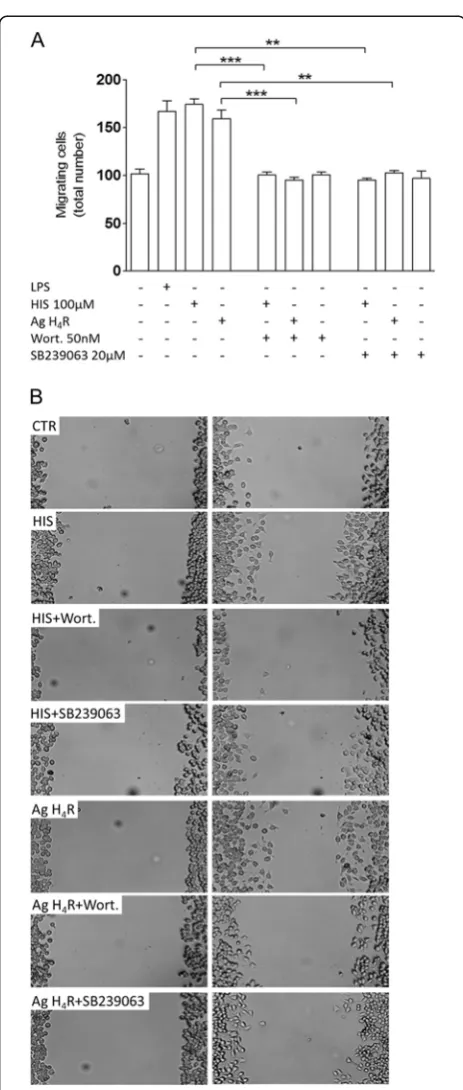
Histamine-induced migration requires p38 and AKT signaling pathways
We also addressed which signaling pathways could be involved in our migration model (Figure 4). Given that we have previously demonstrated the involve-ment of p38 MAPK signaling in LPS-induced migra-tion [16] and that the PI3K/Akt pathway has been correlated with H4R-stimulated mast cell chemotaxis [23] and with microglia migration [24,25], we evalu-ated the effect of SB239063 (p38 inhibitor) and wort-mannin (Akt inhibitor) on histamine-induced motility. Accordingly, in the presence of 50 nM wortmannin, both histamine- and H4R agonist-induced migration
were abolished (meanHIS+wort= 100.4 ± 3.4 cells;
meanH4R ag+wort= 95.1 ± 3.1 cells; p<0.001, n= 3)
(Figure 4A). Similarly, pre-treatment with 20 μM
SB239063 inhibited both histamine- and H4R
agonist-induced migration (meanHIS+SB239063= 95.0 ± 2.1 cells;
meanH4R ag+SB239063= 102.8 ± 2.4 cells; p<0.01, n= 3) (Figure 4A). Neither wortmannin nor SB239063 alone modulated microglia migration. These data suggest that the p38 and AKT signaling pathways can be involved in the migration induced by histamine or H4R agonist per se.
Histamine H4receptor activation modulates LPS-induced
microglia motility
We then decided to evaluate the role of histamine in an inflammatory context and found that, upon LPS and his-tamine co-administration, migration induced by LPS alone was significantly inhibited (meanLPS= 167.0 ± 11.2 cells, meanHIS+LPS= 112.0 ± 5.7cells; p<0.001, n= 7-13)
Figure 4Histamine-induced migration requires p38 and Akt signaling pathways.(A) In scratch wound assays, both histamine-and H4R agonist-induced migration were inhibited in the presence of
(Figure 5A). The co-administration of histamine to-gether with LPS did not induce cell death or prolifera-tion (data not shown). Moreover, cells treated with both histamine and LPS and in the presence of the H4R an-tagonist displayed a motility rate similar to LPS alone-treated microglia. The inhibition of LPS-induced migra-tion by histamine is mediated by H4R activamigra-tion, since in the presence of H4R antagonist, the stimulatory effect
on migration is restored (meanHIS+LPS+H4R ant= 160.8 ±
11.2 cells;n= 3-15;p<0.05,n= 4). Likewise, application of H4R agonist significantly reduced LPS-induced motil-ity (meanH4R Ag+LPS= 114.0 ± 7.7 cells; p<0.01, n= 4). Accordingly, none of the other receptor antagonists interfered with the ability of histamine to reduce LPS-stimulated migration (Figure 5A). Notably,
his-tamine-loaded microparticles (10 μg/ml) also
inhib-ited LPS-induced motility (meanuP10μg/ml
+LPS= 91.3 ± 22.5 cells; p<0.001, n= 4). Representa-tive digital images depict the inhibitory effect of
his-tamine on LPS-induced motility through H4R
activation (Figure 5B).
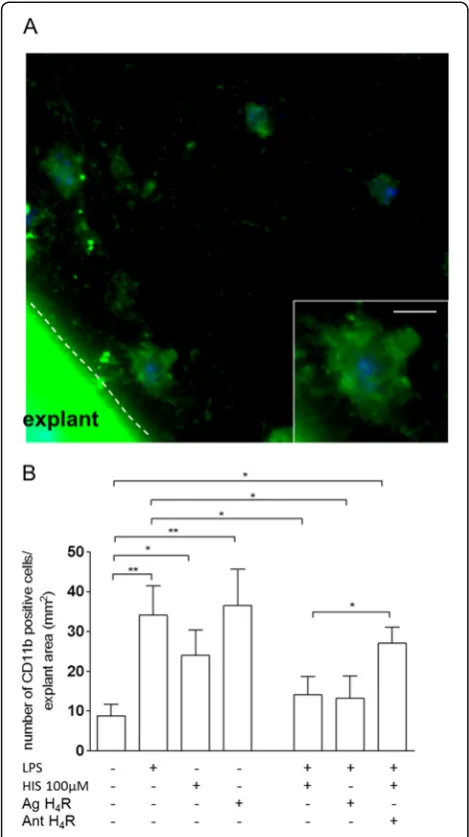
We later explored the role of histamine in a more complex biological model that closely resembles the physiological environment by using murine cortex explants (Figure 6). Similarly to the in vitro cell line model, we observed that histamine alone, acting through H4R, or LPS alone stimulated migration of microglia/
CNS invading macrophages (meanCTR= 8.9 ± 2.8 cells;
meanHIS= 24.0 ± 6.5cells; meanH4R ag= 36.5 ± 9.1 cells; meanLPS= 34.1 ± 7.4 cells; p<0.05, p<0.01, n= 5-9). Moreover, exposure of explants to histamine or H4R agonist, together with LPS, reduced migration, whereas blockade of H4R, in this context, restored migration to levels similar to the ones observed in LPS-treated cells (meanHIS+LPS= 14.2 ± 4.5 cells; meanH4R ag+LPS= 13.2 ± 5.6 cells; meanHIS+H4R ant+LPS= 27.0 ± 3.9 cells; p<0.05, n= 5-8) (Figure 6B). Altogether, these data suggest that histamine, in the presence of a robust inflammatory mi-lieu induced by LPS, acting via H4R activation, drastic-ally reduced microglia migration.
Application of the H4R antagonist restored LPS-stimu-lated motility in the presence of histamine. Data are expressed as mean ± SEM (n= 4-9) and as total number of migrating CD11b-positive cells per explant area (number of migrating CD11b-positive cells/mm2) (*p<0.05, using Bonferroni’s mu comparison test). Scale bar, 10μm.
Histamine modulates proinflammatory cytokine release Another key feature of inflammation is the release of
sig-naling proinflammatory cytokines, such as IL-1β and
tumor necrosis factor-alpha (TNF-α), by microglial cells [26].
Using a quantitative method, ELISA, we analyzed
the release of IL-1β and TNF-α by N9 microglial
Figure 5LPS-induced motility is inhibited by histamine via H4
receptor activation.(A) Upon an inflammatory challenge triggered by LPS (100 ng/ml), 100μM histamine inhibited migration, an effect involving H4R activation. In the presence of histamine, only when
H4R was blocked (Ant H4R: JNJ7777120, 5μM), LPS-induced
migration was restored. (B) Representative photomicrographs depict the inhibitory effect of histamine on LPS-induced motility upon H4R
cell line (Figure 7A) and by hippocampal organoty-pic slice cultures (Figure 7B). Regarding the cell line, we observed that in the presence of LPS, there was a significant release of biologically active IL-1β(mature
form) to the culture media (meanCTR= 138.5 ± 28.3
pg, meanLPS= 410.5 ± 96.2 pg; p<0.01, n= 4-7)
(Figure 7A,left graph). LPS-induced release of IL-1β
was abolished when cells were simultaneously treated
with histamine and LPS (meanHIS+LPS= 119.0 ± 28.0
pg;p<0.01,n= 3). Application of the H4R agonist (20
μM) together with LPS mimicked the inhibitory effect
of histamine, whereas blockade of H4R (5 μM)
abol-ished histamine-induced inhibition of IL-1β release
(meanH4R ag= 72.1 ± 43.8 pg, meanH4R ag+LPS= 101.5 ±
64.4 pg, meanHIS+H4R ant+LPS= 534.1 ± 119.4 pg; p
<0.01, p<0.001, n= 3). Noteworthily, application of
histamine-loaded microparticles (10 μg/ml) had the
same inhibitory effect of histamine under an
inflam-matory challenge (meanuP10μg/ml+LPS= 142.7 ± 29.7
cells;p<0.05,n= 3).
Interestingly, the inhibitory effect of histamine was not
observed over LPS-induced TNF-α release (Figure 7A,
right graph). LPS challenge stimulated TNF-α release; however, histamine or histamine-loaded microparticles had no effect on the release of this cytokine induced by LPS (Figure 7A,right graph).
In hippocampal organotypic slice cultures, LPS signifi-cantly increased IL-1β release (meanCTR= 17.6 ± 5.5 pg/ ml; meanLPS= 200.7 ± 37.4 pg/ml; p<0.001, n= 8-11) (Figure 7B, left graph). However, in the presence of his-tamine or H4R agonist, LPS-induced release was mark-edly reduced (meanHIS+LPS= 41.3 ± 4.2 pg/ml; meanH4R ag
+LPS= 37.1 ± 1.7 pg/ml; p<0.001, n= 4). Further enhan-cing the involvement of H4R on this effect, the use of a H4R antagonist partially restored the full LPS-induced IL-1βrelease (meanHIS+H4R ant+LPS= 68.0 ± 9.4 pg/ml; p <0.05, n= 4) (Figure 7B, left graph). Histamine-loaded
microparticles did not decrease LPS-induced IL-1β
re-lease in this experimental model (data not shown). Since using microwell inserts, where organotypic slices are placed, does not allow an efficient interaction with microparticles (particles will tend to deposit on the bot-tom of the well), we did not pursue their application further. In accordance with the previous data shown in the N9 cell line, in hippocampal organotypic slice
cul-tures, TNF-α release remained unaffected by histamine
treatment (Figure 7B, right graph). LPS challenge
sig-nificantly increased TNF-α release, but histamine did
not reduce this effect.
Discussion
Historically, histamine has been mainly addressed as an important mediator of allergic reactions occurring in per-ipheral tissues. In recent years, with the discovery of new histamine receptors and new sources of histamine in the brain, it has become clear that histamine has an increas-ingly defined role in the CNS. Regarding brain function, histamine is involved in the modulation of biological rhythms, sensory and motor systems, thermoregulation, learning and memory, mood and feeding behavior [27]. However, little is known about the role of histamine in brain inflammation. Most importantly, although microglial cells have been identified as a source of histamine, there
Figure 6Histamine promotes microglia/CNS macrophage migration via H4receptor activation in cortex explants. (A) Representative confocal microscopy micrographs depict CD11b-positive cells (ingreen) migrating from and within the explant, in the presence of LPS treatment (100 ng/ml).Insethighlights microglia/ CNS macrophage morphology.(B)LPS and histamine individually increased motility, while co-administration abolished this effect. Likewise, in the presence of H4R agonist, LPS-induced motility was
are few reports on how the activity/function of these cells is modulated by this amine. As such, given the active immunoregulatory role of microglial cells in the brain par-enchyma, we sought to decipher the modulatory actions of histamine over classical microglial responses, such as migration and inflammatory mediator release. In our
study, we demonstrated that histamine, acting via H4R, showed dual effects on microglia-induced responses. His-tamine per se stimulated microglia motility, as compared with untreated controls, and this migratory effect requires the expression of α5β1 integrin and occurs with the in-volvement of p38 MAPK and Akt signaling pathways. This
Figure 7Histamine inhibits IL-1βrelease induced by LPS administration in N9 microglial cells (a) and in hippocampal organotypic slice cultures (b) measured by ELISA.(a) N9 microglial cells, upon inflammatory challenge induced by LPS (100 ng/ml), are able to release a significantly higher amount of proinflammatory cytokine IL-1βwhen compared to untreated cells. Histamine treatment (100μM) significantly inhibited this effect through the activation of H4R. Accordingly, histamine-loaded microparticles (μP 10μg/ml) mimicked the anti-inflammatory
effect of histamine (left graph). The anti-inflammatory action of histamine appears to modulate specifically the IL-1βsignaling pathway, since it does not alter the release of another relevant proinflammatory cytokine, such as TNF-α, in the presence of 100 ng/ml LPS. Additionally, the application of histamine-loaded microparticles (10μg/ml) did not inhibit LPS-induced TNF-αrelease (right graph). (b) Regarding hippocampal organotypic slice cultures, only soluble histamine significantly decreased IL-1βrelease induced by LPS. Again, H4R activation also mimicked the
effect of histamine, whereas H4R blockade partially recuperated LPS-induced IL-1βrelease (right graph). Data are expressed as mean ± SEM
effect suggests that histamine alone may function similarly to an inflammatory mediator, although it does not change the release of the cytokines IL-1βand TNF-α.
Importantly, it should be noted that microglial activa-tion, a common feature of most brain pathologies, can be coupled to either a pro- or anti-inflammatory profile and exhibits several functionally distinct phenotypes. More-over, since microglia continually surveys their micro-environment, migration cannot be univocally associated with a proinflammatory setting (as reviewed by [28]).
Of note, given an inflammatory context, such as our experimental paradigm mimicked by the administration of LPS, histamine inhibited microglial migration to levels similar to control cultures. Moreover, histamine also inhibited LPS-induced IL-1βrelease, although it did not interfere with LPS-induced TNF-αrelease. Therefore, in the presence of a strong and robust inflammatory con-text, histamine may repress microglia-mediated migra-tion and counteract addimigra-tional tissue damage.
To our knowledge, we are the first to report the detec-tion of H4R expression in both primary microglia cul-tures from the cerebral cortex and in a microglia cell line. To evaluate the role of histamine on microglial cell migration, we adopted two experimental approaches, with different degrees of complexity: scratch wound assays and murine cortex explants. The use of scratch wound assays allowed a complete pharmacological study of the dual effect of histamine on cell migration, while cortex explants offered a more physiological
environ-ment. Using these models, we observed that 100μM
his-tamine and 10 μg/ml histamine-loaded microparticles
stimulated microglia migration. Histamine-loaded
microparticles can promote a more efficient and con-trolled delivery of histamine, without cytotoxic effects [13].
Moreover, we determined that this pro-migratory ef-fect induced by histamine was occurring through H4R activation. H4R is expressed mainly by immune cells whose receptor activation modulates migration or cell recruitment, calcium mobilization, cell differentiation and cytokine production, depending on the cell type. In particular, H4R induces chemotaxis of eosinophils, mast cells, and dendritic and T cells, while reducing monocyte recruitment (reviewed in [8]). In response to injury or inflammation, microglial cells become activated and mi-grate in a process that requires actin polymerization and the upregulation of adhesion molecule CD11b, among other adhesion molecules. In eosinophils, low concentra-tions of histamine (below 10 mM) lead to actin polymerization and significant CD11b upregulation, an effect blocked by thioperamide, an H3R/H4R antagonist [29]. Another key component required for cell move-ment is the expression of integrins, a diverse family of migration-inducing receptors, which are responsible for
cell-cell, cell-extracellular matrix (ECM) and cell-patho-gen interactions [22,30]. Integrins are heterodimeric complexes composed by different combinations of alpha (α)/beta (β) subunits, which later define receptor specifi-city.β1 integrin is the most widespread βsubunit, and it is involved in microglia chemotaxis and proliferation [31]. In our work, we showed that α5β1 integrin block-ade impaired histamine-induced migration, suggesting that this heterodimer is required for microglia migration. We should note that scratch wound assays were per-formed without any substrate covering the bottom of the wells. However, microglial cells are able to secrete fibronectin, an α5β1 ligand (assessed by Western blot-ting; data not shown), therefore enabling cell movement. We further disclosed the mechanisms involved in hista-mine-induced migration by evaluating the participation of p38 mitogen-activated protein kinase and Akt signal-ing pathways. We have previously demonstrated that LPS-induced microglia migration requires p38 phosphor-ylation [16]. In addition, H4R activation may rapidly and transiently induce the phosphorylation of ERK, MEK and Akt in other immune cell types [23-25]. Nevertheless, using selective inhibitors of these pathways, SB239063 (p38 inhibitor) and wortmannin (Akt inhibitor), we blocked histamine-induced migration, suggesting that these pathways are required for cell movement.
To the best of our knowledge, there is only one report suggesting crosstalk between alpha5beta1 integrin expres-sion and p38/Akt pathways in cell migration. α5β1 and
αvβ3 integrin-mediated human umbilical vein endothelial cell (HUVEC) adhesion to fibronectin or vitronectin acti-vates integrin-dependent intracellular signaling cascades, including PI3K/AKT, ERK, p38 and JNK, which subse-quently lead to the stimulation of AP-1-dependent MMP-9 expression in HUVECs. However, the authors only showed that blocking antibodies targetingα5β1 andαvβ3 integrins abolished fibronectin-stimulated c-Jun phosphorylation, while blocking antibodies targeting β1 and αvβ3 reduced vitronectin-stimulated MMP-9 activity [32]. Nevertheless, several studies showed some evidence of crosstalk among integrins/MAPKs/Akt in cell invasion, although invasion and migration are different processes since the latter does not necessarily require invasion to occur [33-37].
observed the same inhibitory action of histamine in the presence of an inflammatory challenge using this model. In that sense, H4R may have therapeutic value in the treat-ment of inflammatory conditions or symptoms, although histamine has been mainly regarded as a proinflammatory agent. In fact, Smits and colleagues have designed and eval-uated the role of several H4R ligands. The group found that 6,7-dichloro-3-(4-methylpiperazin-1-yl)quinoxalin-2(1 H)-one and 2-benzyl-3-(4-methyl-piperazin-1-yl)quinoxaline (two H4R agonists) displayed significant in vivo anti-inflam-matory activity in the rat carrageenan-induced paw edema model [38], validating the use of H4R ligands as anti-inflam-matory agents in vivo. Furthermore, application of H4R agonists has provided a reduction of asthma-like symptoms due to enhanced migration of CD4+/CD25+/FoxP3+ T regulatory cells (Tregs) to the inflammation site, where these accumulated cells release the anti-inflammatory cyto-kine IL-10 [39]. Additionally, Osna and colleagues reported that histamine upregulates IL-10 production by murine splenocytes in a dose-dependent manner; however, this ef-fect was reversed by both H1- and H2-receptor antagonists [40]. In addition, H4R activation prevents the development of reperfusion injury in a model of ischemia-induced liver damage [41]. Hence, our work extends the knowledge of H4R activity and supports new perspectives on the use of H4R agonists in a pathological context. Moreover, micro-particle-mediated delivery of histamine or H4R agonists might provide a new opportunity for the treatment of vari-ous CNS disorders accompanied by microglia-derived in-flammation. Nevertheless, it should be noted that the majority of reports refer to the anti-inflammatory actions of H4R antagonists in conditions such as pruritus, dermatitis, airway inflammation and arthritis [8].
Cytokine signaling is a particularly relevant feature in the local development of the inflammatory reaction, but also in the recruitment of immune cells through the upre-gulation of adhesion molecules and induction of chemo-kines. In our study we evaluated the role of the
pro-inflammatory cytokines IL-1β and TNF-α in
histamine-modulated migration. We have previously described the involvement of IL-1β signaling in LPS-induced microglia migration in which we observed that blocking the IL-1 re-ceptor led to the loss of the LPS pro-migratory effect [16]. However, IL-1 receptor blockade does not impair hista-mine-induced migration, suggesting the involvement of different migration mechanisms. Cells pre-treated with IL-1ra (150 ng/ml) and incubated with histamine were still able to migrate, although not entirely similarly to hista-mine alone (meanHIS + IL-1ra = 148 ± 5.8 cells; mean-HIS = 174.4 ± 5.9 cells, n= 3; data not shown), suggesting that histamine does not require IL-1βsignaling to induce migration, but affects its release, possibly to control a cytotoxic effect caused by the IL-1βrelease induced by a strong LPS inflammatory stimulus [16]. Interestingly,
histamine per se does not modulate IL-1β release;
how-ever, it inhibited IL-1βrelease upon LPS challenge, an ef-fect mimicked by H4R agonist. In organotypic slice cultures, which include more complex cellular interactions and different cell types (including neurons, astrocytes, oli-godendrocytes and microglia), H4R blockade did not fully restore LPS-induced IL-1βrelease in the presence of hista-mine, suggesting the involvement of other receptors or different modulation mechanisms by other types of cells present in the slices. Our results suggest that in a healthy brain parenchyma, exogenous application of histamine would enhance the microglia surveillance, whereas, in an inflammatory context, histamine would restrain microglia motility while maintaining a putative anti-inflammatory profile.
Importantly, it has been reported that in IL-1 receptor 1 (IL-1R1) null mice microglia activation is abrogated when stab wounds are performed. The authors observed that in the absence of IL-1R1, almost no reactive or ameboid microglia are found, and leukocyte infiltration is very reduced. Also, in these knockout mice, the pro-inflammatory cytokine expression was decreased [42].
However, we did not observe TNF-αrelease upon
hista-mine or histahista-mine microparticles in both experimental models used.
Regarding TNF-α release, histamine has been
described to inhibit LPS-stimulated TNF-α release by
human monocytes, human and rat alveolar macro-phages, and human peripheral blood mononuclear cells [43-45]. In the studies performed by Sirois and colleagues [44] and by Morichika and colleagues [45],
the LPS concentration ranged from 1–5 ng/ml, and
the histamine concentration ranged from 10-4-10-7 M;
their co-administration inhibited LPS-induced TNF-α
release. However, in the study developed by Rowe and colleagues [43], human alveolar macrophages and monocytes differed in their responses to histamine.
Histamine (10-5 M) inhibited LPS-stimulated (500 ng/
ml) TNF-α release by monocytes, but had no effect
on alveolar macrophages. These results suggest that the inhibitory effect of histamine on LPS-induced
TNF-α release may depend on the LPS concentration
(we used 100 ng/ml) and on cell type. Noteworthily, work performed by Desai and Thurmond [23] showed that histamine and LPS co-treatment potentiated IL-6 release by mast cells, although it had no effect on
TNF-α release, suggesting that, depending on the
Conclusions
Our results open promising new perspectives for the therapeutic use of histamine and histamine receptor ago-nists to treat or ameliorate inflammation-associated pro-cesses. Histamine has been perceived as a major inflammatory mediator in allergic responses. In accord-ance, we showed that histamine can trigger microglia motility per se, but not the release of the
pro-inflamma-tory cytokines IL-1β and TNF-α. Most importantly, we
have now revealed new anti-inflammatory properties of
histamine H4 receptor agonists that counteract
LPS-mediated inflammatory actions, namely
motility/migra-tion and IL-1β release by microglia/CNS invading
macrophages. Therefore, the use of histamine-loaded microparticles (or ultimately, microparticles loaded with H4R agonists) could provide a rapid, more effi-cient and affordable approach in an inflammatory context.
Abbreviations
ELISA: Enzyme-linked Immunosorbent Assay; H4R: Ristamine Receptor 4;
IL-1β: Interleukin-1beta; LPS: Lipopolysaccharide; MAPK: Mitogen-activated Protein Kinase; qPCR: quantitative Polymerase Chain Reaction; TLR4: Toll-like Receptor 4; RT: Room Temperature; TNF-α: Tumor Necrosis Factor-alpha.
Competing interests
This work is under patent protection (patent application no. 20111000054869).
Acknowledgements
The authors wish to thank Prof. Claudia Verderio from the CNR Institute of Neuroscience and Department of Medical Pharmacology, University of Milan, Italy, for her generous gift of the murine N9 microglial cell line and Prof. Paulo Santos from the Center for Neuroscience and Cell Biology, University of Coimbra, Portugal. This work was supported by FCT Portugal and FEDER, PTDC/SAU-NEU/104415/2008 and PTDC/SAU-NEU/101783/2008, grant no. 96542, from the Calouste Gulbenkian Foundation and L'Oréal-UNESCO Portugal forWomen in Science,MIT-Portugal and PTDC/CTM/099659/2008.
Author details
1CNC - Center for Neuroscience and Cell Biology, University of Coimbra,
Coimbra, Portugal.2Laboratory of Pharmacology and Experimental
Therapeutic, Faculty of Medicine, University of Coimbra, Coimbra, Portugal.
3Institute of Biomedical Research on Light and Image (IBILI), Faculty of
Medicine, University of Coimbra, Coimbra, Portugal.4CICS-UBI - Health
Sciences Research Center, University of Beira Interior, Covilhã, Portugal.
5
Biocant, Centro de Inovação em Biotecnologia, Cantanhede, Portugal.6 CICS-UBI - Health Sciences Research Center, Av. Infante D. Henrique, University of Beira Interior, Covilhã 6200-506, Portugal.
Authors’contributions
RF performed the scratch wound assays, cortex explants, real-time PCR studies, Western blotting, ELISA measurements for IL-1β,
immunocytochemistry studies, statistical analysis and wrote the manuscript. TS participated in the scratch wound assays and cortex explants, real-time PCR studies and acquisition of confocal images. JG performed the ELISA measurements for TNF-α. GB performed the primary microglia cultures. LF developed the PLGA microparticles and participated in coordination of the study. FA participated in the project design, provided financial support and coordinated the study. LB conceived the study, participated in its design, performed primary microglial cultures and organotypic slice cultures, wrote the manuscript, provided financial support and coordinated the project. All authors read and approved the manuscript.
Received: 25 January 2012 Accepted: 8 May 2012 Published: 8 May 2012
References
1. Garden G, Möller T:Microglia biology in health and disease.J Neuroimmune Pharmacol2006,1:127–137.
2. Block ML, Zecca L, Hong J-S:Microglia-mediated neurotoxicity: uncovering the molecular mechanisms.Nat Rev Neurol2007,8:57–69. 3. Rivest S:Molecular insights on the cerebral innate immune system.Brain
Behav Immun2003,17:13–19.
4. Cohen J:The immunopathogenesis of sepsis. Nature2002,420:885–891. 5. Kinet JP:The high-affinity IgE receptor (Fc epsilon RI): from physiology to
pathology.Annu Rev Immunol1999,17:931–972.
6. Katoh Y, Niimi M, Yamamoto Y, Kawamura T, Morimoto-Ishizuka T, Sawada M, Takemori H, Yamatodani A:Histamine production by cultured microglial cells of the mouse.Neurosci Lett2001,305:181–184. 7. Dy M, Schneider E:Histamine-cytokine connection in immunity and
hematopoiesis.Cytokine Growth Factor Rev2004,15:393–410.
8. Zampeli E, Tiligada E:The role of histamine H4 receptor in immune and inflammatory disorders.Br J Pharmacol2009,157:24–33.
9. de Esch IJ, Thurmond RL, Jongejan A, Leurs R:The histamine H4 receptor as a new therapeutic target for inflammation.Trends Pharmacol Sci2005, 26:462–469.
10. Ferreira R, Xapelli S, Santos T, Silva AP, Cristovao A, Cortes L, Malva JO: Neuropeptide Y modulation of interleukin-1 beta (IL-1β)-induced nitric oxide production in microglia.J Biol Chem2010,285:41921–41934. 11. Saura J, Tusell JM, Serratosa J:High-yield isolation of murine microglia by
mild trypsinization.Glia2003,44:183–189.
12. Ferreira LS, Gerecht S, Fuller J, Shieh HF, Vunjak-Novakovic G:Bioactive hydrogel scaffolds for controllable vascular differentiation of human embryonic stem cells.Biomaterials2007,28:2706–2717.
13. Bernardino L, Eiriz MF, Santos T, Xapelli S, Grade S, Rosa A, Cortes L, Ferreira R, Bragança J, Agasse F,et al:Histamine stimulates neurogenesis in the rodent subventricular zone.Stem Cells2012,30:773–784.
14. Valster A, Tran NL, Nakada M, Berens ME, Chan AY, Symons M:Cell migration and invasion assays.Methods2005,37:208–215.
15. Ferreira R, Santos T, Viegas M, Cortes L, Bernardino L, Vieira OV, Malva JO: Neuropeptide Y inhibits interleukin-1beta-induced phagocytosis by microglial cells.J Neuroinflammation2011,8:169.
16. Ferreira R, Santos T, Cortes L, Cochaud S, Agasse F, Silva AP, Xapelli S, Malva JO:Neuropeptide Y inhibits interleukin-1 beta (IL-1β)-induced microglia motility.J Neurochem2012,120:93–105.
17. Bustin S:Absolute quantification of mRNA using real-time reverse transcription polymerase chain reaction assays.J Mol Endocrinol2000, 25:169–193.
18. Strakhova MI, Nikkel AL, Manelli AM, Hsieh GC, Esbenshade TA, Brioni JD, Bitner RS:Localization of histamine H4 receptors in the central nervous system of human and rat.Brain Res2009,1250:41–48.
19. Connelly WM, Shenton FC, Lethbridge N, Leurs R, Waldvogel HJ, Faull RL, Lees G, Chazot PL:The histamine H4 receptor is functionally expressed on neurons in the mammalian CNS.Br J Pharmacol2009,157:55–63. 20. Vetvicka V, Hanikyrova M, Vetvickova J, Ross GD:Regulation of CR3
(CD11b/CD18)-dependent natural killer (NK) cell cytotoxicity by tumour target cell MHC class I molecules.Clin Exp Immunol1999, 115:229–235.
21. Fletcher DA, Mullins RD:Cell mechanics and the cytoskeleton.Nature
2010,463:485–492.
22. Parsons JT, Horwitz AR, Schwartz MA:Cell adhesion: integrating cytoskeletal dynamics and cellular tension.Nat Rev Mol Cell Biol2010,11:633–643. 23. Desai P, Thurmond RL:Histamine H receptor activation enhances
LPS-induced IL-6 production in mast cells via ERK and PI3K activation.Eur J Immunol2011,41:1764–1773.
24. Horvath RJ, DeLeo JA:Morphine enhances microglial migration through modulation of P2X4 receptor signaling.J Neurosci2009, 29:998–1005.
25. Martin S, Vincent JP, Mazella J:Involvement of the neurotensin receptor-3 in the neurotensin-induced migration of human microglia.J Neurosci
2003,23:1198–1205.
27. Haas HL, Sergeeva OA, Selbach O:Histamine in the nervous system.
Physiol Rev2008,88:1183–1241.
28. Perry VH, Nicoll JAR, Holmes C:Microglia in neurodegenerative disease.
Nat Rev Neurol2010,6:193–201.
29. Buckland KF, Williams TJ, Conroy DM:Histamine induces cytoskeletal changes in human eosinophils via the H(4) receptor.Br J Pharmacol2003, 140:1117–1127.
30. Ridley AJ, Schwartz MA, Burridge K, Firtel RA, Ginsberg MH, Borisy G, Parsons JT, Horwitz AR:Cell migration: integrating signals from front to back.
Science2003,302:1704–1709.
31. Nasu-Tada K, Koizumi S, Inoue K:Involvement of beta1 integrin in microglial chemotaxis and proliferation on fibronectin: different regulations by ADP through PKA.Glia2005,52:98–107.
32. Jin YJ, Park I, Hong IK, Byun HJ, Choi J, Kim YM, Lee H:Fibronectin and vitronectin induce AP-1-mediated matrix metalloproteinase-9 expression through integrin alpha(5)beta(1)/alpha(v)beta(3)-dependent Akt, ERK and JNK signaling pathways in human umbilical vein endothelial cells.
Cell Signal2011,23:125–134.
33. Hague A, Jones GE:Cell motility assays.Cell Biol Toxicol2008,24:381–389. 34. Matsuo M, Sakurai H, Ueno Y, Ohtani O, Saiki I:Activation of MEK/ERK and
PI3K/Akt pathways by fibronectin requires integrin alphav-mediated ADAM activity in hepatocellular carcinoma: a novel functional target for gefitinib.Canc Sci2006,97:155–162.
35. Wang HQ, Bai L, Shen BR, Yan ZQ, Jiang ZL:Coculture with endothelial cells enhances vascular smooth muscle cell adhesion and spreading via activation of beta1-integrin and phosphatidylinositol 3-kinase/Akt.Eur J Cell Biol2007,86:51–62.
36. Zhu H, Liu XW, Cai TY, Cao J, Tu CX, Lu W, He QJ, Yang B:Celastrol acts as a potent antimetastatic agent targeting beta1 integrin and inhibiting cell-extracellular matrix adhesion, in part via the p38 mitogen-activated protein kinase pathway.J Pharmacol Exp Ther2010,334:489–499. 37. Furundzija V, Fritzsche J, Kaufmann J, Meyborg H, Fleck E, Kappert K, Stawowy P:
IGF-1 increases macrophage motility via PKC/p38-dependentαvβv3-integrin inside-out signaling.Biochem Biophys Res Commun2010,394:786–791. 38. Smits RA, Lim HD, Hanzer A, Zuiderveld OP, Guaita E, Adami M, Coruzzi G,
Leurs R, de Esch IJ:Fragment based design of new H4 receptor-ligands with anti-inflammatory properties in vivo.J Med Chem2008,51:2457–2467. 39. Morgan RK, McAllister B, Cross L, Green DS, Kornfeld H, Center DM,
Cruikshank WW:Histamine 4 receptor activation induces recruitment of FoxP3+ T cells and inhibits allergic asthma in a murine model.J Immunol
2007,178:8081–8089.
40. Osna N, Elliott K, Khan MM:Regulation of interleukin-10 secretion by histamine in TH2 cells and splenocytes.Int Immunopharmacol2001,1:85–96. 41. Adachi N, Liu K, Motoki A, Nishibori M, Arai T:Suppression of ischemia/
reperfusion liver injury by histamine H4 receptor stimulation in rats.Eur J Pharmacol2006,544:181–187.
42. Basu A, Krady JK, O'Malley M, Styren SD, DeKosky ST, Levison SW:The type 1 interleukin-1 receptor is essential for the efficient activation of microglia and the induction of multiple proinflammatory mediators in response to brain injury.J Neurosci2002,22:6071–6082.
43. Rowe J, Finlay-Jones JJ, Nicholas TE, Bowden J, Morton S, Hart PH:Inability of histamine to regulate TNF-alpha production by human alveolar macrophages.Am J Respir Cell Mol Biol1997,17:218–226.
44. Sirois J, Menard G, Moses AS, Bissonnette EY:Importance of histamine in the cytokine network in the lung through H2 and H3 receptors: stimulation of IL-10 production.J Immunol2000,164:2964–2970. 45. Morichika T, Takahashi HK, Iwagaki H, Yoshino T, Tamura R, Yokoyama M,
Mori S, Akagi T, Nishibori M, Tanaka N:Histamine inhibits
lipopolysaccharide-induced tumor necrosis factor-alpha production in an intercellular adhesion molecule-1- and B7.1-dependent manner.J Pharmacol Exp Ther2003,304:624–633.
doi:10.1186/1742-2094-9-90
Cite this article as:Ferreiraet al.:Histamine modulates microglia function.Journal of Neuroinflammation20129:90.
Submit your next manuscript to BioMed Central and take full advantage of:
• Convenient online submission
• Thorough peer review
• No space constraints or color figure charges
• Immediate publication on acceptance
• Inclusion in PubMed, CAS, Scopus and Google Scholar
• Research which is freely available for redistribution