R E S E A R C H
Open Access
Macrophages inhibit human osteosarcoma cell
growth after activation with the bacterial cell
wall derivative liposomal muramyl tripeptide in
combination with interferon-
γ
Jens HW Pahl
1,6, Kitty MC Kwappenberg
1,6, Eleni M Varypataki
2, Susy J Santos
1,6, Marieke L Kuijjer
3,
Susan Mohamed
1,6, Juul T Wijnen
4, Maarten JD van Tol
1,6, Anne-Marie Cleton-Jansen
3, R Maarten Egeler
5,
Wim Jiskoot
2, Arjan C Lankester
1,6and Marco W Schilham
1,6*Abstract
Background:In osteosarcoma, the presence of tumor-infiltrating macrophages positively correlates with patient survival in contrast to the negative effect of tumor-associated macrophages in patients with other tumors. Liposome-encapsulated muramyl tripeptide (L-MTP-PE) has been introduced in the treatment of osteosarcoma patients, which may enhance the potential anti-tumor activity of macrophages. Direct anti-tumor activity of human macrophages against human osteosarcoma cells has not been described so far. Hence, we assessed osteosarcoma cell growth after co-culture with human macrophages.
Methods:Monocyte-derived M1-like and M2-like macrophages were polarized with LPS + IFN-γ, L-MTP-PE +/− IFN-γor IL-10 and incubated with osteosarcoma cells. Two days later, viable tumor cell numbers were analyzed. Antibody-dependent effects were investigated using the therapeutic anti-EGFR antibody cetuximab.
Results:M1-like macrophages inhibited osteosarcoma cell growth when activated with LPS + IFN-γ. Likewise, stimulation of M1-like macrophages with liposomal muramyl tripeptide (L-MTP-PE) inhibited tumor growth, but only when combined with IFN-γ. Addition of the tumor-reactive anti-EGFR antibody cetuximab did not further improve the anti-tumor activity of activated M1-like macrophages. The inhibition was mediated by supernatants of activated M1-like macrophages, containing TNF-αand IL-1β. However, specific blockage of these cytokines, nitric oxide or reactive oxygen species did not inhibit the anti-tumor effect, suggesting the involvement of other soluble factors released upon macrophage activation. While LPS + IFN-γ–activated M2-like macrophages had low anti-tumor activity, IL-10–polarized M2-like macrophages were able to reduce osteosarcoma cell growth in the presence of the anti-EGFR cetuximab involving antibody-dependent tumor cell phagocytosis.
Conclusion:This study demonstrates that human macrophages can be induced to exert direct anti-tumor activity against osteosarcoma cells. Our observation that the induction of macrophage anti-tumor activity by L-MTP-PE required IFN-γmay be of relevance for the optimization of L-MTP-PE therapy in osteosarcoma patients.
Keywords:Macrophages, Muramyl tripeptide, IFN-γ, Osteosarcoma, Cetuximab
* Correspondence:M.W.Schilham@lumc.nl
1
Department of Pediatrics, Leiden University Medical Center, Leiden, the Netherlands
6
Laboratory for Immunology, P3-P, Department of Pediatrics, Leiden University Medical Center, PO Box 9600, 2300 RC Leiden, the Netherlands
Full list of author information is available at the end of the article
Introduction
Osteosarcoma is the most frequent malignant bone tumor in adolescents and young adults. Of patients with localized, non-metastatic disease, up to 70% achieve persistent remis-sion [1]. In contrast, prognosis of patients with advanced, metastatic and recurrent disease is as low as 20% despite in-tensive chemotherapy and surgery. Thus, novel therapies are needed, especially for patients with chemotherapy-resistant disease [2,3]. Recently, we have demonstrated that the presence of tumor-infiltrating macrophages at the time of diagnosis is positively correlated with a favorable outcome of patients with osteosarcoma [4]. Hence, target-ing tumor-associated macrophages in osteosarcoma with macrophage-activating agents is an attractive option to complement current anti-tumor treatments.
Macrophages are mononuclear phagocytic cells that are involved in homeostatic, pro-inflammatory and immune regulatory responses in the tissue [5,6]. While macrophages can originate from blood monocytes under inflammatory conditions, as in the classical model for macrophage development, it has recently been revealed that under non-inflammatory conditions tissue macrophages primarily originate from the yolk sac and fetal liver and are main-tained independently of hematopoietic precursors [7]. Macrophages possess great functional and phenotypic plasticity which is often simplified by classification in M1 and M2 phenotypes [8]. M1 macrophages are in-volved in host defense through their bactericidal and tumoricidal activity and pro-inflammatory cytokine produc-tion if ‘classically-activated’ by interferon-γ (IFN-γ) and Toll-like receptor ligands such as bacterial lipopolysacchar-ide (LPS) [9,10]. M2 macrophages can exhibit many differ-ent phenotypes in response to diverse stimuli such as polarization with interleukin-10 (IL-10) or LPS. M2 macro-phages are involved in scavenging cell debris and bacteria, antibody-dependent phagocytosis, tissue remodeling, angio-genesis, wound healing and immune regulation. In contrast to ‘classically-activated’ M1 macrophages, macrophages with an M2-like phenotype are often detected in solid tu-mors and considered to promote tumor progression [8-11].
Macrophages constitute the majority of tumor-infiltrating immune cells in solid tumors including osteosarcoma [4,12]. In most tumors, the presence of macrophages repre-sents an unfavorable prognostic factor [13]. In contrast, in osteosarcoma as well as colorectal cancer higher numbers of tumor-infiltrating macrophages correlate with better survival [4,14,15]. In osteosarcoma, there was no clear asso-ciation of good survival with an M1-like or M2-like pheno-typic polarization of macrophages [4].
Monocytes and macrophages activated with LPS have been implicated in anti-tumor responses for a long time [16-21]. But while canine macrophages have been reported to have anti-tumor activity against canine osteosarcoma cells, comparable evidence for anti-tumor activity of human
macrophages against human osteosarcoma cells is not available. The anti-tumor activity of canine macrophages was shown to be dependent on stimulation with LPS or another bacterial cell wall constituent, i.e. muramyl dipep-tide (MDP) or the lipophilic derivative muramyl tripepdipep-tide phoshatidylethanolamine (MTP-PE) [22]. Application of
liposome-encapsulated MTP-PE (L-MTP-PE)in vivo
im-proved survival of dogs with osteosarcoma [23]. This observation encouraged the addition of L-MTP-PE to the treatment of osteosarcoma patients as a macrophage-activating agent but did not increase event-free survival of non-metastatic or metastatic osteosarcoma patients [1,24]. Therefore, we set out to investigate the anti-tumor ac-tivity of human macrophages against human osteosar-coma cells and determine whether this activity can be
manipulated. We set up an in vitromodel in which the
effect of human macrophages on the growth of osteosar-coma cells can be directly assessed by counting residual tumor cells after a two-day co-culture with macrophages. Using this model we demonstrate how anti-tumor activity of M1-like macrophages and M2-like macrophages can be induced by bacterial stimuli like L-MTP-PE and the thera-peutic anti-EGFR antibody cetuximab, respectively.
Materials and methods
Cell lines
The osteosarcoma cell lines HOS, HOS-143b, OHS, OSA, SAOS-2 and U2OS were obtained from the EuroBoNeT cell line repository (2007) [25]. Cell line identity was con-firmed by short tandem repeat DNA fingerprinting in 2012. All cell lines were maintained in RPMI 1640 (Invitrogen, Carlsbad, CA, USA) supplemented with 10% fetal calf serum (Invitrogen) and 100 U/ml penicillin and 100 ug/ml streptomycin (Invitrogen). All cell lines were negative for mycoplasma infection as regularly tested by RT-PCR.
Preparation of liposomal MTP-PE
Liposomes (multi-lamellar vesicles) were prepared from a mixture of the synthetic phospholipids 1-palmitoyl-2-oleoyl-sn-glycero-3-phosphocholine (POPC, 850457P) and 1,2-dioleoyl-sn-glycero-3-phospho-L-serine (DOPS, 840035P) (both from Avanti Polar Lipids, Alabaster, Al, USA) at a 7:3 molar ratio in chloroform by mechanical agitation on a vortex mixer. MTP-PE (Mr1237.5 g/mol; Mifamurtide; Sigma-Aldrich, St. Louis, MO, USA) was dissolved in chloroform:methanol:water 60:36:4 (v/v/v).
5 mg of liposomes (Mr 775 g/mol) were loaded with
0.02 mg of MTP-PE (1:250 ratio). The organic solution was dried in a rotary evaporator under reduced pressure for one hour to obtain a dry lipid film. Afterwards, the lipid film was rehydrated in 2.5 ml sterile PBS, resulting in a final concentration of 6.45 nmol MTP-PE per
2 μmol/ml liposome preparation (L-MTP-PE). The
polycarbonate filter (Nuleopore). Empty control lipo-somes (L-PBS) were prepared by the same procedure ex-cept without MTP-PE addition. The z-average diameter of the liposomes was ~350 nm with a mean zeta poten-tial of−97 mV as measured on a Zetasizer (version 6.01) (Malvern Instruments, Worcestershire, UK).
Monocyte Isolation and differentiation to macrophages
PBMC were separated from buffy coats of healthy adult donors (Sanquin Blood bank, Region Southwest, Rotter-dam, the Netherlands) by Ficoll-Hypaque density gradient centrifugation. Monocytes were isolated from PBMC by positive selection using anti-CD14 MicroBeads (Miltenyi Biotech, Bergisch Gladbach, Germany). For M1-like and M2-like macrophage differentiation, monocytes (1,5 × 10⁶ per well per 3 ml of a 6-well tissue culture plate) were incu-bated with GM-CSF (80 ng/ml; Peprotech, Rocky Hill, NJ, USA) and M-CSF (20 ng/ml, R&D Systems, Minneapolis, MN, USA) for seven days as previously established [10,26]. In some conditions, M1-like and M2-like macrophages were additionally stimulated during the last day of differen-tiation with combinations of LPS (10 ng/ml; E. coli strain 0111:B4; Sigma-Aldrich), IFN-γ (100 U/ml; Boehringer, Mannheim, Germany), empty control liposomes (250 nmol) (L-PBS) or liposomes (250 nmol) containing MTP-PE (0.8 nmol, i.e., 1μg) (L-MTP-PE) per 3 ml culture medium. M2-like macrophages were alternatively stimulated with IL-10 (10 ng/ml; Peprotech) during the last two days of dif-ferentiation. The phenotype of macrophage populations was tested in each experiment. Macrophages were devoid of the monocyte-derived dendritic cell marker CD1a (data not shown).
Macrophage-tumor cell co-cultures
After seven-day differentiation, culture supernatants of macrophages were collected. Adherent macrophages were washed in cold PBS, detached by incubation in accutase (Sigma-Aldrich) for 30 min at 37°C and combined with the non-adherent cell fraction. Cell scraping of firmly ad-herent macrophages was avoided to maximize macro-phage viability. Macromacro-phages were seeded in 96-well flat-bottom plates in RPMI medium at 3,000 (cell conjugate formation assay) or 30,000 cells (tumor cell survival assay) per well (four wells per condition) and incubated for cell attachment. After two hours 3,000 osteosarcoma cells were added and macrophage-tumor cell co-cultures were incubated for two hours in cell conjugate formation assays at a 1:1 ratio in 50μl medium and for two hours, one day and two days in tumor cell survival assays at a 10:1 ratio
in 100 μl medium. In some experiments, tumor cells
were coated with the chimeric monoclonal antibody cetuximab (anti-epidermal growth factor receptor, 1μg/ml final concentration in co-cultures; Erbitux; Merck KGaA, Darmstadt, Germany) or with the non-binding anti-CD20
antibody rituximab (1 μg/ml; MabThera; Roche, Basel, Switzerland) prior to the co-culture. In blocking experi-ments, co-cultures were performed in the presence of the soluble tumor-necrosis factor-α (TNF-α) receptor
etanercept (10 μg/ml; Enbrel; Wyeth; Madison, NJ,
USA) and TNF-α neutralizing antibody adalimumab
(10 μg/ml; Humira; Abbot; North Chicago, IL, USA),
the IL-1 receptor antagonist anakinra (10 μg/ml; Kineret; Amgen; Thousand Oaks, CA, USA), nitric oxide species
inhibitor Nω-Nitro-L-arginine methyl ester (10 μM;
L-NAME; Sigma-Aldrich), reactive oxygen species inhibi-tors catalase (186μg/ml; Sigma-Aldrich) and superoxide dismutase (4.2μg/ml; Sigma-Aldrich).
Anti-tumor activity assay
The effect of macrophages on tumor cell survival was assessed by enumerating tumor cells by flow cytometry [15,27]. Adherent and non-adherent cells were harvested after co-culture using accutase (if necessary supported by cell scraping) and stained with CD56 and anti-CD32 to distinguish tumor cells and macrophages, re-spectively. The complete tumor cell-macrophage suspen-sion was analyzed by flow cytometry. Live-gated tumor cells present at the end of the co-culture were quantified and in each experiment compared to the number of tumor cells grown in the absence of macrophages. In some experiments viable tumor cell numbers were mea-sured after their incubation in medium with 50% (v/v) of macrophage cell-free supernatant or after their incubation with inhibitors in the presence of macrophages. Single mea-surements from multiple independent experiments were combined as indicated in figure legends.
Cell conjugate formation
Tumor cell lines were labeled with CFSE (1μM; Invitro-gen) and incubated overnight to allow leakage of excess
CFSE. IL-10–stimulated M2-like Macrophages were
co-cultured with CFSE-labeled HOS-143b cells for two hours at 1:1 ratio. All cells were harvested from the culture by cell scraping and macrophages were labeled with APC-labeled anti-CD32 antibodies. Cell conjugate formation between macrophages and tumor cells was analyzed by flow cytometry, assessing the percentage of
CD32+ macrophages acquiring high CFSE fluorescence
from tumor cells.
For an indication of phagocytosis, after the cell
conju-gate formation assay, CD32+ macrophages which have
acquired the fluorescent signal of CFSE+ tumor cells
CA, USA). Cell conjugates were examined with a Leica DM5000 fluorescence microscope and LAS-AF acquisi-tion program (Leica, Solms, Germany), detecting nuclei
in blue, HLA-DR+macrophages in red and CFSE+tumor
cells in green.
Flow cytometry
The following fluorochrome-labeled mouse anti-human monoclonal antibodies were used: CD32APC(clone FLI8.26), CD86PE(FUN-1), CD163PE(GHI/61), HLA-DRFITC (L243) (BD Biosciences, Franklin Lakes, NJ, USA); CD56PE
(NKH-1) (Beckman Coulter, Brea, CA, USA); CD16FITC (3G8),
CD64FITC (22) (IOTEST Immunotech, Marseille, France). Measurements were performed with the FACSCalibur (BD Biosciences) and analyzed with the BD Cell Quest ProTM software (version 5.2.1). Fold-expression data indicated in histogram plots were calculated by dividing the geometric mean fluorescence intensity (geoMFI) of antibodies by the geoMFI of the PBS control.
Luminex assay
Cytokine production in cell-free supernatants of macro-phage cultures was measured using the Bio-Plex Pro Human Cytokine 27-plex group 1 panel according to the manufacturer’s description (Bio-Rad Laboratories, Hercules, CA, USA).
Statistical analysis
Paired student t-tests were performed to compare the means two samples. One-way analysis of variance (ANOVA) was performed to compare the means of
three or more samples followed by Dunnett’s or
Bon-ferroni’s multiple comparison post test to compare
samples of interest with a control sample as described in the figure legends. Error bars represent the standard error of the mean. A P-value of <0.05 was considered statistically significant. All statistical analyses were per-formed using Graphpad Prism version 5.04 (La Jolla, CA, USA).
Results
M1-like macrophages inhibit osteosarcoma cell growth if activated with LPS + IFN-γ
The potential of human macrophages to inhibit osteo-sarcoma cell growthin vitro was investigated. M1-like and M2-like macrophages were differentiated from blood monocytes with or without the polarization stimuli LPS + IFN-γ(for M1 and M2) or IL-10 (for M2) as previously established [10,26,28]. The various macro-phage subtypes were co-cultured with osteosarcoma cell lines and after two days the residual number of viable tumor cells was assessed by flow cytometry [15,27]. In par-ticular M1-like macrophages pre-stimulated with LPS + IFN-γwere able to significantly reduce tumor cell numbers
of HOS-143b and OHS cells to as low as 50% and to lesser extend of four other osteosarcoma cell lines (Figure 1, panel A-C). The inhibition of tumor cell growth as a consequence of macrophage addition was not yet apparent after 2 and 24 hours of co-culture but became pronounced after two days of co-culture (Figure 1, panel D). The inhibitory effect of activated M1-like macrophages could be titrated and was near-maximal at >6:1 ratio (Additional file 1: Figure S1, panel A).
M2-like macrophages stimulated with LPS + IFN-γ
showed less anti-tumor activity than LPS + IFN-γ–
stimulated M1-like macrophages, while IL-10–
polar-ized M2-like macrophages were not able to inhibit tumor cell growth (Figure 1, panel A and B). Incuba-tion of tumor cells with LPS + IFN-γalone had no inhi-biting effect (data not shown).
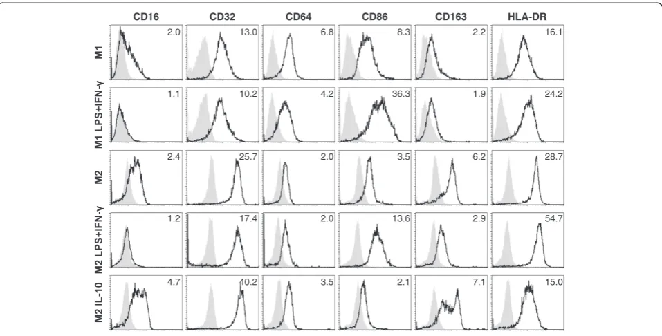
Induction of anti-tumor activity by M1-like macro-phages after stimulation with LPS + IFN-γwas associated with a more activated phenotype as indicated by the up-regulation of CD86 and HLA-DR expression (Figure 2). In contrast to M1-like macrophages, M2-like macro-phages expressed CD163, a marker frequently described for tumor-associated M2-like macrophages. LPS +
IFN-γ–stimulated M2-like macrophages showed phenotypic
similarities to LPS + IFN-γ–stimulated M1-like macro-phages, exhibiting reduced levels of CD163 and increased levels of CD86 and HLA-DR expression. Notably, in par-ticular when stimulated with IL-10, M2-like macrophages displayed high levels of FcγRII (CD32) in addition to FcγRI (CD64) and FcγRIIIa (CD16), suggesting that IL-10– polar-ized M2-like macrophages could exert antibody-dependent functions as described below.
Overall, of the different macrophage populations tested, LPS + IFN-γ–activated M1-like macrophages, resembling
‘classically-activated’M1-like macrophages [8-10], were the most capable of inhibiting osteosarcoma cell growth.
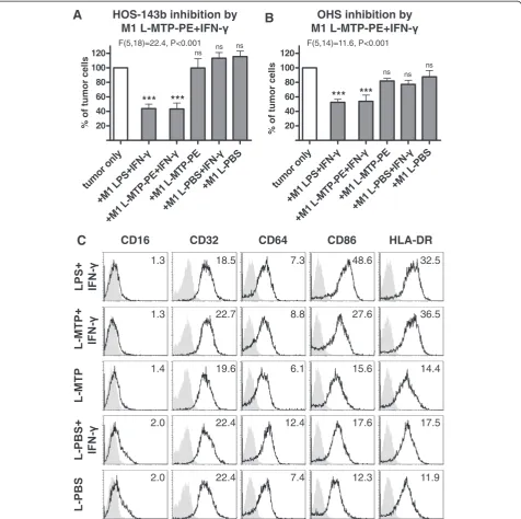
Liposomal muramyl tripeptide only induces anti-tumor activity of M1-like macrophages in the presence of IFN-γ
inhibition by L-MTP-PE + IFN-γ–stimulated M1-like
mac-rophages was as potent as by LPS + IFN-γ–activated
M1-like macrophages. In contrast, M1-like macrophages stimulated with MTP-PE-loaded liposomes alone, empty liposomes (L-PBS) or empty liposomes in combination with IFN-γ failed to inhibit tumor cell growth. Moreover, only when co-stimulated with IFN-γ, L-MTP-PE–stimulated M1-like macrophages exhibited an activated phenotype by CD86 and HLA-DR up-regulation similar to LPS + IFN-γ– activated M1-like macrophages (Figure 3, panel C). In conclusion, L-MTP-PE stimulation induced substantial anti-tumor activity of M1-like macrophages but only after co-stimulation with the pro-inflammatory cytokine IFN-γ.
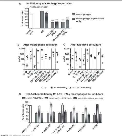
Soluble factors produced by M1-like macrophages after LPS + IFN-γand L-MTP-PE + IFN-γactivation inhibit tumor cell growth
Next, the mechanisms involved in the strong anti-tumor effect of LPS + IFN-γ–activated and L-MTP-PE + IFN-γ–
activated M1-like macrophages were investigated. Incu-bation of osteosarcoma cells in medium with cell-free supernatant of activated M1-like macrophages reduced tumor cell growth to similar levels as activated M1-like macrophages themselves (Figure 4, panel A). In contrast, supernatant from non-activated M1-like macrophages did not reduce tumor cell growth. The inhibitory effect of macrophage supernatant was dose-dependent (Additional file 1: Figure S1, panel B). These data indicate that both LPS + IFN-γ–activated and L-MTP-PE + IFN-γ–activated M1-like macrophages produced soluble factors that inhib-ited osteosarcoma cell growth. Therefore we measured the levels of soluble factors produced by activated macrophages alone and after two-day co-culture with the tumor cells. Activation of M1-like macrophages with LPS + IFN-γ en-hanced the production of the pro-inflammatory cytokines IL-1β, IL-6, IL-12p70, TNF-α, CXCL10 (IP-10) and CCL5 (Rantes), while CCL2 (MCP-1), CCL3 (MIP-1α) and CCL4
(MIP-1β) remained unchanged amongst a panel of 27
A
C
B
% of tumor cells
OHS inhibition by M1/M2
***
Inhibition of osteosarcoma cells
20 40 60 80 100
HOS-143b
OHS OSA HOS U2OS
SAOS-2
% of tumor cells
HOS-143b inhibition by M1/M2
% of tumor cells
***
HOS-143b counts
D
Growth inhibitionhours
tumor only +M1
2 24 48
*** ***
0 20000 40000 60000
**
20 40 60 80 100 120 F(5,41)=31.6, P<0.001
ns ns
ns
F(5,48)=25.3, P<0.001
ns
ns
* ns
ns
F(2,10)=24.4, P<0.001 20
40 60 80 100 120
ns
*
n=2 n=2
cytokines and chemokines tested (Figure 4, panel B). L-MTP-PE + IFN-γ–activated M1-like macrophages displayed a similar cytokine profile except for lower levels of IL-12p70.
Since TNF-α was reported to be able to confer anti-tumor effects [29] and was also produced by both LPS + IFN-γ–activated and L-MTP-PE + IFN-γ–activated M1-like macrophages during the co-culture (Figure 4, panel C), a role for TNF-α in the inhibition of osteosarcoma
cell growth was examined. Blocking of TNF-α during
the co-culture of macrophages and tumor cells by the soluble TNF receptor etanercept combined with the
TNF-α neutralizing antibody adalimumab did not
pre-vent the inhibition of cell growth of HOS-143b and
OHS cells by LPS + IFN-γ–activated M1-like
macro-phages or supernatants derived from these macromacro-phages
(Figure 4, panel D and data not shown). Blocking
of TNF-α did also not prevent the inhibiting effects
of L-MTP-PE + IFN-γ–activated M1-like macrophages
(data not shown). Moreover, blocking of IL-1 receptor,
combined blocking of TNF-α and IL-1 receptor, or
in-hibition of nitric oxide and reactive oxygen species did not significantly interfere with the inhibition of tumor cell growth by activated macrophages (Figure 4, panel D). None of the tested inhibitors affected tumor cell growth as compared to tumor cells incubated in the ab-sence of inhibitors.
These results indicate that the inhibition of osteosar-coma cell growth by activated M1-like macrophages was
mediated by soluble factors induced by macrophage acti-vation in a TNF-α/IL-1–independent manner.
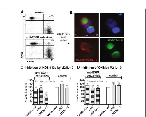
IL-10–stimulated M2-like macrophages can inhibit growth of osteosarcoma cells in an antibody-dependent manner
Both M1-like and M2-like macrophages have been detected in osteosarcoma lesions [4]. Hitherto, IL-10–stimulated M2-like macrophages were unable to inhibit osteosarcoma cell growth. In a previous study it has been shown that IL-10-polarized M2-like macrophages internalized antibody-coated B cell lymphoma cells [28]. Since IL-10–stimulated M2-like macrophages exhibited the highest expression of FcγR in our experiments, we investigated whether these macrophages are able to form cell conjugates with and internalize osteosarcoma cells in an antibody-dependent manner as a potential anti-tumor mechanism. After two-hour co-culture of IL-10–stimulated M2-like macrophages and CFSE-labeled HOS-143b cells, CD32+M2-like macro-phages acquiring the fluorescent signal of HOS-143b cells were analyzed by flow cytometry, as similarly described be-fore [28,30]. If HOS-143b cells were coated with the anti-EGFR antibody cetuximab, the percentage of CD32+CFSE+ macrophages was increased, indicative for cell conjugate formation between macrophages and tumor cells (Figure 5, panel A). Furthermore, FACS-sorted CD32+CFSE+ macro-phages (upper right quadrant) were analyzed by immuno-fluorescence microscopy and contained tumor cells bound to macrophages as well as tumor cells internalized by mac-rophages (Figure 5, panel B).
CD16
2.0
1.1
2.4
1.2
4.7
CD32
13.0
10.2
25.7
17.4
40.2
CD64
6.8
4.2
2.0
2.0
3.5
CD86
8.3
36.3
3.5
13.6
2.1
CD163
2.2
1.9
6.2
2.9
7.1
HLA-DR
16.1
24.2
28.7
54.7
15.0
Next, it was examined whether the antibody-dependent interaction between tumor cells and macrophages can re-sult in inhibition of tumor cell growth. Indeed, IL-10– stim-ulated M2-like macrophages substantially inhibited growth of two osteosarcoma cell lines (2/6) such as HOS-143b to as low as 50% if coated with cetuximab (Figure 5 panel C and data not shown). There was not such an inhibiting
effect when HOS-143b cells were treated with the isotype-matched, non-tumor-binding anti-CD20 antibody rituxi-mab. In contrast, IL-10–stimulated M2-like macrophages failed to inhibit cell growth of (or form cell conjugates with) cetuximab-coated OHS cells despite high levels of EGFR expression [31], indicating that additional cell char-acteristics play a role in determining the sensitivity of
A
HOS-143b inhibition by% of tumor cells
OHS inhibition by
% of tumor cells
B
20 40 60 80 100 120
*** ***
20 40 60 80 100 120
*** ***
CD16 CD32 CD64 CD86 HLA-DR
1.3 22.7 8.8 27.6 36.5
1.4 19.6 6.1 15.6 14.4
2.0 22.4 12.4 17.6 17.5
2.0 22.4 7.4 12.3 11.9
1.3 18.5 7.3 48.6 32.5
C
1 0 0 . 0 < P , 6 . 1 1 = ) 4 1 , 5 ( F 1
0 0 . 0 < P , 4 . 2 2 = ) 8 1 , 5 ( F
ns ns
ns
ns ns ns
B After macrophage activation
pg/ml
C After two days co-culture
M1
A Inhibition by macrophage supernatant
% of tumor cells
pg/ml
macrophage supernatant only
macrophages
D
% of tumor cells
tumor only + inhibitors 1
10 100 1000 10000
1 10 100 1000 10000 20
40 60 80 100 120 140
*** *** *** ***
20 40 60 80 100 120
F(6,35)=45.7, P<0.001
ns *
(See figure on previous page.)
Figure 4Tumor growth inhibition by activated M1-like macrophages is mediated by soluble factors. (A)HOS-143b cells were incubated with M1-like macrophages (pre-activated +/−LPS + IFN-γor L-MTP-PE + IFN-γ) (no pattern) or with cell culture supernatant from these macrophages (hatched pattern) for two days. Tumor cell numbers were analyzed relative to the control (ANOVA and Dunnett’s post test n = 3–8). The cytokine/chemokine profile of M1-like macrophages was assessed in cell-free supernatants obtained(B)after macrophage activation or(C)
after macrophage activation and subsequent two-day co-culture with HOS-143b cells. Data of IL-1β, IL6, IL-12p70, TNF-α, CXCL10 (IP-10), CCL2 (MCP-1), CCL3 (MIP-1α), CCL4 (MIP-1β) and CCL5 (Rantes) were acquired by Luminex assays (n = 2–3). There was no cytokine/chemokine production by tumor cells alone. Compared to activated macrophages alone the co-culture with tumor cells did not enhance cytokine/chemokine production (data not shown).(D)HOS-143b cells were incubated with/without LPS + IFN-γ-activated M1-like macrophages in the presence or absence of inhibitors against TNF-α(by neutralizing antibody and soluble TNF receptor), IL-1 receptor (by IL-1Ra), TNF-αand IL-1 receptor, nitric oxide (by L-NAME) or reactive oxygen species (by catalase and SOD). For each set of inhibitor experiments (n = 3–6) tumor cell numbers of the following conditions are depicted: after co-culture with activated macrophages (filled bar), tumor cells alone with inhibitors (white bar hatched pattern), after co-culture with activated macrophages with inhibitors (filled bar hatched pattern), analyzed relative to tumor cells only without inhibitors (white bar). There was no significant difference between co-cultures with and without inhibitors, or between tumor cells alone and co-cultures with inhibitors, whereas differences between tumor cells alone and co-cultures with/without inhibitors were statistically significant (P < 0.05, ANOVA and Bonferroni’s post test).
% of tumor cells
+M2
+M2 IL-10 tumor only
+M2
+M2 IL-10 tumor only
+M2
+M2 IL-10 tumor only
+M2
+M2 IL-10 tumor only
Inhibition of HOS-143b by M2 IL-10
D
Inhibition of OHS by M2 IL-10C
3.7% 0.7%
upper right FACS sorted
anti-EGFR cetuximab
control
B
A
CFSE
CD32
DAPI
HLA-DR+ M2 IL-10
CFSE+ tumor
***
% of tumor cells
anti-EGFR
cetuximab control
anti-EGFR
cetuximab control
20 40 60 80 100 120 140
20 40 60 80 100 120
140 F(5,30)=1.8,P=0.125
1 0 0 . 0 < P , 9 . 2 1 = ) 0 3 , 5 ( F
ns
ns ns
ns ns ns ns
Figure 5Antibody-dependent tumor cell growth inhibition by IL-10–polarized M2-like macrophages. (A)CFSE-labeled HOS-143b cells coated with anti-EGFR cetuximab or non-binding anti-CD20 rituximab were incubated with IL-10–stimulated M2-like macrophages for two hours. Cell conjugate formation was evaluated by flow cytometry, assessing CD32+macrophages acquiring high CFSE fluorescence of the tumor cells.
Representative data of two experiments are depicted.(B)In one experiment, CD32+CFSE+cells (upper right quadrant in lower panel A) were
sorted by flow cytometry and examined by Immunofluorescence microscopy, detecting HLA-DR-stained macrophages in red (lower left), CFSE+
tumor cells in green (lower right) and DAPI-stained cell nuclei in blue (upper right) and composites (upper left).(C)HOS-143b (n = 4–7) and(D)
osteosarcoma cell lines to antibody-dependent anti-tumor activity (Figure 5, panel D and data not shown). In the ab-sence of macrophages, there was no inhibitory effect by cetuximab on osteosarcoma cells (Figure 5, panel C and D) [31]. Inhibition of tumor cell growth by LPS + IFN-γ– stim-ulated M1-like macrophages was not further increased by cetuximab (data not shown).
Hence, at least for some cell lines, IL-10–stimulated M2-like macrophages have the potential to inhibit osteo-sarcoma cell growth in an antibody-dependent manner with similar efficacy as antibody-independent inhibition by activated M1-like macrophages.
Discussion
In this report, we describe for the first time that human macrophages can interfere with the growth of human osteosarcoma cells. Significant induction of anti-tumor activity of human M1-like macrophages by liposomal muramyl tripeptide required co-stimulation with
pro-inflammatory IFN-γ. Inhibition of osteosarcoma cell
growth by activated M1-like macrophages was mediated by soluble factors which were induced upon macro-phage activation before interaction with tumor cells. In addition, we report that IL-10–polarized M2-like macro-phages could exert anti-tumor activity against some osteo-sarcoma cell lines in an antibody-dependent manner.
More than 100 years ago it has been observed by Busch, Fehleisen, Bruns and others that bacterial infections can result in tumor regression accompanied by febrile inflam-matory responses which presumably mediated the anti-tumor effects [32,33]. These findings pioneered the first ex-tensive immunotherapy of bone sarcoma patients by Coley, administering heat-inactivated bacterial preparations with considerable but disputed remission rates. The anti-tumor effect was probably, at least in part, linked to the pro-inflammatory response of innate immune cells such as macrophages to bacterial constituents like LPS [33,34]. An-other bacterial cell wall component, muramyl dipeptdide (MDP) has originally been discovered as the minimal (synthetically-derived) moiety of peptidoglycan which can substitute for mycobacteria in Freund’s complete adjuvant [35]. MTP-PE is a lipophilic, synthetic derivate of MDP which has low toxicity and enhanced macrophage-activating properties if incorporated in liposomes (L-MTP-PE) [21]. To mimic bacterial infections and trigger macrophage activation, L-MTP-PE has been included in the treatment of osteosarcoma patients [1]. Our
ob-servation that the anti-tumor effect of L-MTP-PE–
stimulated macrophages was dependent on IFN-γ is
noteworthy in this respect. IFN-γ was originally de-scribed as macrophage-activating factor [36].‘Priming’
of macrophages by IFN-γ may enhance liposome
up-take and improve the response to bacterial compo-nents by, for instance, intracellular NOD2, which is the
receptor for MDP and presumably MTP-PE [21,37-39]. The significance of IFN-γobserved in our experiments reproduces previous studies using different tumor cells which showed that activation of human/murine mono-cytes/macrophages by L-MTP-PE was enhanced by simul-taneous or preceding stimulation with IFN-γ [17,21,38]. Furthermore, addition of IFN-γto L-MTP-PE was reported to improve survival and inhibit metastases in murine renal adenocarcinoma [40]. Altogether, the clinical efficacy of L-MTP-PE addition in the treatment of osteosarcoma pa-tients may be improved by the inclusion of a macrophage-priming signal like IFN-γ.
This raises the question how such a macrophage-priming factor could be safely introduced in the osteosarcoma microenvironment. IFN-γlevels could be increased by local or systemic IFN-γtherapy as applied in patients with can-cers or mycobacterial infections [41,42]. To target the same macrophages with IFN-γ as with L-MTP-PE, IFN-γcould be incorporated in MTP-PE-containing liposomes which are then both efficiently internalized by phagocytic cells such as tumor-resident or tumor-infiltrating macrophages. This approach is supported by murine studies in which the incorporation of IFN-γinto MTP-PE–containing liposomes enhanced the tumoricidal activity of macrophages as com-pared to liposomal MTP-PE alone [21,43]. Alternatively, lymphocytes such as NK cells activated to secrete IFN-γ and recruited to tumor sites might enhance local IFN-γ production [44].
Inhibition of osteosarcoma cell growth was mediated by soluble factors which were produced by activated M1-like macrophages before interaction with the tumor cells. It is noteworthy that the macrophages themselves as well as their secreted factor reached a maximal effect on inhibiting tumor cell numbers to about 50%. Inhib-ition of cell growth required time and became only evi-dent after more than one day. Altogether these data suggest that the inhibitory factor may either limit growth of the tumor cells to a certain maximal cell density or that this factor has a delayed cytotoxic effect. However, because a cytotoxic effect is expected to become evident sooner, an anti-proliferative effect of this factor is more likely. Similar to our results, inhibition of colorectal cancer cells by an unidentified soluble factor of macro-phages has recently been reported [45].
be negatively influenced by infiltrating M2-like macro-phages [46,47]. Therefore, several studies have considered depleting macrophage numbers or inhibiting macrophage recruitment to the tumor [11,48,49]. Instead, in our experi-ments, IL-10–polarized M2-like macrophages could be in-duced to inhibit osteosarcoma cell growth if the tumor cells were coated with the therapeutic anti-EGFR antibody cetuximab. Antibody-dependent cell conjugate formation and inhibition of tumor cell growth were only observed for half of the osteosarcoma cell lines despite significant EGFR expression [31]. Hence, to improve antibody-dependent anti-tumor activity by M2-like macrophages, it would be re-quired to elucidate additional parameters besides surface antigen expression that determine inhibition of tumor cell growth by macrophages. Expression of CD47 on tumors cells has been described to block phagocytic function by binding to SIRP1αexpressed on phagocytic cells [50]. How-ever, CD47 gene expression was not significantly different between the cell lines (inhibited or not inhibited by M2-like macrophages), as concluded from previously published genome-wide gene profiling data of osteosarcoma cell lines [4] (data not shown).
The potential of antibody-dependent anti-tumor activity by macrophages has been shown to mediate anti-tumor re-sponses in murine lymphoma models [51,52]. In humans, the addition of rituximab therapy to patients with follicular lymphoma can counteract the non-favorable prognostic factor of high macrophage counts in the tumor [53]. We have previously demonstrated that the cytotoxic activity of NK cells can be enhanced and directed to osteosarcoma cells by anti-EGFR cetuximab [31]. Since macrophages abundantly infiltrate osteosarcoma le-sions, antibody-dependent inhibition of osteosarcoma cell growth by macrophages may be an additional anti-tumor mechanism of cetuximab.
The recent finding that anti-CD40 therapy can induce anti-tumor activity in mice and humans independently of T cells but presumably via activating macrophages has revived the role of macrophages in anti-tumor responses [54]. Overall, activation of macrophages by e.g.
L-MTP-PE in the presence of IFN-γ, and/or treatment with
tumor-reactive antibodies may in particular be advanta-geous in tumors like osteosarcoma that have a high con-tent of infiltrating macrophages.
Additional file
Additional file 1: Figure S1.Inhibition of tumor cell growth by activated M1-like macrophages is dose-dependent. (A) HOS-143b and OHS cells were incubated with increasing numbers of LPS+IFN-γ–activated M1-like macrophages as indicated by the macrophage:tumor ratios from 0 to 20. (B) HOS-143b and OHS cells were incubated with increasing amounts of cell-free culture supernatant of LPS+IFN-γand L-MTP-PE +IFN-γ–activated M1-like macrophages as indicated by the percentage of culture supernatant present during tumor cell culture. Of note,
supernatant from LPS+IFN-γ-activated M1-like macrophages was slightly more potent in tumor growth inhibition than supernatant of L-MTP-PE+IFN-γ activated M1-like macrophages.
Competing interests
The authors declare that they have no competing interests.
Authors’contributions
JP, KK, EV, SS, SM, MK performed experiments and analyzed data. EV and WJ generated and provided liposome preparations. JW verified cell line identities. JP, KK, MT, AC, RE, WJ, AL and MS participated in study conception and data interpretation. MT, RE, AL and MS coordinated this study. JP, AL and MS wrote the manuscript. All authors read and approved the final manuscript.
Acknowledgments
This work was financially supported by a grant from the foundation“Quality of Life Gala 2007”, the European Commission projects“EuroSarc”(No 278742) and the Dutch Foundation Children Cancer Free (grant 2009–052).
Author details
1Department of Pediatrics, Leiden University Medical Center, Leiden, the
Netherlands.2Division of Drug Delivery Technology, Leiden Academic Center for Drug Research, Leiden University, Leiden, the Netherlands.3Department
of Pathology, Leiden University Medical Center, Leiden, the Netherlands.
4Department of Clinical Genetics, Leiden University Medical Center, Leiden,
the Netherlands.5Division of Hematology/Oncology, Hospital for Sick Children/University of Toronto, Toronto, Canada.6Laboratory for
Immunology, P3-P, Department of Pediatrics, Leiden University Medical Center, PO Box 9600, 2300 RC Leiden, the Netherlands.
Received: 23 October 2013 Accepted: 3 March 2014 Published: 10 March 2014
References
1. Meyers PA, Schwartz CL, Krailo MD, Healey JH, Bernstein ML, Betcher D, Ferguson WS, Gebhardt MC, Goorin AM, Harris M, Kleinerman E, Link MP, Nadel H, Nieder M, Siegal GP, Weiner MA, Wells RJ, Womer RB, Grier HE:
Osteosarcoma: the addition of muramyl tripeptide to chemotherapy improves overall survival-a report from the Children's Oncology Group. J Clin Oncol2008,26:633–638.
2. Zhou Y, Huang Z, Wu S, Zang X, Liu M, Shi J:miR-33a is up-regulated in chemoresistant osteosarcoma and promotes osteosarcoma cell resistance to cisplatin by down-regulating TWIST.J Exp Clin Cancer Res2014,33:12. 3. Yang J, Zhang W:New molecular insights into osteosarcoma targeted
therapy.Curr Opin Oncol2013,25:398–406.
4. Buddingh EP, Kuijjer ML, Duim RA, Burger H, Agelopoulos K, Myklebost O, Serra M, Mertens F, Hogendoorn PCW, Lankester AC, Cleton-Jansen AM:
Tumor-infiltrating macrophages are associated with metastasis suppression in high-grade osteosarcoma: a rationale for treatment with macrophage activating agents.Clin Cancer Res2011,17:2110–2119.
5. Nathan CF, Murray HW, Cohn ZA:The macrophage as an effector cell. N Engl J Med1980,303:622–626.
6. van Furth R:Macrophage activity and clinical immunology. Origin and kinetics of mononuclear phagocytes.Ann N Y Acad Sci1976,278:161–175. 7. Wynn TA, Chawla A, Pollard JW:Macrophage biology in development,
homeostasis and disease.Nature2013,496:445–455.
8. Mosser DM, Edwards JP:Exploring the full spectrum of macrophage activation.Nat Rev Immunol2008,8:958–969.
9. Ricardo SD, van GH, Eddy AA:Macrophage diversity in renal injury and repair.J Clin Invest2008,118:3522–3530.
10. Sica A, Schioppa T, Mantovani A, Allavena P:Tumour-associated macrophages are a distinct M2 polarised population promoting tumour progression: potential targets of anti-cancer therapy.Eur J Cancer2006,
42:717–727.
11. Qian BZ, Li J, Zhang H, Kitamura T, Zhang J, Campion LR, Kaiser EA, Snyder LA, Pollard JW:CCL2 recruits inflammatory monocytes to facilitate breast-tumour metastasis.Nature2011,475:222–225.
13. Ruffell B, Affara NI, Coussens LM:Differential macrophage programming in the tumor microenvironment.Trends Immunol2012,33:119–126. 14. Edin S, Wikberg ML, Dahlin AM, Rutegard J, Oberg A, Oldenborg PA,
Palmqvist R:The distribution of macrophages with a M1 or M2 phenotype in relation to prognosis and the molecular characteristics of colorectal cancer.PLoS One2012,7:e47045.
15. Forssell J, Oberg A, Henriksson ML, Stenling R, Jung A, Palmqvist R:High macrophage infiltration along the tumor front correlates with improved survival in colon cancer.Clin Cancer Res2007,13:1472–1479.
16. Fidler IJ, Jessup JM, Fogler WE, Staerkel R, Mazumder A:Activation of tumoricidal properties in peripheral blood monocytes of patients with colorectal carcinoma.Cancer Res1986,46:994–998.
17. Galligioni E, Quaia M, Spada A, Favaro D, Santarosa M, Talamini R, Monfardini S:Activation of cytolytic activity in peripheral blood monocytes of renal cancer patients against non-cultured autologous tumor cells.Int J Cancer1993,55:380–385.
18. Kleinerman ES, Erickson KL, Schroit AJ, Fogler WE, Fidler IJ:Activation of tumoricidal properties in human blood monocytes by liposomes containing lipophilic muramyl tripeptide.Cancer Res1983,43:2010–2014. 19. Sone S, Utsugi T, Tandon P, Yanagawa H, Okubo A, Ogura T:Tumor
cytotoxicity and interleukin 1 production of blood monocytes of lung cancer patients.Cancer Immunol Immunother1990,30:357–362. 20. Sone S, Fidler IJ:In vitro activation of tumoricidal properties in rat
alveolar macrophages by synthetic muramyl dipeptide encapsulated in liposomes.Cell Immunol1981,57:42–50.
21. Nardin A, Lefebvre ML, Labroquere K, Faure O, Abastado JP:Liposomal muramyl tripeptide phosphatidylethanolamine: Targeting and activating macrophages for adjuvant treatment of osteosarcoma.Curr Cancer Drug Targets2006,6:123–133.
22. Kurzman ID, Shi F, Vail DM, MacEwen EG:In vitro and in vivo enhancement of canine pulmonary alveolar macrophage cytotoxic activity against canine osteosarcoma cells.Cancer Biother Radiopharm 1999,14:121–128.
23. Kurzman ID, MacEwen EG, Rosenthal RC, Fox LE, Keller ET, Helfand SC, Vail DM, Dubielzig RR, Madewell BR, Rodriguez CO Jr:Adjuvant therapy for osteosarcoma in dogs: results of randomized clinical trials using combined liposome-encapsulated muramyl tripeptide and cisplatin. Clin Cancer Res1995,1:1595–1601.
24. Chou AJ, Kleinerman ES, Krailo MD, Chen Z, Betcher DL, Healey JH, Conrad EU III, Nieder ML, Weiner MA, Wells RJ, Womer RB, Meyers PA:Addition of muramyl tripeptide to chemotherapy for patients with newly diagnosed metastatic osteosarcoma: a report from the Children's Oncology Group. Cancer2009,115:5339–5348.
25. Ottaviano L, Schaefer KL, Gajewski M, Huckenbeck W, Baldus S, Rogel U, Mackintosh C, de AE, Myklebost O, Kresse SH, Meza-Zepeda LA, Serra M, Cleton-Jansen AM, Hogendoorn PCW, Buerger H, Aigner T, Gabbert HE, Poremba C:Molecular characterization of commonly used cell lines for bone tumor research: a trans-European EuroBoNet effort.Genes Chromosomes Cancer2010,49:40–51.
26. Verreck FA, de Boer T, Langenberg DM, van der Zanden L, Ottenhoff TH:
Phenotypic and functional profiling of human proinflammatory type-1 and anti-inflammatory type-2 macrophages in response to microbial antigens and IFN-gamma- and CD40L-mediated costimulation.J Leukoc Biol2006,79:285–293.
27. Munn DH, Cheung NK:Antibody-dependent antitumor cytotoxicity by human monocytes cultured with recombinant macrophage colony-stimulating factor. Induction of efficient antibody-mediated antitumor cytotoxicity not detected by isotope release assays.J Exp Med1989,170:511–526.
28. Leidi M, Gotti E, Bologna L, Miranda E, Rimoldi M, Sica A, Roncalli M, Palumbo GA, Introna M, Golay J:M2 macrophages phagocytose rituximab-opsonized leukemic targets more efficiently than m1 cells in vitro.J Immunol2009,182:4415–4422.
29. Decker T, Lohmann-Matthes ML, Gifford GE:Cell-associated tumor necrosis factor (TNF) as a killing mechanism of activated cytotoxic macrophages. J Immunol1987,138:957–962.
30. Oflazoglu E, Stone IJ, Brown L, Gordon KA, van RN, Jonas M, Law CL, Grewal IS, Gerber HP:Macrophages and Fc-receptor interactions contribute to the antitumour activities of the anti-CD40 antibody SGN-40.Br J Cancer 2009,100:113–117.
31. Pahl JH, Ruslan SE, Buddingh EP, Santos SJ, Szuhai K, Serra M, Gelderblom H, Hogendoorn PC, Egeler RM, Schilham MW, Lankester AC:Anti-EGFR
antibody cetuximab enhances the cytolytic activity of natural killer cells toward osteosarcoma.Clin Cancer Res2012,18:432–441.
32. Coley WB:II. Contribution to the Knowledge of Sarcoma.Ann Surg1891,
14:199–220.
33. Wiemann B, Starnes CO:Coley’s toxins, tumor necrosis factor and cancer research: a historical perspective.Pharmacol Ther1994,64:529–564. 34. Carswell EA, Old LJ, Kassel RL, Green S, Fiore N, Williamson B:An
endotoxin-induced serum factor that causes necrosis of tumors.Proc Natl Acad Sci USA1975,72:3666–3670.
35. Merser C, Sinay P, Adam A:Total synthesis and adjuvant activity of bacterial peptidoglycan derivatives.Biochem Biophys Res Commun1975,
66:1316–1322.
36. Schroder K, Sweet MJ, Hume DA:Signal integration between IFNgamma and TLR signalling pathways in macrophages.Immunobiology2006,
211:511–524.
37. Totemeyer S, Sheppard M, Lloyd A, Roper D, Dowson C, Underhill D, Murray P, Maskell D, Bryant C:IFN-gamma enhances production of nitric oxide from macrophages via a mechanism that depends on nucleotide oligomerization domain-2.J Immunol2006,176:4804–4810. 38. Sone S, Tandon P, Utsugi T, Ogawara M, Shimizu E, Nii A, Ogura T:
Synergism of recombinant human interferon gamma with liposome-encapsulated muramyl tripeptide in activation of the tumoricidal properties of human monocytes.Int J Cancer1986,38:495–500. 39. Girardin SE, Hugot JP, Sansonetti PJ:Lessons from Nod2 studies: towards a
link between Crohn’s disease and bacterial sensing.Trends Immunol2003,
24:652–658.
40. Dinney CP, Utsugi T, Fidler IJ, von Eschenbach AC, Killion JJ:
Immunotherapy of murine renal adenocarcinoma by systemic administration of liposomes containing the synthetic macrophage activator CGP 31362 or CGP 19835A in combination with interleukin 2 or gamma-interferon.Cancer Res1992,52:1155–1161.
41. Jarvis JN, Meintjes G, Rebe K, Williams GN, Bicanic T, Williams A, Schutz C, Bekker LG, Wood R, Harrison TS:Adjunctive interferon-gamma immunotherapy for the treatment of HIV-associated cryptococcal meningitis: a randomized controlled trial.AIDS2012,26:1105–1113.
42. Kleinerman ES, Kurzrock R, Wyatt D, Quesada JR, Gutterman JU, Fidler IJ:
Activation or suppression of the tumoricidal properties of monocytes from cancer patients following treatment with human recombinant gamma-interferon.Cancer Res1986,46:5401–5405.
43. Goldbach P, Dumont S, Kessler R, Poindron P, Stamm A:In situ activation of mouse alveolar macrophages by aerosolized liposomal IFN-gamma and muramyl tripeptide.Am J Physiol1996,270:L429–L434.
44. O’Sullivan T, Saddawi-Konefka R, Vermi W, Koebel CM, Arthur C, White JM, Uppaluri R, Andrews DM, Ngiow SF, Teng MW, Smyth MJ, Schreiber RD, Bui JD:Cancer immunoediting by the innate immune system in the absence of adaptive immunity.J Exp Med2012,209:1869–1882.
45. Engstrom A, Erlandsson A, Delbro D, Wijkander J:Conditioned media from macrophages of M1, but not M2 phenotype, inhibit the proliferation of the colon cancer cell lines HT-29 and CACO-2.Int J Oncol2014,
44:385–392.
46. Suriano F, Santini D, Perrone G, Amato M, Vincenzi B, Tonini G, Muda A, Boggia S, Buscarini M, Pantano F:Tumor associated macrophages polarization dictates the efficacy of BCG instillation in non-muscle invasive urothelial bladder cancer.J Exp Clin Cancer Res2013,32:87.
47. Brandau S, Suttmann H:Thirty years of BCG immunotherapy for non-muscle invasive bladder cancer: a success story with room for improvement.Biomed Pharmacother2007,61:299–305.
48. Luo Y, Zhou H, Krueger J, Kaplan C, Lee SH, Dolman C, Markowitz D, Wu W, Liu C, Reisfeld RA, Xiang R:Targeting tumor-associated macrophages as a novel strategy against breast cancer.J Clin Invest2006,116:2132–2141. 49. Germano G, Frapolli R, Belgiovine C, Anselmo A, Pesce S, Liguori M, Erba E,
Uboldi S, Zucchetti M, Pasqualini F, Nebuloni M, van RN, Mortarini R, Beltrame L, Marchini S, Fuso NI, Sanfilippo R, Casali PG, Pilotti S, Galmarini CM, Anichini A, Mantovani A, D'Incalci M, Allavena P:Role of macrophage targeting in the antitumor activity of trabectedin.Cancer Cell2013,23:249–262.
50. Majeti R, Chao MP, Alizadeh AA, Pang WW, Jaiswal S, Gibbs KD Jr, van RN, Weissman IL:CD47 is an adverse prognostic factor and therapeutic antibody target on human acute myeloid leukemia stem cells.Cell2009,
138:286–299.
macrophages promote invasion while retaining Fc-dependent anti-tumor function.J Immunol2012,189:5457–5466.
52. Minard-Colin V, Xiu Y, Poe JC, Horikawa M, Magro CM, Hamaguchi Y, Haas KM, Tedder TF:Lymphoma depletion during CD20 immunotherapy in mice is mediated by macrophage FcgammaRI, FcgammaRIII, and FcgammaRIV.Blood2008,112:1205–1213.
53. Canioni D, Salles G, Mounier N, Brousse N, Keuppens M, Morchhauser F, Lamy T, Sonet A, Rousselet MC, Foussard C, Xerri L:High numbers of tumor-associated macrophages have an adverse prognostic value that can be circumvented by rituximab in patients with follicular lymphoma enrolled onto the GELA-GOELAMS FL-2000 trial.J Clin Oncol2008,
26:440–446.
54. Beatty GL, Chiorean EG, Fishman MP, Saboury B, Teitelbaum UR, Sun W, Huhn RD, Song W, Li D, Sharp LL, Torigian DA, O'Dwyer PJ, Vonderheide RH:
CD40 agonists alter tumor stroma and show efficacy against pancreatic carcinoma in mice and humans.Science2011,331:1612–1616.
doi:10.1186/1756-9966-33-27
Cite this article as:Pahlet al.:Macrophages inhibit human osteosarcoma cell growth after activation with the bacterial cell wall derivative liposomal muramyl tripeptide in combination with interferon-γ.Journal of Experimental & Clinical Cancer Research201433:27.
Submit your next manuscript to BioMed Central and take full advantage of:
• Convenient online submission
• Thorough peer review
• No space constraints or color figure charges
• Immediate publication on acceptance
• Inclusion in PubMed, CAS, Scopus and Google Scholar
• Research which is freely available for redistribution