Original Article
J Reprod Infertil. 2012;13(1):13-19
The Effect of Macromolecule Source and Type of Media During in vitro
Maturation of Sheep Oocytes on Subsequent Embryo Development
Abolfazl Shirazi 1,2, Mohammad Ansari Ardali 2, Ebrahim Ahmadi 2,Hassan Nazari 2, Morteza Mamuee 3, Banafsheh Heidari 1*
1- Department of Embryology and Andrology, Reproductive Biotechnology Research Center, Avicenna Research Institute, ACECR, Tehran, Iran
2- Department of Gametes and Cloning, Research Institute of Animal Embryo Technology, Shahrekord University, Shahrekord, Iran 3- Faculty of Agriculture and Natural Resources, Ahvaz University, Ahvaz, Iran
Abstract
Background: Oocyte maturation and subsequent in vitro production (IVP) of em-bryos are affected by diverse groups of chemicals in maturation medium which are needed for successful mammalian oocyte maturation during which the dramatic cytoplasmic and nuclear reprogramming events take place. This study was designed to evaluate the effects of protein source (fetal bovine serum, FBS, and bovine serum albumin, BSA) as well as two different maturation media during in vitro maturation of ovine oocytes on subsequent embryo development.
Methods: Cumulus oocyte complexes were recovered from ovaries obtained from slaughter house and cultured for 24 hr in either TCM-199 or SOFaa maturation me-dium supplemented with 10% (v/v) FBS or 0.8% (w/v) BSA. Data were analyzed by one-way ANOVA using Sigma Stat (Ver. 2). A p-value smaller than 0.05 was con-sidered statistically significant.
Results: The proportions of cleavage and total blastocyst (evaluated on days 3 and 6, respectively) were significantly higher in FBS than BSA supplemented groups, though no differences were observed between the two used different maturation media. The cryotolerance of blastocysts was negatively influenced by the presence of FBS rather than BSA during IVM. The quality of produced embryos, however, was affected neither by the source of macromolecules nor the maturation medium in terms of hatching rate, total blastocyst cells and inner cell mass/total cell ratio. Conclusion: The rate of oocyte development was improved by the presence of FBS, though the cryosurvival of resulting blastocysts was negatively influenced by the presence of the serum during in vitro production of sheep oocytes.
Keywords: Bovine serum albumin, Differential staining, Sheep, Vitrification.
To cite this article: Shirazi A, Ansari Ardali M, Ahmadi E,Nazari H, Mamuee M, Heidari B. The Effect of Macromolecule Source and Type of Media During in vitro Maturation of Sheep Oocytes on Subsequent Embryo Development. J Reprod Infertil. 2012;13(1):13-19.
Introduction
uring in vitro maturation, prior to the re-sumption of nuclear maturation, oocytes undergo a series of cytoplasmic changes, leading to variable competence of the resulting embryos (1). Alteration of maturation media can significantly affect oocyte competence as reflect-ed by the morula and blastocysts yield after in vitro fertilization (IVF) (2, 3). The synthesis and storage of certain forms of mRNA and protein during IVM and early embryonic development
that are necessary for further development are in- fluenced by the composition of maturation media (4−6). Despite the undefined and variable nature of serum composition, serum and BSA are among the most common components of media in mam-malian oocyte and embryo culture systems.
There are several known functions of different kinds of sera in maturation media such as chelat-ing of heavy metals, some pH bufferchelat-ing, scav- enging of reactive oxygen species, and growth
* Corresponding Author:
Banafsheh Heidari, Department of Embry-ology and AndrEmbry-ology, Reproductive Bio-technology Research Center, Avicenna Research Institute, ACECR, Tehran, Iran, P.O. Box: 19615-1177.
E-mail:
Received: Oct. 29, 2011 Accepted: Jan. 3, 2012
14 J Reprod Infertil, Vol 13, No 1, Jan/Mar 2012 The Effect of Media Components on IVM Outcome
JRI
stimulating (7).
In bovine, the presence of fetal bovine serum (FBS) during in vitro maturation (IVM) using tissue culture medium-199 (TCM-199) signify-cantly enhanced oocyte maturation and subse-quent embryonic development when compared with the use of synthetic oviductal fluid (SOF) supplemented with serum. Indeed, the effect of FBS as a protein supplement during IVM of bo-vine oocytes was dependent on the maturation medium used (6, 8−10). Moreover, it has been demonstrated that FBS is a superior protein sup-plement compared with the often used 0.8% BSA for IVM of hamster and cow oocytes (11).
It has been shown that the kinetics of bovine em-bryos produced in vitro could be affected by serum supplementation during IVM–IVF and em-bryo cultures (12). Accordingly, in early emem-bryo the first and fourth cell cycles were prolonged by 4–5 h in the absence of serum during IVM–IVF, whereas the presence of serum during embryo culture decreased the duration of the fourth cell cycle leading to premature blastulation (12). In another study, the possibility of using chemically defined-synthetic serum substitute (SSS) in place of fetal calf serum (FCS) during maturation of bovine oocytes showed that the SSS supplemented group had higher apoptotic nuclei as compared to the FCS group. Additionally, expression of sev-eral gene transcripts such as heat shock protein 70 (Hsp70), interferon tau (IF-tau), DNA methyl-transferase 3a (Dnmt3a), desmosomal glycopro-tein desmocollin III (DcIII) and insulin-like growth factor II receptor (Igf-2r) were altered in accordance to different culture conditions (6). Despite several reports regarding the beneficial effects of serum supplementation during IVM on mammalian oocytes developmental potential, there are some controversial reports showing the deleterious effects of serum supplementation dur-ing IVM in some animal species (9, 13−18). Over-all, since the presence of serum macromolecules were found to be necessary for in vitro maturation of oocytes, with an aim to improve the in vitro production of embryos, the present study was designed to compare the effects of FBS and BSA, as well as maturation medium used during IVM on ovine oocyte developmental competence.
Methods
Except where otherwise indicated, all chemicals were obtained from Sigma (St. Louis, MO, USA).
Oocyte collection: Sheep ovaries, mainly from Lori-Bakhtiari breed, were collected from slaugh-terhouse during nonbreeding season (May− Au-gust) and transported to the laboratory in saline (30–35 ºC), within 1–3 hr following collection. All visible follicles with a diameter of 2–6 mm
were aspirated using gentle vacuum (30 mm Hg) via a 20 −gauge short− beveled needle. Prior to aspiration, the collecting tube was filled with 2 ml
preincubated Hepes-modified TCM, supplement-ed with 50 IU/ml heparin. After aspiration, only oocytes surrounded by more than three layers of unexpanded cumulus cells (COCs: cumulus oocyte complexes) were selected for IVM.
Experiment design: The selected oocytes were randomly subjected to the following IVM media: I) TCM 199+10% FBS; II) TCM + 8 mg/ml BSA; III) SOFaa+10% FBS; and IV) SOFaa+8 mg/ml
BSA. After IVM, the oocytes were fertilized with fresh semen and cultured for 8 days. The cleav-age, blastocyst, and hatching rates were detected on days 3, 6, and 7, respectively (Day 0 defined as the day of fertilization). To evaluate the effects of type of macromolecule source and media on cryotolerance of resulting embryos, the blasto-cysts were vitrified and after at least one week, they were warmed and cultured for 2 more days. The survival and hatching rates of vitrified/ warmed blastocysts were then evaluated. In each group, the rest of resulting blastocysts were subjected to differential cell staining.
In vitro maturation: Prior to maturation and in accordance to the IVM medium, the oocytes were randomly washed (3 times) in four washing media: I) Hepes-buffered TCM199 (H-TCM199) plus 2 mM glutamine supplemented with 10% FBS (Gibco 10270); II) H-TCM199 plus 2 mM
glutamine supplemented with 8 mg/ml BSA; III) Hepes-buffered SOFaa (H-SOFaa, 19) plus 2 mM
glutamine supplemented with 10% FBS, and IV) Hepes-buffered SOFaa (H-SOFaa) plus 2 mM
glutamine supplemented with 8 mg/ml BSA. The oocyte maturation media were consisted of either bicarbonate-buffered TCM 199 or SOFaa supple-mented with 2 mM L-glutamine, 0.05 U/ml FSH (F8174), 0.2 mM Na–Pyruvate, 100 U/ml peni-cillin, 100 µg/ml streptomycin and serum or BSA according to the experiment design. The medium osmolarity was adjusted to 275 mOsm. The COCs were randomly distributed in maturation droplets (10 oocytes in 50 µl) and covered by sterile paraffin oil in a 60 mm Petri dish (Falcon 1008;
Shirazi A, et al.
JRI
J Reprod Infertil, Vol 13, No 1, Jan/Mar 2012 15
Becton Dickinson, Lincoln Park, NJ) and were then incubated under an atmosphere of 5% CO2, 95% air with 100% humidity at 39 ºC for 24 hr.
Preparation of sperm and in vitro fertilization: Before transfer to fertilization drops, the oocytes were washed four times in Hepes-Synthetic Ovi-duct Fluid (H-SOF) and once in the fertilization medium. For preparing H-SOF, 20 mM of NaHCO3 was substituted with 20 mM Hepes (10
mM free acid and 10 mM Na salt). Both media were supplemented with 100 U/ml penicillin and 100 µg/ml streptomycin.
Fresh semen was collected from a Lori-Bakhtiari breed ram of proven fertility. For swim up, 80– 100 µl of semen was kept under 1 ml of BSA-HSOF in a 15 ml conical Falcon tube at 39 ºC for up to 45 min. After swim up, 700–800 µl of the supernatant was added to 3 ml of BSA-HSOF, centrifuged twice at 200 g for 3 min and the final pellet was resuspended with BSA-HSOF. Insem-ination was carried out by adding 1.0×10⁶ sperm/ml to the fertilization medium. The fertil-ization medium was SOF enriched with 20% heated and inactivated estrous sheep serum. A 5 µL aliquot of sperm suspension, containing 1.0×10⁶ sperm/ml, was transferred into
fertil-ization medium that included 10 oocytes per 45µL
fertilization drop. Fertilization was carried out by co-incubation of sperm and oocytes in an atmos-phere of 5% CO2 in humidified air at 39 ºC for 22 hr.
In vitro culture: After IVF, presumptive zygotes were vortexed for 2–3 min to remove the cumulus cells and then washed in H-SOF to remove sperm-atozoa and cellular debris. They were then allo-cated to 20 µL drop of IVC-SOF (five to six em-bryos/drop) consisting of SOF supplemented with 2% (v/v) BME-essential amino acids, 1% (v/v) MEM nonessential amino acids, 1 mM glutamine and 8 mg/ml fatty acid−free BSA. The incubation conditions were humidified by 7% O2, 5% CO2, and 88% N2 at 39 ºC. On the third and fifth day of culture (Day 0 defined as the day of fertilization) 10% charcoal stripped fetal bovine serum (FBS) was added to the medium. The culture was con-tinued until 8 days post-fertilization.
Vitrification and warming procedures: The em-bryos were vitrified according to Shirazi et al. (20). Briefly, the basic media for preparation of all vitrification solutions was DMEM supplemented by 5.5 mM glucose, 19 mM NaHCO3, 25 mM
Hepes, 100 IU/ml penicillin, and 20% (v/v) FCS. All equilibration and dilution steps, as well as
warming was performed at room temperature (ap-proximately 25 ºC).
The blastocysts were placed into a 100 µL drop of basic medium (20–30 s) and were then trans-ferred to the equilibration medium. For equilibr-ation the embryos were placed into a 100 µL drop of equilibration solution (1.35 M ethylene glycol+ 1.05 M DMSO) for 8 min, and then transferred to a 100 µL drop of vitrification solution (2.7 M
ethylene glycol+2.1 M DMSO+0.5 M sucrose) for 30 s. The cryodevice was prepared according to Shirazi et al. (20). Briefly, the embryos were load-ed with a fine bore pasture pipette onto the inner surface of the tip of sharpened 0.25 ml straw1 with
a the minimum volume of vitrification medium (<0.1 µl). After loading, almost all the solution was removed with a fine bore pasture pipette and the straw was quickly immersed in liquid nitrogen. The time limit from the time the embryos were being transferred to the tip of the straw until the immersion of straw into LN2 was 45 s. For warming, the tip of the straw was directly immersed into the 100 µL drop of dilution solution containing 0.5 M sucrose for 5 min and then washed (twice) in basic medium. The cryo-preserved-warmed blastocysts were cultured in IVC-SOF medium for 2 days.
Cell counting: For differential staining of the inner cell mass (ICM) and TE cell compartments the blastocysts which had been stained with PI were incubated in Triton X-100 prepared in the base medium for 20 s. The blastocysts were then stained in the base medium containing 30 µg/ml
PI for 1 min followed by two washes in the base medium. The blastocysts were then transferred into ice-cold ethanol containing 10 µg/ml Ho-echst 33342 for 15 min. The blastocysts were directly mounted into a small droplet of glycerol on a glass slide and examined under an epifluores-cent microscope. ICM nuclei appeared blue, caused by DNA labeling with Hoechst 33342, while TE cells appeared red due to staining of nuclear DNA with the membrane impermeable PI.
Statistical analysis: Data was collected over at least five replicates. The blastocyst cell counts were analyzed by one-way ANOVA using Sigma-Stat software (Ver. 2). When ANOVA revealed a significant effect, the experimental groups were compared by Tukey's method. When equal vari-ance test failed, the treatments were compared by
1- The straw was cut at an angle with the scalpel blade to resemble the tip of a long-beveled injection needle
16 J Reprod Infertil, Vol 13, No 1, Jan/Mar 2012 The Effect of Media Components on IVM Outcome
JRI
Student–Newman–Keuls method. When normality test failed, the Kruskal–Wallis one-way ANOVA by ranks was applied. The values were presented as Mean±SEM and a p <0.05 was considered as significant.
Results
There were significant differences in cleavage rate among treatments (Table 1). The cleavage rate was significantly higher when IVM medium was supplemented with FBS than BSA. Similarly, the overall blastocyst rates were significantly higher in FBS supplemented groups. No signifi-cant differences were observed in the rates of early, expanded, and hatched blastocysts among groups. The type of medium (TCM or SOF) dur-ing IVM, however, had no difference on
subse-quent embryo development (Table 1).
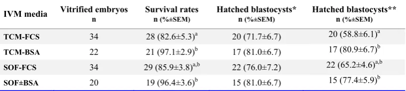
The blastocyst cell numbers, inner cell mass (ICM), trophectoderm (TE) and total cells, had been influenced neither by the type of IVM me-dium nor by the type of serum supplements. Moreover, the ICM/TE ratio was not influenced by the type of media or serum supplements (Table 2). The survival rate of vitrified-warmed blastocysts derived from oocytes matured in vitro in the pres-ence of FBS was lower than those matured in the presence of BSA. The difference in hatching rate of vitrified-warmed blastocysts, however, was insignificant among experiment groups (Table 3).
Discussion
Serum supplement is routinely added to the in vitro maturation and fertilization medium of
sev-Table 1. Developmental competence of ovine oocytes matured in different maturation media
Type of Media Oocytes n n (%±SEM)Cleavage Blastocysts n (%±SEM)
Early Expanded Total Hatched
TCM-FCS 244 199 (81.6±2.1) a 21 (27.9±3.0) 46 (60.0±3.2) 77 (31.7±1.8) a 10 (12.2±3.2)
TCM-BSA 237 157 (66.1±2.3) b 14 (27.4±3.5) 35 (63.6±5.3) 51 (21.4±0.9) b 2 (4.2±2.7)
SOF-FCS 238 189 (79.2±2.4) a 19 (24.9±4.8) 48 (64.2±5.4) 76 (32.1±1.6) a 9 (10.9±3.8)
SOF-BSA 236 152 (67.5±3.1) b 14 (29.7±3.9) 32 (66.7±4.0) 48 (20.4±0.7) b 2 (3.6±2.4)
a, b: Numbers with different superscripts in the same column differ significantly (p< 0.001).
The rates of cleavage and blastocysts (early and expanded) were evaluated on days 3 and 6 after fertilization, respectively.
Table 2. Cell allocation of ovine blastocysts derived from oocytes matured in different maturation media
IVM media Blastocysts* n Total cell M±SEM M±SEM ICM 1 M±SEM TE 2 ICM/TE %±SEM
TCM-FCS 19 54.6±4.6 12.6±1.0 41.9±3.7 23.6±0.7
TCM-BSA 14 53.1±4.5 12.7±1.1 40.4±3.4 23.8±0.9
SOF-FCS 20 57.8±3.9 12.5±0.9 45.3±3.2 22.0±1.1
SOF-BSA 13 57.1±4.5 13.1±1.0 44.0±3.8 23.2±1.2
* 6-day old blastocysts
1:Inner cell mass; 2:Trophectoderm cells
Table 3. Cryotolerance of vitrified ovine blastocysts derived from oocytes matured in different maturation media
IVM media Vitrified embryos n Survival rates n (%±SEM) Hatched blastocysts* n (%±SEM) Hatched blastocysts** n (%±SEM)
TCM-FCS 34 28 (82.6±5.3)a 20 (71.7±6.7) 20 (58.8±6.1)a
TCM-BSA 22 21 (97.1±2.9)b 17 (81.0±6.7) 17 (80.9±6.7)b
SOF-FCS 34 29 (85.9±3.8)a,b 22 (76.0±7.2) 22 (65.2±4.6)a,b
SOF±BSA 20 19 (96.4±3.6)b 15 (81.0±6.7) 15 (77.4±5.9)b
a, b: Numbers with different superscript in the same column differ significantly (p< 0.05). The survival and hatching rates of vitrified-warmed blastocysts were evaluated 24 hr and 72 hr after warming. * The hatching rate was calculated based on the number of survived blastocysts; ** The hatching rate was calculated based on the number of vitrified blastocysts
Shirazi A, et al.
JRI
J Reprod Infertil, Vol 13, No 1, Jan/Mar 2012 17
eral animal species (11, 14, 16, 17, 21−23). The beneficial effect of serum supplement during IVM may be due to the presence of some components such as hormones, catecolamines, vitamins, lipids, proteins and growth factors which are effective on resumption of oocyte meiosis and cytoplasmic maturation (22, 24). Additionaly, there evidence indicating the effect of serum on relative abun-dance of mRNAs of cumulus cells (25) and the reduced amount of apoptosis in cumulus cells treated with serum during IVM in which the latter may be attributed to the action of factors such as insulin-like factor I, epidermal and basic fiand the growth factors (26).
Among different types of sera, FBS and BSA are typically added to the medium as a protein supple-ment to improve culture efficiency (11); though different, a lot of these proteins can produce high-ly variable effects during culture, ranging from highly stimulatory to highly inhibitory (27).
In the present study, the ability of ovine oocytes to develop to the blastocyst stage was improved after maturation in the presence of serum com-pared with PVA (data not shown), confirming the results obtained by Pinyopummintr and Bavister (28), who demonstrated that embryo development to the blastocyst stage was reduced by replacing BSA or serum with PVA. Though, our finding was in contrast to what was reported by Ali and Sirard (3) in bovine and Herrick et al. in goat oocytes. In Ali and Sirard study, when serum and BSA-V were replaced by synthetic macromol-ecules such as PVP-40 (but not PVP-360) more embryos developed to the morula and blastocyst stages compared with IVM medium supplemented with serum or BSA.
Our results clearly demonstrated that the type of protein supplemented to the maturation medium of ovine oocytes can influence the subsequent de-velopment of resulting embryos. Indeed, inde-pendent of the culture medium used (TCM or SOF), supplementation of IVM medium with FBS was superior to that of BSA in terms of cleavage and blastocyst rates evaluated on day 3 and 6 post−fertilization, respectively. The results were in agreement with Leibfried-Rutledge et al. (11), who demonstrated that FCS was a superior protein supplement compared with BSA for IVM of cow and hamster oocytes. Contrarily, more blastocysts were developed in goats per cleaved embryo fol-lowing maturation in SOF with BSA than TCM 199 with goat serum (14). In goat study, however,
the effect of BSA on post−fertilization develop-ment was dependent on the proportion of BSA used in maturation medium as such SOF with 2.5 or 8.0 mg/ml BSA yielded more blastocysts than SOF with 20.0 mg/ml BSA or TCM199 with 10% goat serum (14). In our study, one explanation for the higher cleavage and blastocyst rates in FBS supplemented groups might be related to the entity of FBS as a non-defined than a semi-de-fined serum. In this sense, the presence of fetuin (an FBS component, inhibiting ZP hardening dur-ing oocyte maturation), hormones, growth factors, proteins and some other components in FBS may exert different effects on oocyte competence com-pared with BSA (29). Additionally, the synthesis and storage of certain forms of mRNA and pro-teins during IVM and early embryonic develop-ment which are necessary for further developdevelop-ment may be influenced by the type of serum used (4−6, 30).
Regarding the time of blastulation, it has been shown that the kinetics of bovine embryo devel-opment is affected by serum supplementation dur-ing IVM–IVF and embryo culture as the first and fourth cell cycles are prolonged by 4–5 hr in the absence of serum during IVM–IVF which in turn can influence the time of blastulation (12). It has also been shown that the expression of genes in early preimplantation embryos, including genes involved in compaction and blastulation could be substantially affected by the culture medium in general and serum in particular (31).
In our study, the time of blastulation was accel-erated in FBS supplemented groups compared with those supplemented by BSA during IVM (data not shown). In this context, replacement of BSA with FCS in SOF medium 120 hr post- insemination significantly accelerated the time of blastulation in bovine embryos (13). Contrary to Lonergan et al. (8) that showed higher oocyte maturation and subsequent embryonic develop-ment by using serum suppledevelop-mented TCM-199 compared with serum supplemented SOF, no such difference or even tendency was observed in the current study. Indeed, in the present study the ef-fect of FCS and BSA as a protein supplement during IVM of ovine oocytes was independent of the used maturation medium. On the other hand, ovine oocytes are not sensitive to the alteration of basic maturation medium as much as bovine oocytes (2). The reasons behind these discrep-ancies between our findings and the results of
18 J Reprod Infertil, Vol 13, No 1, Jan/Mar 2012 The Effect of Media Components on IVM Outcome
JRI
other investigators in different animal species or even between same species, apart from a probable species-specific differences, might have been arisen from some other variables such as culture conditions including use of different basic culture media, co-culture systems, CO2 and O2 pressures,
types of gonadotrophin, or some other additives such as hormones, growth factors, and different batches of commercially available sera.
The total blastocyst cell numbers and cell alloca-tions were affected neither by the basic maturation medium nor serum supplements. Our finding was in contrast to what was reported in bovine in which the IVM medium that was supplemented with serum resulted in blastocysts with a larger number of cells allocated to the inner cell mass (ICM) compared with those supplemented with fatty acid-free serum albumin (9).
The cryotolerance of blastocysts derived from different IVM culture conditions was dependent on serum supplements used during IVM. The blastocysts derived from IVM medium supple-mented with BSA showed higher survival rates compared with those supplemented with FBS (p <0.05). Though, despite a tendency toward BSA supplemented groups, the hatching rate was not significantly influenced by either maturation me-dium or serum supplements. There is large body of evidence indicating the inferior cryosurviv-ability of IVM/IVF derived embryos cultured in serum supplemented media (32). In the current study, it seemed that the accumulation of cyto-plasmic lipid droplets in oocytes after maturation in FBS supplemented groups was higher than those matured in BSA supplemented groups which in turn may affect the cryotolerance of resulting blastocysts. Therefore, at least in TCM-FBS group, the significantly lower survival rate (p <0.05) of vitrified-warmed blastocysts might be due to the excess accumulation of cytoplasmic lipid droplets in resulting blastocysts compared to the other groups. Though, the alteration of basic maturation medium could not affect oocyte com-petence as reflected by indifferent survival and hatching rates of vitrified-warmed blastocyst de-rived from TCM and SOF groups.
Conclusion
In our study the, the rate of oocyte development was improved by the presence of FBS, though the cryosurvivability of resulting blastocysts was negatively influenced by the presence of serum during IVM of sheep oocytes.
Acknowledgement
The authors would like to thank the Research Institute of Animal Embryo Technology for tech-nical and financial supports, Shahrekord Univer-sity, and Shahrekord’s slaughterhouse staff for their cooperation.
References
1. Moor RM, Mattioli M, Ding J, Nagai T. Maturation of pig oocytes in vivo and in vitro. J Reprod Fertil Suppl. 1990;40:197-210.
2. Rose TA, Bavister BD. Effect of oocyte maturation medium on in vitro development of in vitro fertil-ized bovine embryos. Mol Reprod Dev. 1992;31(1): 72-7.
3. Ali A, Sirard MA. Effect of the absence or presence of various protein supplements on further develop-ment of bovine oocytes during in vitro maturation. Biol Reprod. 2002;66(4):901-5.
4. Motlík J, Fulka J. Factors affecting meiotic compe-tence in pig oocytes. Theriogenology. 1986;25(1): 87-96.
5. Thibault C, Szöllösi D, Gérard M. Mammalian oocyte maturation. Reprod Nutr Dev. 1987;27(5): 865-96.
6. Sagirkaya H, Misirlioglu M, Kaya A, First NL, Par-rish JJ, Memili E. Developmental potential of bo-vine oocytes cultured in different maturation and culture conditions. Anim Reprod Sci. 2007;101(3-4):225-40.
7. Natsuyama S, Noda Y, Narimoto K, Mori T. Role of protein supplements in the culture of mouse em-bryos. Theriogenology. 1993;40(1):149-57.
8. Lonergan P, Carolan C, Mermillod P. Development of bovine embryos in vitro following oocyte matur-ation under defined conditions. Reprod Nutr Dev. 1994;34(4):329-39.
9. Korhonen K, Kananen K, Ketoja E, Matomäki J, Halmekytö M, Peippo J. Effects of serum-free in vitro maturation of bovine oocytes on subsequent embryo development and cell allocation in two de-velopmental stages of day 7 blastocysts. Reprod Domest Anim. 2010;45(1):42-9.
10. Gómez E, Rodríguez A, Muñoz M, Caamaño JN, Hidalgo CO, Morán E, et al. Serum free embryo culture medium improves in vitro survival of bo-vine blastocysts to vitrification. Theriogenology. 2008;69(8):1013-21.
11. Leibfried-Rutledge ML, Critser ES, First NL. Ef-fects of fetal calf serum and bovine serum albumin on in vitro maturation and fertilization of bovine and hamster cumulus-oocyte complexes. Biol Reprod. 1986;35(4):850-7.
Shirazi A, et al.
JRI
J Reprod Infertil, Vol 13, No 1, Jan/Mar 2012 19 12. Holm P, Booth PJ, Callesen H. Kinetics of early in
vitro development of bovine in vivo- and in vitro-derived zygotes produced and/or cultured in chem-ically defined or serum-containing media. Repro-duction. 2002;123(4):553-65.
13. Yoshioka K, Othman AM, Taniguchi T, Yamanaka H, Sekikawa K. Differential patterns of blastulation in bovine morulae cultured in synthetic oviduct fluid medium containing FCS or BSA. Theriogen-ology. 1997;48(6):997-1006.
14. Herrick JR, Behboodi E, Memili E, Blash S, Eche-lard Y, Krisher RL. Effect of macromolecule sup-plementation during in vitro maturation of goat oocytes on developmental potential. Mol Reprod Dev. 2004;69(3):338-46.
15. Gil L, Saura S, Echegaray A, Martinez F, de Blas I, Akourki A, et al. Effect of the in vitro maturation medium on equine oocytes: comparison of follicu-lar fluid and oestrous mare serum. Acta Vet Hung. 2005;53(2):241-8.
16. Mingoti GZ, Castro VS, Méo SC, Sá Barretto LS, Garcia JM. The effects of macromolecular and serum supplements and oxygen tension during bo-vine in vitro procedures on kinetics of oocyte maturation and embryo development. In Vitro Cell Dev Biol Anim. 2011;47(5-6):361-7.
17. Marco-Jiménez F, Vicente JS, Viudes-de-Castro M P. Effect of lanosterol on the in vitro maturation in semi-defined culture system of prepubertal ewe oocytes. Zygote. 2011:1-8.
18. Bavister BD. Culture of preimplantation embryos: facts and artifacts. Hum Reprod Update. 1995;1(2): 91-148.
19. Tervit HR, Whittingham DG, Rowson LE. Suc-cessful culture in vitro of sheep and cattle ova. J Reprod Fertil. 1972;30(3):493-7.
20. Shirazi A, Soleimani M, Karimi M, Nazari H, Ah-madi E, Heidari B. Vitrification of in vitro pro-duced ovine embryos at various developmental stages using two methods. Cryobiology. 2010;60 (2):204-10.
21. Willis P, Caudle AB, Fayrer-Hosken RA. Equine oocyte in vitro maturation: influences of sera, time, and hormones. Mol Reprod Dev. 1991;30(4):360-8.
22. Saeki K, Hoshi M, Leibfried-Rutledge ML, First NL. In vitro fertilization and development of bo-vine oocytes matured in serum-free medium. Biol Reprod. 1991;44(2):256-60.
23. Zheng YS, Sirard MA. The effect of sera, bovine serum albumin and follicular cells on in vitro maturation and fertilization of porcine oocytes. Theriogenology. 1992;37(4):779-90.
24. Shirazi A, Shams-Esfandabadi N, Ahmadi E, Hei-dari B. Effects of growth hormone on nuclear mat-uration of ovine oocytes and subsequent embryo development. Reprod Domest Anim. 2010;45(3): 530-6.
25. Calder MD, Caveney AN, Sirard MA, Watson AJ. Effect of serum and cumulus cell expansion on marker gene transcripts in bovine cumulus-oocyte complexes during maturation in vitro. Fertil Steril. 2005;83 Suppl 1:1077-85.
26. Ikeda S, Imai H, Yamada M. Apoptosis in cumulus cells during in vitro maturation of bovine cumulus-enclosed oocytes. Reproduction. 2003;125(3):369-76.
27. McKiernan SH, Bavister BD. Different lots of bo-vine serum albumin inhibit or stimulate in vitro de-velopment of hamster embryos. In Vitro Cell Dev Biol. 1992;28A(3 Pt 1):154-6.
28. Pinyopummintr T, Bavister BD. In vitro-matured/ in vitro-fertilized bovine oocytes can develop into morulae/blastocysts in chemically defined, protein-free culture media. Biol Reprod. 1991;45(5):736-42.
29. Schroeder AC, Schultz RM, Kopf GS, Taylor FR, Becker RB, Eppig JJ. Fetuin inhibits zona pellu-cida hardening and conversion of ZP2 to ZP2f during spontaneous mouse oocyte maturation in vitro in the absence of serum. Biol Reprod. 1990; 43(5):891-7.
30. Wrenzycki C, Herrmann D, Keskintepe L, Martins A Jr, Sirisathien S, Brackett B, et al. Effects of cul-ture system and protein supplementation on mRNA expression in pre-implantation bovine embryos. Hum Reprod. 2001;16(5):893-901.
31. Niemann H, Wrenzycki C. Alterations of expres-sion of developmentally important genes in preim-plantation bovine embryos by in vitro culture con-ditions: implications for subsequent development. Theriogenology. 2000;53(1):21-34.
32. Abe H, Yamashita S, Satoh T, Hoshi H. Accumu-lation of cytoplasmic lipid droplets in bovine em-bryos and cryotolerance of emem-bryos developed in different culture systems using serum-free or serum containing media. Mol Reprod Dev. 2002;61(1): 57-66.