*Corresponding author:Marwa Adel Mahmoud ISSN: 0976-3031
Research Article
SERUM LIPIDS AND OTHER RISK FACTORS FOR DIABETIC RETINOPATHY
IN TYPE 2 DIABETIC PATIENTS
*Marwa Adel Mahmoud
1., Soher Abdel Wahab
2., Abdelmoneim Abdelsalam Makhlouf
3.,
Hadeer Mohamed Baker
4and Iman Abdel Fattah Fahmy
51
Biochemistry Specialist, El-Haram Hospital
2
Medical Biochemistry Department, Research Institute of Ophthalmology
3,4Chemistry Department, Faculty of Science, Fayoum University
5
Ophthalmic Medicine and Surgery Department, Research Institute of Ophthalmology
DOI: http://dx.doi.org/10.24327/ijrsr.2017.0806.0347
ARTICLE INFO ABSTRACT
To estimate the risk factors of diabetic retinopathy in type 2 diabetes, fasting blood glucose, glycosylated haemoglobin, serum lipids, serum urea, creatinine and urine albumin creatinine ratio have been measured in 45 diabetic patients (15 cases without diabetic complications, 15 cases with preproliferative diabetic retinopathy, 15 cases with proliferative diabetic retinopathy) and 15 healthy subjects. This study has tried to show the relation between aspirin therapy taken and progression of diabetic retinopathy. Levels of fasting glucose, total cholesterol, triglyceride, low density lipoprotein, urea, creatinine and urine albumin creatinine ratio were statistically higher in proliferative diabetic retinopathy compared to diabetics without complications and those with preproliferative diabetic retinopathy. The current study concluded that hyperglycemia, duration of diabetes > 9-10 years, hyperlipidemia, microalbuminurea and persons with diabetes who were on aspirin were significantly associated with proliferative diabetic retinopathy. Whether these findings are a possible complication of aspirin intake or whether they reflect severe diabetes already present among patients on aspirin, needs further clinical studies.
INTRODUCTION
Diabetic retinopathy is a potentially sight-threatening micovascular complication of diabetes (Calderon, et al., 2017) and important cause of preventable blindness in the world (American Diabetic Association 2008), it is an essential cause of blindness in adult aged 20-74 years. Retinopathy is responsible for approximately 8% of cases of vision loss in the world (American Diabetes Association 2006). Diabetic retinopathy can be divided into: no apparent diabetic retinopathy (no DR), non-proliferative diabetic retinopathy (NPDR), proliferative diabetic retinopathy (PDR) and advanced diabetic eye disease (ADED) (Malaysia MoH., 2011).
PDR is the commonest cause of vision loss among diabetic patients (Klein R., et al., 1992).
Diabetic retinopathy frequently has no early warning signs before diagnosis, therefore an annual eye examination by the ophthalmologist is recommended for patients with diabetes
mellitus to prevent the progression of the disease (Fong DS,
et al., 2003). At any time during the progression of DR, diabet ic patients can also develop diabetic maculare dema (DME), which characterized by retinal thickening in the macular area
(Thomas A., et al., 2003).
The prevalence of diabetic retinopathy depends on multiple risk factors including hyperglycemia and the level of glycosylated haemoglobin (HbA1c) (Ding, J.et al., 2012; Klein R., et al., 1989). The prescence of diabetic retinopathy increases with the duration of diabetes (Tapp R., et al., 2006).
Voutilainen-Kaunisto et al (Voutilainen-Kaunisto, et al., 2001)
indicated that when diabetes is diagnosed at an ageless than 30 years, after 10 years approximately 50% of these diabetic patients would have diabetic retinopathy and after 30years, this rate would increase to 95% compared to those who presented with a shorter duration of diabetes. The duration of diabetes is an independent, and perhaps one of the most critical risk factors for retinopathy (SalwaS elim & Ayman S.A., 2015).
Recent Scientific
Research
International Journal of Recent Scientific Research
Vol. 8, Issue, 6, pp. 17403-17409, June, 2017
Copyright © Marwa Adel Mahmoud et al, 2017, this is an open-access article distributed under the terms of the Creative Commons Attribution License, which permits unrestricted use, distribution and reproduction in any medium, provided the original work is properly cited.
DOI: 10.24327/IJRSR
CODEN: IJRSFP (USA)
Article History:
Received 15th March, 2017
Received in revised form 25th April, 2017
Accepted 28th May, 2017 Published online 28th June, 2017
Key Words:
In Type 2 Diabetic Patients
Hypercholestoremia (Ajith, et al., 2015), glycosylated haemoglobin (HbA1c) and microalbuminurea are also known to be important risk factors for retinopathy (Muhammad, et al.,
2014).
HbA1c refers to glycated haemoglobin. It formed when haemoglobin (Hb), a protein within red blood cells that responsible for carrying the oxygen through the body, attaches non-enzymatically with blood glucose, becoming “glycated’. By measuring glycated haemoglobin (HbA1c) allowing clinicians to get an overall picture of the average blood glucose levels through a period of 3 months (John, W. G.1997;
Diabetes.co.uk ; karly, et al., 2016). The higher the HbA1c, the
greater the risk of developing diabetes related complications
(Elisa, et al., 2016)
When our body processes sugar, blood glucose spontaneously joins with Hb. The amount of combined glucose with this protein is directly proportional to the overall amount of sugar that is in our body at that time. Since red blood cells survive for 8-12 weeks before renewal in the body, measuring HbA1c is considered an useful indicator for serum blood glucose on long term (Diabetes.co.uk; karly, et al., 2016). HbA1c can indicate people with prediabetes or diabetes as follow (American Diabetes Association. 2017).
Two large-scale studies-the Diabetes Control and Complications Trial (DCCT) and the UK Prospective Diabetes Study (UKPDS) - showed that enhancing HbA1c by 1%(or 11 mmol/mol) for diabetic patients with either type 1 or type 2 reduces the risk of micovascular complications by 25%.
Research has also indicated that type 2 diabetic patients who reduce their HbA1c level by1% are (Association of glycaemia with macrovascular and micovascular complications of Type 2 diabetes: 2000). 19% less likely to develop, cataracts 16%less likely to develop heart, failure 43% less likely to develop amputation or mortality due to peripheral vascular disease There is a growing body of evidence that one of the risk factor associated with diabetes is hyperlipidemia which contributes to the development and severity of DR. High content of lipid in diabetic patients increases the risk of DR and particularly diabetic macular edema. Still, it is unclear how altered lipid levels affect the onset and progression of DR, may be through alterations in the levels of compounds suchas ketone bodies, acylcarnitine, oxidized fatty acids, polyunsaturated fatty acids and sphingolipids. The Early Treatment of DR study demonstrated that elevated serum lipid levels are associated with an increased risk of retinal hard exudates, accompanying diabetic macular edema with an increased risk of visual impairment. The presence of hard exudates in DR patients has been shown to be associated with increased serum cholesterol levels. In diabetes, a high-fat diet may increase oxidative stress and contribute to the inflammatory response and alters metabolite pools in the retina. On the opposite side, treatment with a lipid lowering agent like fenofibrate reduced the need for laser treatment and reduced the progression of DR (Bobeck,
et al., 2016; Gunjan, et al., 2017).
Healthy kidneys filter waste from our blood and hang on the healthy components, including proteins such as albumin. Kidney damage can cause protein to leak via our kidneys and exit our body in the urine. Albumin is one of the first proteins to leak when kidneys become damaged. Urine microalbuminurea test is a test to detect very small levels of a blood protein (albumin) in our urine. Microalbuminurea test is recommended for people with an increased risk of kidney disease, such as those with diabetes or high blood pressure
(William, 2016; Muhammed, et al., 2014; Pagana, et al., 2010).
Increased blood glucose level induces injury in the kidney through various pathways, including increased formation of reactive oxygen species (ROS), as well as advanced glycation end products, (AGEs) which play a critical role in the development of diabetic nephropathy (Ha H., et al.,2008;
Heilig, et al., 2013). Engagement of AGEs to their receptors
(RAGE) has been shown to play a critical role in diabetic complications, including DN (Ishibashi, et al., 2012). This is because activation f (RAGE) induces oxidative stress, vascular inflammation and thrombosis, thus playing a crucial role in the pathogenesis of vascular complications in diabetes (Yamagishi,
et al., 2007; Yamagishi, et al., 2009).
Aspirin is one of the most widely used medications in the world, with anti-inflammatory, analgesic, and antiplatelet properties (klein, et al., 2012). It has a crucial role in reducing the risk for cardiovascular disease among the old people
(Hennekens & Dalen, 2013).
Aspirin's effects and specific mechanisms of action differ with dose (Steven, 2016; Vane JR1& Botting RM, 2003; Rainsford KD, 2007):
Low doses (typically 75 to 81 mg/day) are sufficient to irreversibly acetylate serine 530 of cyclooxygenase (COX)-1. Which prevent platelet generation of thromboxane A2, leading to an antithrombotic effect.
Intermediate doses (650 mg to 4 g/day) causing inhibition to COX-1 and COX-2, blocking prostaglandin (PG) production, resulting in analgesic and antipyretic effects The trial use of aspirin therapy taken was depended on clinical observation and aspirin's respective mechanisms of action. Preceding data of patients with diabetes mellitus who were taking high doses of aspirin for rheumatoid arthritis indicated that the prevalence of retinopathy in this group was less than the expected prevalence in the diabetic patients. Evidence showed that diabetic patients have altered platelet aggregation and disaggregation, which perhaps related to the capillary closure seen in retinopathy. This abnormality is reversed by aspirin in vitro. (ETDRS, 2006).
In 2009, prescribing 75-162 mg of aspirin daily was recommended by the American Diabetes Association (ADA) in diabetic patients older than 40 years or with those who were more susceptible for CVD, having family history of CVD, hypertension, dyslipidemia, smoking or albuminuria (American Diabetes Association, 2009), also aspirin was considered as a risk factor for the age-related macular degeneration (ARMD), which characterized by central visual impairment (Bird,
Bressler N.M., et al.,1995), due to its antiplatelet properties
(Liew, Mitchell, Wong, Rochtchina & Wang, 2013; De Jong,
HbA1c mmol/mol %
Normal Below 39mmol/mol Below 5.7%
Prediabetes 39 to 47 mmol/mol 5.7% to 6.4%
et al., 2012). The aim of this study was to determine the risk factors associated with the development of diabetic retinopathy among people with type 2DM.
MATERIAL AND METHODS
The medical records were studied directly from the diabetes clinic of (RIO) after the patients consulted he doctors. The selected patients were type 2 diabetic outpatients, aged from 45 to 60 years, with active follow-up at the diabetic clinic. Patients were asked to fill a questionnaire form including (age, duration of diabetes, daily aspirin dose as protection against CVD and any other diseases)
This study included 60 subjects. All of diabetic patients had controlled blood pressure. Any disease other than diabetes mellitus with retinopathy had been excluded.
These subjects were divided into the following groups:
Control group: involved15 healthy subjects.
Group (1): involved 15 diabetic patients without retinopathy. Group(2): involved 15 diabetic patients with pre-proliferative diabetic retinopathy (Pre-PDR).
Group (3): involved 15 diabetic patients with Proliferative diabetic retinopathy (PDR).
Patient diagnosis
Retinopathy was assessed by direct and indirect ophthalmoscopy and documented by color photography and fluoresce in angiography. A modified version of early treated diabetic retinopathy study (ETDRS) grading system was used to grade the photographs. Pre-proliferative diabetic retinopathy was diagnosed if microaneurisms, dot hemorrhages, exudates or venous changes were present in any field. Proliferative retinopathy was diagnosed if new vessels were present on the disk or elsewhere on the retina by fluorescence fundus angiography (FFA) and ocular computerized tomography (OCT).
BLOOD SAMPLE COLLECTION
On the day of the study, subjects reported to our laboratory in the morning after an overnight fasting of 6-8 hours. One ml of venous blood was collected in vacutainer containing EDTA for estimation of glycosylated Heamoglobin. Two ml were collected in vacutainer containing fluoride for glucose estimation.
Another sample after an overnight fasting of 12-14 hours was taken, collected in plain vacutainers, centrifuged at 5000g for 5
minutes and stored at -80 OC till used for the estimation of total cholesterol, HDL-C, triglycerides, creatinine and urea
Urine Sample Collection
Randommid stream urine samples were collected in sterile bottles (preferably plastic disposable containers with cover). The fresh urine samples collected from patients and control groups were used for the estimation of both creatinine and albumin.
Kits used in biochemical analysis of samples
Fasting plasma glucose (hexokinase method) was measured using kits supplied by Roche Diagnostics (Mannheim, Germany) Glycated haemoglobin (HbA1c) using Lobona Check A1C (Hemoglobin A1C Analyzer) (American Diabetes Association, 2010). Serum total cholesterol (cholesterol oxidase - peroxidase-amidopyrine method), serum triglycerides (glycerol phosphate oxidase-peroxidase-amidopyrine method), HDLcholesterol (direct method-polyethylene glycol- pretreated enzymes), serum urea and creatinine using Hitachi-912 Autoanalyser (Hitachi, Mannheim, Germany).
Low-densitylipoprotein (LDL) cholesterol was calculated using the Friedewald formula (Friedewald, Levy, et al.,1972). Microalbumin concentration was measured using immunoturbidometric assay. Microalbuminurea was de ned as aurine albumin excretion of 30-299 g/mg of creatinine
(Pradeepa et al., 2010) using Hitachi-912 Autoanalyser
(Hitachi, Mannheim, Germany).
Statistical analysis of results
Statistics (Statistical Package for Social Sciences) software version 22.0, IBM Corp., Chicago, USA, 2013. Descriptive statistics were done for quantitative data as minimum & maximum of the range as well as mean±SD (standard deviation) for quantitative normally distributed data.
Inferential analyses were done for quantitative variables using independent t-test in cases of two independent groups with normally distributed data, ANOVA test with post hoc Bonferroni test for more than two independent groups with normally distributed data. The level of significance was taken at P value < 0.05 is significant, otherwise is non-significant.
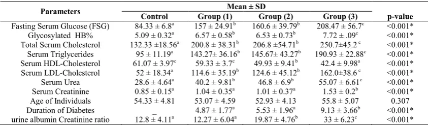
Table 1 Levels of all parameters in all studied groups
Parameters Mean ± SD
Control Group (1) Group (2) Group (3) p-value
Fasting Serum Glucose (FSG) 84.33 ± 6.8a 157 ± 24.91b 160.6 ± 39.79b 208.47 ± 56.7c <0.001*
Glycosylated HB% 5.09 ± 0.32a 6.57 ± 0.58b 6.53 ± 0.73b 7.72 ± .09c <0.001*
Total Serum Cholesterol 132.33 ±18.56a 200.8 ± 38.31b 206.8 ±54.71b 250.7±45.2 c <0.001*
Serum Triglycerides 95 ± 11.19a 143.27± 36.16b 145.67± 43.27b 190.93 ± 22.88c <0.001*
Serum HDL-Cholesterol 61.07 ± 3.97c 59.33 ± 3.7c 49.93 ± 9.41b 42.4 ± 9.98a <0.001*
Serum LDL-Cholesterol 52 ± 18.34a 114.6 ± 35.19b 124.6 ± 45.12b 162.0±38.6 c <0.001*
Serum Urea 28.6 ± 4.64a 40.2 ± 9.81b 46.8 ± 6.9b 55.07 ± 6.61c <0.001*
Serum Creatinine 0.85 ± 0.15a 1.04 ± 0.35a 1.01 ± 0.37a 1.53 ± 0.2b <0.001*
Age of Individuals 54.33 ± 4.81 53.07 ± 4.59 52.93 ± 4.13 55.8 ± 5.07 0.307
Duration of Diabetes _ 4.87 ± 1.77a 5.53 ± 1.96a 9.13 ± 3.66b <0.001*
urine albumin Creatinine ratio 12.8 ± 4.11a 12.27 ± 6.04a 19.87 ± 4.76b 33 ± 6.23c <0.001*
In Type 2 Diabetic Patients
RESULTS
There was high significant increase in the levels of fasting serum glucose in groups 1, 2 and 3 when compared to the control group (P < 0.001)
A statistically significant increase in the levels of fasting serum glucose in group 3 was detected when compared its values in (group 2 and group 1), but there was non-significant increase in group 1 when compared with group 2 (P < 0.001).
There was high significant increase in glycosylated HB% in (group1, 2 and 3) in comparison with the control group (P < 0.001) and also on comparing its percentage in group (3)with groups (1 and 2) (P < 0.001), but there was non-significant increase in group (2) when compared with group (1) (P < 0.001)
There was a significant increase of total serum cholesterol in groups (1, 2, 3) when compared to control group (p < 0.001), but there was a non-significant increase of total serum cholesterol in the diabetic group with retinopathy (group 2) when compared to diabetic group without retinopathy group (1) (p < 0.001). The cholesterol levels in group (3) was increased when compared with its corresponding levels in groups (2, 1) this increase was statistically significant (P < 0.001).
With respect to serum triglycerides, statistical- significant differences were observed in comparing all groups of diabetes (group 1, 2 and 3) with the control group (P < 0.001). Values of serum triglycerides showed significant increase in group (3) when compared with its corresponding values in groups (1, 2) and this increase was statistically significant (P< 0.001). But the levels of serum triglycerides in group (1) and (2) were increased but this increase was non-significant (P < 0.001).
There was significant decrease in HDL-cholesterol levels in diabetic groups with retinopathy (3, 2) when compared to its corresponding levels in the Control group (P < 0.001), but this decrease was non-significant on comparing HDL-cholesterol levels between group (1) and the control group, also there was a significant decrease in HDL-cholesterol levels in group (3) when compared to its levels in group (1, 2) (P < 0.001).
A statistical significant increase in LDL-cholesterol was observed in groups (1, 2, 3) when compared to control group (P < 0.001). The LDL- cholesterol levels in group (3) showed high significant increase when compared with its corresponding levels in group (1,2). This increase was statistically significant (P < 0.001). But there was a non-significant increase in LDL-cholesterol in group (2) of diabetics retinopathy when compared with group (1) of diabetics without retinopathy of (P < 0.001).
Values of serum urea in groups (1,2,3) showed high significant increase when compared to control group (P < 0.001). There was a significant increase in the levels of serum urea in group (3) when compared to groups (1, 2) (P < 0.0001), however there was non-significant increase in the levels of serum urea in group (2) when compared to group (1) (P < 0.001).
With respect to serum creatinine, a significant increase in the values of serum creatinine in group (3) was observed when compared to the control group, groups (1, 2) (P < 0.001), but there was non-significant difference between the levels of serum creatinine in group (1) and its corresponding level in the group (2) (P < 0.001). And also non-significant difference between the levels of serum creatinine in group (1, 2) when compared to the control group.
There was non-significant increase between the levels of urine albumin/creatinine ratio in group (1) and the control group (P < 0.001), but there was a high significant increase in group (3) when compared to groups (1, 2) and control group (1) (P < 0.001), also there was a high significant increase between the levels of urine albumin/creatinine ratioin group (1) and (2) (P < 0.001).
The duration of diabetes was increased in group (3) when compared to groups (1, 2) (P < 0.001), also the duration of diabetes was increased in group (2) over (1), but this increase was non-significant (P < 0.001).
Table 2 Comparison between (aspirin and non-aspirin) takers regarding different variables in uncomplicated
diabetic group.
Factors Takers(n=5) Non-takers(n=10) P
FBG 185.0±21.0 143.0±10.8 <0.001*
HbA1c 6.7±0.5 6.5±0.6 0.630
Cholesterol 202.6±43.9 199.9±37.7 0.903
TG 155.4±42.3 137.2±33.4 0.378
LDL 108.2±29.9 117.8±38.7 0.636
HDL 59.4±4.2 59.3±3.7 0.963
Urea 44.6±10.4 38.0±9.2 0.232
Creatinine 1.16±0.40 0.98±0.33 0.373
Ratio 14.4±8.5 11.2±4.6 0.352
Independent t-test, *Significant In uncomplicated diabetic group, aspirin takers had significantly higher FBG than non-aspirin takers.
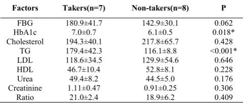
Table 3 Comparison between (aspirin and non-aspirin) takers regarding different variables in Pre-PDR group
Factors Takers(n=7) Non-takers(n=8) P
FBG 180.9±41.7 142.9±30.1 0.062
HbA1c 7.0±0.7 6.1±0.5 0.018*
Cholesterol 194.3±40.1 217.8±65.7 0.428
TG 179.4±42.3 116.1±8.8 <0.001*
LDL 118.6±34.5 129.9±54.6 0.646
HDL 46.7±10.4 52.8±8.1 0.228
Urea 49.4±8.2 44.5±5.0 0.176
Creatinine 1.11±0.47 0.91±0.25 0.306
Ratio 21.0±2.4 18.9±6.2 0.409
Independent t-test, *Significant In Pre-PDR group, aspirin takers had significantly higher HbA1c and TG than non- aspirin takers.
Table 4 Comparison between aspirin and non-aspirin takers regarding different variables in PDR group
Factors Takers(n=10) Non-takers(n=5) P
FBG 238.0±43.7 149.4±20.2 <0.001*
HbA1c 8.1±0.7 6.9±0.7 0.011*
Cholesterol 267.8±45.5 216.4±17.0 0.032*
TG 198.9±22.2 175.0±15.7 0.049*
LDL 179.0±32.5 128.0±25.9 0.009*
HDL 40.5±10.1 46.2±9.7 0.315
Urea 57.9±6.0 49.4±3.4 0.012*
Creatinine 1.63±0.16 1.32±0.08 <0.001*
Ratio 32.0±6.8 35.0±4.9 0.400
Independent t-test, *Significant
As regards the age, there was non-significant increase between all groups because the age of control group and diabetics groups were very near to each other.
DISCUSSION
Diabetic retinopathy is a potentially sight-threatening micovascular complication of diabetes (Williams, et al., 2004). Diabetic retinopathy is aprogressive disorder of the retinal microcirculation (klein, 2007) and it is a leading cause of vision-loss globally (Masliza, et al., 2016).The present study evaluated the factors affecting diabetic retinopathy. The most important risk factors identi ed for retinopathy were elevated glycosylated hemoglobin A1c (HbA1c), hyperlipidemia, microalbuminurea, duration of diabetes and special pharmacological treatment of DM. Our study revealed that the duration of DM of more than 9-10 years is significantly associated with PDR as confirmed in other studies (Masliza,
et al., 2016; Tung, Liu, Lee, Chen, Li & Chou, 2006). Therefore the duration of diabetes considered to be the strongest factor associated with the progression of retinopathy, similar to results reported in other studies (Mallika, et al.,
2012; Maberley, et al., 2002). Type 2 DM has almost always
been present for several years before presentation. Therefore, patients with type 2 DM must be screened for complications from the time of presentation, as these patients are already at risk.
In this study we agreed with studies that reported a signi cant association between retinopathy and triglyceride levels (Ajith,
et al., 2015). In contrast with Dornan’s study (Dornan, et al., 1982), which didnot nd any association between triglyceride levels in patients with proliferative retinopathy and normal patients. Many studies showed the association between serum lipids and diabetic retinopathy (Milijanovic, Glynn, Nathan,
Manson & Schaumberg, 2004), but types of lipids were not the
same. Several studies reported a relation between
hyperlipidemia and the risk of diabetic retinopathy (Ajith,
et al., 2015). The present study showed that healthy (i.e. high) HDL levels are significantly associated with a reduction in DR risk, while the opposite is true for low HDL levels, and this agreed with the Diabetes Control and Complication Trial (DCCT), higher HDL levels were also inversely associated with increased risk of retinopathy (Lyons, et al., 2004).
The results of this study reported that Patients with high HbA1c % are prone to a worsening of proliferative diabetic retinopathy compared with patients with low HbA1c % and agreed with the fact that HbA1c is one of the most important risk factors for the development and progression of diabetic retinopathy (Waked, Nacouzi & Haddead, 2006). Also disagreed with another study that showed there was no signi cant difference between retinopathy and HbA1c (Salwa
Selim & Ayman S.A., 2015). In the current study, there was
significant association between high LDL cholesterol, total serum cholesterol level and diabetic retinopathy and this agreed with (Ajith, et al., 2015). Others have reported no significant difference between retinopathy and high LDL cholesterol and total serum cholesterol level (Salwa Selim & Ayman S.A., 2015). In the current study, we observed an association between hyperglycemia and the severity of diabetic retinopathy, as descried previously in the UKPDS (Stratton,
et al., 2006), in contrast with another study that reported
non-significant difference between retinopathy and fasting plasma glucose (FPG), (Pradeepa, Mohan, Ganesan & Rema, 2008). Ourstudy reported that presence of microalbuminurea was a strong risk factor with the incidence of proliferative diabetic retinopathy as stated elsewhere which agrees with (Newman,
et al., 2005;Klein, et al., 2005). The association between microalbuminuria and DR observed in the present study could be explained by the fact that microalbuminuria might represent a state of generalized vascular dysfunction. Enzymes involved in the metabolism of anionic components of the extracellular matrix (e.g. heparansulphate proteoglycan) vulnerable to hyperglycemia, seem to constitute the primary cause of albuminuria and its associated complications. Genetic polymorphism of such enzymes, as well as of several candidate genes, has been hypothesized to be the main reason forthe variation in susceptibility.
The current study replicates non-significant relation between retinopathy and age similar to the study (Salwa Selim I.A., &
Ayman S.A., 2015). There have been both concerns that aspirin
use might worsen diabetic retinopathy, as well as hopes that aspirin might be beneficial in treating it. All previous trials showed that aspirin alone or in combination with dipyridamole neither lowered nor increased the risk of the development of diabetic retinopathy. The results suggest that there are no ocular contraindications for taking aspirin if required as part of a treatment for cardiovascular diseases or other medical indications (Samuel. L., R Joel Welch & Diana. V. Do, (2015).
Yuan Shi, et al, have showed the cross-sectional association
between aspirin therapy taken and DR among diabetic patients in a population based study. Furthermore vision threatening diabetic retinopathy (VTDR) was defined as the presence of severe non-proliferative DR, proliferative DR or clinically significant macular edema. The relation between aspirin use and the occurrence of either DR or VTDR was showed using multi variable logistic regressions. (Yuan Shi, et al., 2016).
This current study concluded that persons with diabetes who were on aspirin were more likely to have DR, particularly PDR. Whether these findings pointed to a possible complication of taking aspirin or whether it reflects increased severity of diabetes among patients on aspirin requires, further clinical studies are required and cardiovascular benefits of daily low dose aspirin for certain adults are significant and life-saving.
CONCLUSION
The current study concluded that hyperglycemia, duration of diabetes > 9-10 years, hyperlipidemia, presence of microalbuminurea and persons with diabetes who were on aspirin are significantly associated with PDR.
References
Ajith V. L., Gilsa E. S., Sudha V., Pushpalatha M., GeethaDamodaran K.,& M .A. Andrews.(2015).
Serum Lipids and Apolipoproteins in Diabetic Retinopathy: A Case Control Study. IOSR Journal of Dental and Medical Sciences, 14 (2): 70-73.
American Diabetes Association. (2006). Standards of medical care in diabetes, Diabetes Care 2005, 29:S4-2.
In Type 2 Diabetic Patients
American Diabetes Association. (2009). Standards of medical care in diabetes. Diabetes care, 32 (Supplement 1), S13:S61.
American Diabetes Association (2010).clinical practice recommendation, 33 (supplement 1).
American Diabetes Association. (2017). Classification and Diagnosis of Diabetes. Diabetes Care, 40 (Supplement 1):S11-S24.
Association of glycaemia with macrovascular and micovascular complications of Type 2 diabetes: prospective observational study. (2000). British Medical Journal, 321 405-412.
Bird AC., Bressler NM., Bressler SB., et al.(1995). An international classification and grading system for age-related maculopathy and age-related macular degeneration. The International ARM Epidemiological Study Group. SurvOphthalmol, 39:367-74.
Bobeck S., Modjtahedi, Namrata Bose, Thanos D., Papakostas, Lawrence Morse, D.G.V., & Amar U. Kishan.(2016). Lipids and Diabetic Retinopathy. Seminars in Ophthalmology journal, 31: 1-2.
Calderon, OH Juarez, GE Hernandez, SM Punzo and ZD De la Cruz(2017).Oxidative stress and diabetic retinopathy: development and treatment, Eye, 1-9.
De Jong PT, Chakravarthy U, Rahu M, et al (2012). Associations between aspirin use and aging macula disorder: the European Eye Study. Ophthalmology. 2012; 119-112-8. Diabetes. co.uk, http://www. diabetes.co.uk.
Ding, J. & Wong, T. Y. 2012. Current epidemiology of diabetic retinopathy and diabetic macular edema. Curr.Diab. Rep. 12, 346-354.
Dornan T., Carter R., Bron A., Turner R., & Mann J.(1982) . Low density lipoprotein cholesterol: an association with the severity of diabetic retinopathy. Diabetologia, 22:167-70.
Early treatment diabetic retinopathy study (ETDRS). (2006). Elisa Martín-Merino, Joan Fortuny, Elena Rivero-Ferrer,
Marcus Lind & Luis Alberto Garcia-Rodriguez. (2016). Risk factors for diabetic retinopathy in people with Type 2 diabetes: A case-control study in a UK primary care setting. primary care diabetes 1 0 : 300-308.
Fong DS, Aiello L., Gardner TW, King GL, Blankenship G., Cavallerano JD., et al.(2003).Diabetic retinopathy. Diabetes Care, 26:S99-S10.
Friedewald, W. T., Levy, R. I., & Fredrickson, D. S. (1972). Estimation of the concentration of low-density lipoprotein cholesterol in plasma, without use of the preparative ultracentrifuge. Clinical Chemistry, 18, 499-502.
Gunjan Prakash, Rachit Agrawal, Tanie Natung. (2017). Role of Lipids in Retinal Vascular and Macular Disorders. Ind J ClinBiochem, 32(1):3-8.
Ha H., Hwang IA., Park JH., & Lee HB.(2008). Role of reactive oxygen species in the pathogenesis of diabetic nephropathy. Diabetes Res ClinPract, 1:S42-S45. Heilig CW, Deb DK, Abdul A., Riaz H., James LR, Salameh J.,
Nahman Jr NS. (2013). GLUT1 regulation of the pro-sclerotic mediators of diabetic nephropathy. Am J Nephrol, 38:39-49.
Hennekens CH, & Dalen JE. (2013). Aspirin in the treatment and prevention of cardiovascular disease: past and current perspectives and future directions. Am J Med, 126-3738.
Ishibashi Y, Yamagishi S, Matsui T, Ohta K, Tanoue R, Takeuchi M, Ueda S, Nakamura K, Okuda S.(2012). Pravastatin inhibits advanced glycation end products (AGEs)-induce dproximal tubular cell apoptosis and injury by reducing receptor for AGEs (RAGE) level. Metabolism, 61:1067_1072.
John, W. G. (1997). Glycated haemoglobin analysis. Annals of clinical biochemistry, 34, 17-31.
Karly Pippitt, & Marlana li, Holly e., Gurgle. (2016).Diabetes Mellitus: Screening and Diagnosis. Am Fam Physician, 93(2):103-109.
Klein, B. Zinman, R. Gardiner, S. Suissa, S.M. Donnelly, et al,. (2005). The relationship of diabetic retinopathy to preclinical diabetic glomerulopathy lesions in Type 1 diabetic patients. The renin-angiotensin system study, Diabetes, 54: 527-533.
Klein B. (2007). Overview of epidemiologic studies of diabetic retinopathy. Ophthalmic Epidemiol, 14:179-83.
Klein BE, Howard KP, Gangnon RE, et al.(2012). Long-term use of aspirin and age-related macular degeneration. JAMA, 308:2469-78.
Klein R., Klein B., & Moss S. (1989). The Wisconsin Epidemiologic Study of Diabetic Retinopathy: a review. Diab Met Rev 5:559-70.
Klein R., Klein BE & Moss SE.(1992). Epidemiology of proliferative diabetic retinopathy. Diabetes Care, 15(12): 1875-1891.
Liew G., MitchellP., Wong TY., RochtchinaE., & Wang J J.(2013). The association of aspirin use with age-related macular degeneration. JAMA Int Med. 173:258-64. 18. Lyons, A.J., Jenkins, D. Zheng, Garvey, et al.(2004). Diabetic
retinopathy and serum lipoprotein subclasses in the DCCT/EDIC cohort, Invest. Ophthalmol. Vis. Sci, 45: 910-918.
Maberley D., King W., Cruess A., et al.(2002). Risk factors for diabetic retinopathy in the Cree of James Bay. Ophthalmic Epidemiol ,9:153-67.
Malaysia MoH.(2011). Clinical practise guideline on screening of diabetic retinopathy.
Mallika PS, Aziz S., GohP.,et al.(2012). Diabetic retinopathy in native and non-native sar- awakians-findings from the diabetic eye registry. Med J Malays, 67(4): 369.
Masliza H., Mohd Ali, MMed, NaniDraman, et al. (2016).Predictors of proliferative diabetic retinopathy among patients with type 2 diabetes mellitus in Malaysia as detected by fundus photography. Journal of Taibah University Medical Sciences, 11(4): 353-358.
Miljanovic B., Glynn R., Nathan D., Manson J., Schaumberg D. (2004). A prospective study of serum lipids and risk of diabetic macular edema in type 1 diabetes. Diabetes, 53:2883-92.
.Newman, M.B. Mattock, A.B. Dawnay, et al.(2005). Systematic review on urine albumin testing for early detection of diabetic complications, Health Technol. Assess. 9: iii-vi, xiii-163.
Pagana KD., Pagana TJ. Mosby’s Manual of Diagnostic and Laboratory Tests, 4th ed. St. Louis: Mosby Elsevier. (2010).
Pradeepa, R., Anjana, R. M., Unnikrishnan, R., Ganesan, A., et al. (2010). Risk factors for microvascular complications of diabetes among South Indian subjects with type 2 diabetes: The Chennai Urban Rural Epidemiology Study (CURES) Eye Study-5. Diabetes Technology & Therapeutics, 12, 755-761.
Pradeepa B., Mohan V., & Ganesan A., Rema M.(2008). Complications risk factors for diabetic retinopathy in a South Indian Type 2 diabetic population, the Chennai Urban Rural Epidemiology Study (CURES) Eye Study 4R. J Complication Diabetic Med UK, 25:536-42. Rainsford KD. (2007). Inflammatory drugs in the 21st century.
PubMed, SubcellBiochem, 42:3-27.
Salwa Selim Ibrahim Abougalambou, Ayman S. Abougalambou.(2015). Risk factors associated with diabetic retinopathy among type 2 diabetes patients at teaching hospital in Malaysia. Diabetes & Metabolic Syndrome: Clinical Research & Reviews, 9:98-103. Samuel. L. Thomsen, R Joel Welch, Diana. V. Do. (2015).
Medical management of diabetic retinopathy In :R.P.Singh (ed), Managing Diabetic Eye Disease in Clinical Practice, Switzerland, pp.39-47.
Stratton, C.A. Cull, A.I. Adler, D.R. Matthews, et al,.(2006). Additive effects of glycaemia and blood pressure exposure on risk of complications in type 2 diabetes: a prospective observational study (UKPDS 75), Diabetologia, 49: 1761-1769.
Tapp R., Zimmet P., Harper C., Mccarty D., Chitson P., Tonkin A., et al,.(2006). Six-year incidence and progression of diabetic retinopathy: results from the Mauritius diabetes complication study. Diabetes Res ClinPract, 73:298-303.
Thomas A., Ciulla., Armando G., Amador & Bernard Zinman.(2003). Diabetic retinopathy and Diabetic macular edema. Diabetes Care, 26:2653-2664.
Tung T., Liu J., Lee F., Chen S., LI A., & Chou P.(2006). Population-based study of non- proliferative diabetic retinopathy among type 2 diabetic patients in Kinmen, Taiwan. JpnJ Ophthalmol, 50(1):44-52.
Vane JR1, Botting RM.(2003).The mechanism of action of aspirin. PubMed, Thromb Res; 110(5-6):255-8.
Voutilainen-Kaunisto R., Terasvirta M., Uusitupa M., Niskanen L.(2001). Occurrence and predictors of retinopathy and visual acuity in type 2 diabetic patients and control subjects. 10-year follow-up from the diagnosis. J Diabetes Complications, 15:24-33.
Waked N., Nacouzi R., Haddead N.(2006). Epidemiology of diabetic retinopathy in Lebanon. J. Fr. Ophtalmol, 29: 289-295.
William Blahd. (2016). Diabetic Nephropathy. http://www.webmd.com/diabetes/guide/diabetes-kidney-disease.
Williams R., Airey M., Baxter H., et al. (2004) Epidemiology of diabetic retinopathy and macular oedema: a systematic review. Eye, 18:963-83.
Yamagishi S., Matsui T., Nakamura K. (2007). Curr Drug Targets, 8: 1138-43.
Yamagishi S., Nakamura K., Matsui T.(2009), Pharmacol Res, 60, 174-8.
Yuan Shi, Yih-Chung Tham, Robyn J. Tapp, Gavin Tan1,Paul Mitchell, Jie Jin Wang, Yin-Bun Cheung, Ching-Yu Cheng, Tien Yin Wong. (2016). Aspirin and Diabetic Retinopathy: the Singapore Epidemiology of Eye Disease (SEED) study. Invest Ophtalmol.Vis.Sci, 57(12):1595.
*******
How to cite this article: