*
Cor
Tami
1 Depa
Unive Bhara Camp
2 Cent
Resea Missio
Intr
In th a lot demo vario treat deve prod area Plant tanni have Cipa is a brow white glabr oppo smal ThisIn vitr
Thirun
rresponding
ilarasan M
artment of Biotec rsity College of E athidasan Institut pus, Trichy 620
tre for Genetic S arch, The Madra on, Chennai 60
oduction
e recent years, t of attention gl onstrate the pro ous traditional,
ment of huma eloped between
ucts and they s of infectious d ts are rich in a w ins, terpenoids, e been found in v adessa bacifera
shrub, usually 1 wn, ribbed, cover e lenticels. Leav rous or covered osite; leaflet bla ller basally than
s work is licen
tro
antimic
phytoche
navukarasu T
g author:
chnology, Engineering, te of Technology 0024, India.
Studies & as Medical
00037. India.
researches on m obally. Evidence omising potentia complementary n diseases [1] n 1981 and 2 have been very isease and canc wide variety of s alkaloids, flavo vitro to have anti belongs to the fa 1- 4 m tall whic red with yellow p ves are 8-30 cm d with yellow tr ades ovate to o
apically on rach
sed under a C
http
crobial, a
emical an
1
, Santhana L
y
A b s
To stud thrombo The thr antimicr antioxid haemoly activity Among activitie (Sphing faecalis to incre 100.92μ be high % clot l The phy This stu bioactiv compou .Keywo haemoly medicinal plants es have been a al of medicinal y, and alternativ
]. About 61% 002 were bas y successful es cer [2].
secondary meta onoids, glycosid imicrobial prope amily of Meliace h has young br pubescence and m; petiole and r richomes; leaflet ovoid oblong, 3 his. Secondary ve
Creative Comm
p://www.arjo
Origin
ntioxidan
alysis of
Lakshmi K
1, T
s t r a c t
dy the leaves olytic activities a ree extracts (me robial activity ag dant activity of e
ytic activity was by clot disruptio
the different ext s. The most s gomonas, Klebs s) and fungai (Ca ase with increas μ0.41 øg/mL wh er in chloroform lytic whereas sta ytochemical eva udy shows that vity but further und.
rds: Antimicrob ysis.
s have attracted accumulated to plants used in ve systems of of new drugs ed on natural specially in the abolites such as des, etc., which
rties [3, 4]. eae; C. bacifera
anches grayish sparse grayish rachis cylindric, ts usually 9-13 .5-10*1.5-5 cm eins are 8-10
mons Attributi
ournals.org/in
nal Researc
nt, haemo
Cipadess
Tamilarasan M
of Cipadessa and to perform ph
ethanol, cyclohe gainst eight pat extract was stud determined usin n and phytochem tracts tested, the susceptible mic bsiella pneumon Candida albicans sing concentratio hich was found in m extract than m
andard streptok luation indicates
the methanol a compound iso bial activity, an
on ea white with s The p haemo extrac
Mate
Colle
The f collect Tamiln and lo are ge identif
ion 3.0 Licens
ndex.php/ijpm
ch Article
olytic, thro
ssa bacife
M
1*, Sivamani
a bacifera for t hytochemical ev exane, chloroform
thogenic microo died using hydro ng agar diffusion mical potential b e methanol extr cro-organisms w niae, Citrobacte s). H2O2 scaveng
on of the extract n cyclohexane e methanol, cyclohe inase shows 30 s the presence o and chloroform lation is neces ntioxidant, phyto
ch side of mid v or yellow, linear parse appressed purpose of this w olytic, thromboly ct of C. bacifera
erials and m
ection of plant
fresh and heal ted from Pac nadu, India. The ongitudes 78 21 enerally classifie fied and authen
se.
m/index
e
ombolytic
era
leaves
S
2, Sangeeth
their antimicrob valuation.
m) of Cipadessa organisms by w ogen peroxide ra n techniques on by qualitative ana
act of leaves sh were found to er), Gram posi ging activity of C t. IC50 values of
extract. The ha exane and the m
.86 % clot lytic of chemical cons extract of leave ssary to confirm ochemical evalu
vein. Flowers ar r to oblong-ellipt d pubescence. work is to evalu
ytic activities an leaf.
methods
materials
thy leaves of chaimalai hills e area falls with
′ 0″ E. These a ed as rural area nticated by pla
c activities
s extracts
ha D
1, Rajesh
bial, antioxidant sa bacifera were well diffusion me
adical scavengin blood agar plate alysis. howed significan
be Gram nega itive bacteria ( Cipadessa bacife H2O2 scavengin
emolytic activity methanol extract activity in throm tituents. es of Cipadessa m the activities
uation, thrombo
re 3-4 mm in dia tic, 2-3.5 mm ou ate antimicrobia nd phytochemic
the plant C. b region of Sa in the latitudes areas consist of
a. The plant spe nt taxonomist,
s and
s
h TP
1t, haemolytic, e screened for
ethod. In vitro ng assay. The e, thrombolytic t antimicrobial ative bacteria (Enterococcus fera was found ng activity was y was found to t shows 14.63 mbolytic assay. a bacifera has of individual olysis activity,
ameter. Petals utside covered al, antioxidant, cal analysis of
bacifera were alem district, 11 10′ 48″ N villages which ecimens were Dr. G. V. S.
PAGE |
110
|
Murthy, Botanical Survey of India, Southern Regional Centre,Coimbatore.
Extract preparation
The extraction of the C. bacifera leaves was carried out using known standard procedures [5]. The plant leaves were dried in shade and powdered in a mechanical grinder. The powder (20.0 g) of the plant leaves was soaked with 100 mL of chloroform, methanol and cyclohexane by using a Soxhlet extractor for 72 hours at a temperature not exceeding the boiling point of the solvent. The extracts were filtered using Whatman filter paper (No.1) while hot, concentrated in vacuum under reduced pressure using rotary flask evaporator, and dried in a desiccator. More yields of extracts were collected by this method of extractions. The extracts were then kept in sterile bottles, under refrigerated conditions, until further use. The dry weight of the plant extracts was obtained by the solvent evaporation and used to determine concentration in mg/ml. The extract was preserved at 2 to 4 C. This extract was used for further investigation.
Preliminary phytochemical screening
The extracts were subjected to preliminary phytochemical testing to detect for the presence of different chemical groups of compounds. C. bacifera leaves extract were screened for the presence of alkaloids, flavonoids, carbohydrates, glycosides, phenolic compound, tannins, triterpenoids, cardinolides, anthraquinones as described in literatures [6, 7, 8].
Test microorganisms and growth media
The following microorganisms Sphingomonas, Pseudomonas aeuroginosa, Klebsiella pneumoniae, Enterococcus faecalis, Citrobacter and Proteus mirabilis and fungal strains Candida albicans, Aspergillus niger were chosen based on their clinical and pharmacological importance [9]. All microbial cultures were obtained from Plant Biotechnology Laboratory, Department of Biotechnology, University College of Engineering BIT campus, Trichy. The bacterial and fungal stock cultures were incubated for 24 hours at 37 C on nutrient agar and potato dextrose agar (PDA) medium respectively, following refrigeration storage at 4 C. The bacterial strains were grown in Mueller-Hinton agar (MHA) plates at 37 C (the bacteria were grown in the nutrient broth at 37 C and maintained on nutrient agar slants at 4 C, whereas the fungal strains were grown in PDA media, respectively, at 28 C. The stock cultures were maintained at 4 C.
Antimicrobial activity
In vitro antibacterial and antifungal activities were examined for chloroform, methanol and cyclohexane extracts. Antibacterial and antifungal activities of plant part extracts against six pathogenic bacteria (one Gram positive and six Gram negative) and two pathogenic fungi were investigated by the well diffusion methods.
Agar plates were inoculated with 100 μL of standardized inoculums (1.5 x 10⁸ CFU/mL) of each selected bacterium (in triplicates) and
spread with sterile swabs. Wells of 6 mm size were made with sterile borer into agar plates containing the bacterial inoculums and the lower portion was sealed with a little molten agar medium. The sets of three dilutions (10, 25 and 50 μg/mL) of plant leaves extracts (chloroform, methanol and cyclohexane solvent) were poured into a different well of inoculated plates. Control experiments were carried out under similar condition by using cefotaxime for antibacterial activity and nystatin for antifungal activity as standard drugs. Chloroform, methanol and cyclohexane were used as a negative control which was introduced into a well instead of plant extract. The zones of growth inhibition around the well were measured after 18 to 24 hours of in incubation at 37 C for bacteria and 48 to 96 hours for fungi at 28 C. The sensitivities of the microorganism species to the plant extracts were determined by measuring the sizes of inhibitory zones (including the diameter of disk) on the agar surface around the disks, and values <8 mm were considered as not active against microorganisms.
Antioxidant activity
The ability of the extracts to scavenge hydrogen peroxide was determined according to Ruch et al., [10]. A solution of hydrogen peroxide (40 mM) was prepared in phosphate buffer (pH 7.4). The concentration of hydrogen peroxide was determined by absorption at 230 nm using a spectrophotometer. Different concentrations of chloroform, methanol and cyclohexane leaf extracts (2-10 øg/ mL) were added to a hydrogen peroxide solution (0.6 mL, 40 mM). The total volume was made up to 3 mL with phosphate buffer. The absorbance of hydrogen peroxide at 230 nm was determined after ten minutes against a blank solution containing phosphate buffer without hydrogen peroxide. The percentage of hydrogen peroxide scavenging by the extracts and standard compounds was calculated as follows: % Scavenged [H2O2] = [(AoA1)/Ao] x 100
where Ao was the absorbance of the control and A1 was the
absorbance in the presence of the sample of extract and standard.
Haemolytic assay
The haemolytic activity of the extract was determined using agar diffusion technique on blood agar plate [11]. Blood agar was prepared and well measuring 5 mm were made on the agar using cork borer. The wells were filled with 20 øL of different concentration of plant extracts solution. The plates were then incubated at 37 ÀC for 5 hours.
Thrombolytic assay
PAGE |
111
|
without disturbing the clot and the tubes were again weighed todetermine the clot weight (clot weight = weight of the tube containing clot weight of the empty tube). To the each Eppendorf tube containing pre weighed clot, 20 øL, 40 øL, 60 øL, 80 øL and 100 øL of chloroform, methanol and cyclohexane extract were added. For negative control, 50 øL of sterile distilled water was used. All the tubes were incubated at 37 ÀC for 18 hrs and observed for clot lysis. The fluid obtained after the incubation was removed carefully and the tubes were weighed again to observe the difference in weight after clot disruption. Difference in the weight taken before and after clot lysis was expressed as percentage of clot lysis.
Results
Preliminary phytochemical investigation
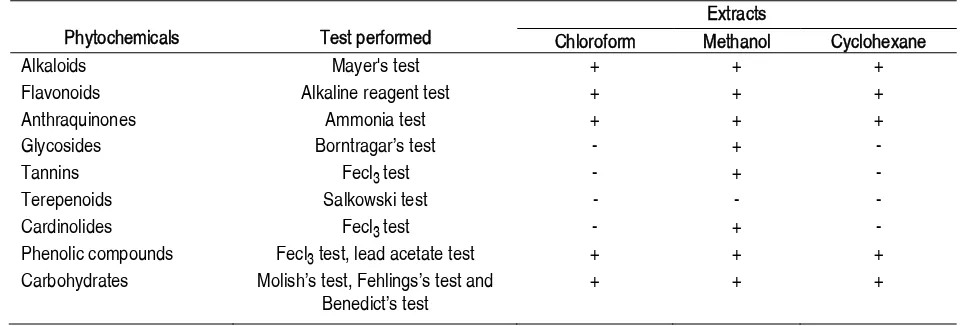
The preliminary phytochemical investigation of the methanolic extract of C. bacifera showed that it contains alkaloids, flavonoids, anthraquinones, glycosides, tannins, cardinolides, phenolic compound, carbohydrates. Triterpenoids were not present in methanol extract. Glycosides, tannins, triterpenoids, cardinolides
were not detected in both chloroform and cyclohexane extracts (Table 1).
Microbial activity
The antimicrobial activity of the leaf extracts of C. bacifera were tested in different concentrations (10, 25 and 50 μg/mL) against six pathogenic bacterial strains, one Gram positive (Enterococcus faecalis) and five Gram negative (Sphingomonas, Pseudomonas aeuroginosa, Klebsiella pneumoniae, Citrobacter and Proteus mirabilis), and two fungal strains (Candida albicans, Aspergillus niger). The extracts showed varying degrees of antimicrobial activity against tested microorganisms, values which are presented in Table 2. Methanol extracts exhibited higher degrees of antimicrobial activity than the other extracts. Among the three extract, cyclohexane extracts showed least inhibition of growth of microorganisms. The inhibitory effects of the extracts were compared with the standard antibiotics such as cefotaxime for bacteria and nystatin for fungal strain.
Table 1 Results of preliminary phytochemical tests for the presence of active constituents in C. bacifera leaves
Phytochemicals Test performed
Extracts
Chloroform Methanol Cyclohexane
Alkaloids Mayer's test + + +
Flavonoids Alkaline reagent test + + +
Anthraquinones Ammonia test + + +
Glycosides BorntragarÊs test - +
-Tannins Fecl3 test - +
-Terepenoids Salkowski test - -
-Cardinolides Fecl3 test - +
-Phenolic compounds Fecl3 test, lead acetate test + + +
Carbohydrates MolishÊs test, FehlingsÊs test and BenedictÊs test
+ + +
Scavenging of hydrogen peroxide
The ability of C. bacifera leaf extract to scavenge hydrogen peroxide was determined according to the method of Ruch et al., [10] and is shown in Table 3 and compared with that of ascorbic acid as standard and the highest IC50 was estimated as 100.92μ0.41 øg/mL. There was a statistically significant correlation between those values and the control (p < 0.01).
Haemolytic activity
PAGE |
112
|
Table 2 Antimicrobial activity of leaves of C. bacifera+ve control for bacteria: cefotaxime; +ve control for fungai: nystatin (μg/ml); Results represented as means μ standard deviation (n = 3); NA: No activity.
Table 3 Antioxidant activity of C. bacifera leaf extract Percentage of H2O2scavenging activity
Concentration (øg/mL) Methanol Cyclohexane Chloroform
20 14.4μ0.2 8.8μ0.4 10μ0.1
40 23.3μ0.2 17.1μ0.2 31μ0.2
60 31μ0.2 30μ0.0 29μ0.3
80 46μ0.1 42μ0.0 48μ0.6
100 56μ0.4 57μ0.7 66μ0.2
IC50 øg/mL 97.832μ0.35 100.925μ0.41 79.799μ0.05
Results represented as means μ standard deviation (n = 3).
Table 4 Zones of haemolysis (mm) of C. bacifera leaf extract at different concentration Extract
Zones of haemolysis (mm) Concentration of crude extract (mg/mL)
25 50 100
Methanol 6μ0.2 6μ0.1 6μ0.4
Cyclohexane 6μ0.1 6μ0.1 6μ0.3
Chloroform 7μ0.2 6μ0.3 7μ0.1
Results represented as means μ standard deviation (n = 3). Microorganism
Zone of inhibition (mm)
Methanol (øg/mL) Cyclohexane (øg/mL) Chloroform (øg/mL)
10 25 50 +ve 10 25 50 +ve 10 25 50 +ve
Bacteria Gram Negative
Sphingomonas 15μ0.2 16μ0.1 19μ0.4 23μ0.4 4μ0.3 9μ0.3 11μ0.3 34μ0.6 NA NA NA 30μ0.6 Pseudomonas
aeuroginosa
14μ0.1 14μ0.3 17μ0.1 27μ0.3 12μ0.2 11μ0.3 12μ0.4 30μ0.2 10μ0.3 13μ0.1 13μ0.3 27μ0.2 Klebsiella
pneumonia
14μ0.3 14μ0.2 15μ0.3 28μ0.3 10μ0.4 11μ0.2 13μ0.3 30μ0.3 13μ0.3 13μ0.2 14μ0.4 25μ0.6 Citrobacter 14μ0.1 16μ0.3 12μ0.6 24μ0.2 9μ0.1 11μ0.0 12μ0.1 35μ0.2 12μ0.1 12μ0.1 14μ0.1 30μ0.2 Proteus
mirabilis
10μ0.3 13μ0.3 15μ0.2 25μ0.6 11μ0.4 14μ0.1 12μ0.6 30μ0.1 10μ0.6 14μ0.4 16μ0.2 27μ0.0 Gram Positive
Enterococcus faecalis
13μ0.1 14μ0.4 20μ0.3 26μ0.2 12μ0.6 11μ0.2 11μ0.1 25μ0.4 10μ0.6 12μ0.0 15μ0.2 29μ0.1 Fungai
Candida albicans
NA 2μ0.1 4μ0.3 6μ0.0 6μ0.2 8μ0.1 10μ0.2 16μ0.3 5μ0.2 6μ0.1 7μ0.4 12μ0.2 Aspergillus
niger
PAGE |
113
|
Table 5 Effect of C. bacifera leaf extracts on in vitro clot lysisExtract
% of clot lysis
Concentration of crude extract (øg/mL)
20 40 60 80 100
Methanol 5 7.5 11.9 14.29 14.63
Cyclohexane 2.63 5.26 7.89 10 12.19
Chloroform 2.44 4.88 7.32 7.69 12.82
Thrombolytic activity
The in vitro thrombolytic activity study revealed that methanol, cyclohexane and chloroform extract showed 14.63%, 12.19%, and 12.82% clot lysis respectively for 100 øg/mL and compared with the negative control (methanol, cyclohexane, chloroform solvent). Statistical representation of the effective clot lysis percentage is tabulated in Table 5.
Discussion
Natural products are in great demand owing to their extensive biological properties and bioactive components which have proved to be useful against large number of diseases. It is proved that present extracts of C. bacifera leaves showed wide array of activities like antimicrobial, antioxidative, antihaemolytic and anti thrombolytic.
In the present work, the extracts obtained from C. bacifera show strong activity against most of the tested bacterial and fungal strains. The results were compared with standard antibiotic drugs. In this screening work, extracts of C. bacifera were found to be active against Gram-positive, Gram-negative, and fungal strains were resistant to all the extracts of C. bacifera except Aspergillus niger. There was no activity was found in chloroform extract against Sphingomonas.
Hydrogen peroxide can be formed in vivo by many oxidase enzymes such as superoxide dismutase. It can cross membranes and may slowly oxidize a number of compounds. The ability of C. bacifera leaves extract to scavenge hydrogen peroxide was determined according to the method of Ruch et al. The C. bacifera chloroform extracts were capable of scavenging hydrogen peroxide in a concentration-dependent manner. IC50 for scavenging of H2O2
were 79.79μ0.05 μg/mL for chloroform extract, 97.83μ0.35 μg/mL for methanol extract and 100.92μ0.41 μg/mL for cyclohexane respectively. The IC50 values for ascorbic acids were 21.4μ0.12
μg/mL. The effectiveness of the leaves might be due to the hydroxyl groups existing in the phenolic compounds chemical structure [12] that can provide the necessary component as a radical scavenger. A potent scavenger of free radicals may serve as a possible preventive intervention for the diseases [13].
This study also shows the presence of different phytochemicals with biological activity that can be of valuable therapeutic index. The result of phytochemicals in the present investigation showed that the plant contains more or less same components like alkaloids, flavonoids, anthraquinones, glycosides, tannins,
cardinolides, phenolic compounds and carbohydrates. Results show that plant rich in anthraquinones and phenolic compounds have been shown to posseÊs antimicrobial activities against a number of microorganisms.
The chloroform extract of C. bacifera shows antihaemolytic activity. The activity of the extract to lyse the blood cell can be linked with bioactive components. It is well documented that flavonoids and the polyphenolic compounds which showed potential beneficial effects on human health and posseÊs antiviral, anti-inflammatory, antitumor, antihaemolytic and antioxidative activity [14]. The zone of haemolysis was directly proportional to concentration of the extract.
Now-a-days, about 61% of the pharmaceuticals are prepared from plants worldwide [2]. A number of studies have been conducted by various researchers to find out the herbs and natural food sources and their supplements having antithrombotic (anticoagulant and antiplatelet) effect and there is evidence that consuming such food leads to prevention of coronary events and stroke [15, 16, 17, 18]. There are several thrombolytic drugs obtained from various sources. Some are modified further with the use of recombinant technology in order to make these thrombolytic drugs more site specific and effective [17]. Side effects related to these drugs have been reported that lead to further complications [18]. Sometimes the patients die due to bleeding and embolism [19, 20, 21, 22]. In our study the in vitro thrombolytic activity results revealed that methanol, cyclohexane and chloroform extracts showed 14.63%, 12.19%, and 12.82% clot lysis respectively for 100 mg/mL and compared with the negative control (methanol, cyclohexane and chloroform solvent).
Conclusion
PAGE |
114
|
References
[1].Borchers AT, Hackman RM, Keen CL, Stern JS, Gershwin ME. Complementary medicine: a review of immunomodulatory effects of Chinese herbal medicines. Am J Clin Nutr. 1997; 66(6):1303-12.
[2].Cragg GM, Newman DJ. Biodiversity: A continuing source of novel drug leads. Pure Appl Chem. 2005;77:7-24.
[3].Cowan MM. Plant products as anti-microbialagents. Clin Microbiol Rev. 1999;12:564-82.
[4].Dahanukar SA, Kulkarni RA, Rege NN. Pharmacology of medicinal plants and natural products. Indian J Pharmacol. 2000;32:S81 S118.
[5].Harborne JB. Phytochemical methods: A guide to modern techniques of plant analysis. Chapman and Hall. Newyork: 1973.
[6].Kokate CK. Practical Pharmacognosy. Delhi: New Gyan Offset Printers; 2000.
[7].Khandelwal KR. Practical Pharmacognosy. Pune: Nirali Prakashan. 2nd ed. 2009.
[8].Kumar A, Ilavarasan R, Jayachandran, Decaraman M, Aravindhan P. Phytochemicals Investigation on a tropical plant in South India. Pak J Nutr. 2009;8:83-5.
[9].McCracken WA, Cowsan RA. Clinical and Oral Microbiology. New York: Hemispher Publishing Corporation; 1983.
[10].Ruch RJ, Cheng SJ, Klaunig JE. Prevention of cytotoxicity and inhibition of intracellular communication by antioxidant catechins isolated from Chinese green tea. Carcinogenesis. 1989;10:1003-1008.
[11].Ahmed F, Islam MA, Rahman MM. Antimicrobial activity of Leonurus sibiricus aerial parts. FITOTERAPIA. 2006;77:316-317.
[12].Pourmoard F, Hosseinimehr SJ, Shahabimajd V. Antioxidant activity, phenol and flavonoid contents of some selected Indian medicinal plants. Asian J Biotech. 2006;6:1197-201.
[13].Gyamfi MA, Yonamine M, Aniya Y. Free radical scavenging action of medicinal herbs from Ghana Thonningla sanguine on experimentally induced liver injuries. General Pharmacol. 1999;32:661- 67. [14].Buhler DR and Miranda C. A net article:
Antioxidant activities of Flavonoids, Department of Environmental and Molecular Toxicology Oregon State University. 2000.
[15].Gillman MW, Cupples LA, Gagnon D, Posner BM, Ellison RC, Castelli WP, Wolf PA. Protective effect of fruits and vegetables on development of stroke in men. JAMA 1995;273:1113-1117. [16].Joshipura KJ, Ascherio A, Manson JE,
Stampher MJ, Rimm EB, Speizer FE. Fruit and vegetable intake in relation to
risk of ischemic stroke. JAMA 1999;282:1233-39.
[17].Liu S, Manson JE, Lee I-M, Cole SR, Hennekens CH, Willett WC, Buring JE. Fruit and vegetable intake and risk of cardiovascular disease: the WomenÊs Health Study. Am J Clin Nutr. 2000;72:922-28.
[18].Bazzano LA, He J, Ogden LG, Loria CM, Vupputuri S, Myers L, Whelton PK. Fruit and vegetable intake and risk of cardiovascular disease in US adults: the first National Health and Nutrition Examination Survey Epidemiologic Follow-up Study. Am J Clin Nutr. 2002;76:93-99.
[19].Gallus AS. Thrombolytic therapy for venous thrombosis & pulmonary embolism. Bailliere Clin Haem. 1998;11:663-73.
[20].Verstraete M. Third generation thrombolytic drugs. Am J Med. 2000;109:52-58.
[21].Wardlaw JM, Berge E, del Zoppo G, Yamaguchi T. Thrombolysis for acute ischemic stroke. Stroke. 2004;35:2914-15.
[22].Capstick T, Henry MT. Efficacy of thrombolytic agents in the treatment of pulmonary embolism. Eur Respir J. 2005;26:864-74.