E X T E N D E D G E N O M E R E P O R T
Open Access
Draft genome sequence and
characterization of commensal
Escherichia
coli
strain BG1 isolated from bovine
gastro-intestinal tract
Audrey Segura
1*†, Pauline Auffret
1†, Christophe Klopp
2, Yolande Bertin
1and Evelyne Forano
1Abstract
Escherichia coliis the most abundant facultative anaerobic bacteria in the gastro-intestinal tract of mammals but can be responsible for intestinal infection due to acquisition of virulence factors. Genomes of pathogenicE. colistrains are widely described whereas those of bovine commensalE. colistrains are very scarce. Here, we report the genome sequence, annotation, and features of the commensalE. coliBG1 isolated from the gastro-intestinal tract of cattle. Whole genome sequencing analysis showed that BG1 has a chromosome of 4,782,107 bp coding for 4465 proteins and 97 RNAs.E. coli BG1 belonged to the serotype O159:H21, was classified in the phylogroup B1 and possessed the genetic information encoding“virulence factors”such as adherence systems, iron acquisition and flagella synthesis. A total of 12 adherence systems were detected reflecting the potential ability of BG1 to colonize different segments of the bovine gastro-intestinal tract.E. coliBG1 is unable to assimilate ethanolamine that confers a nutritional advantage to some pathogenic E. coliin the bovine gastro-intestinal tract. Genome analysis revealed the presence of i) 34 amino acids change due to non-synonymous SNPs among the genes encoding ethanolamine transport and assimilation, and ii) an additional predicted alpha helix inserted in cobalamin adenosyltransferase, a key enzyme required for ethanolamine assimilation. These modifications could explain the incapacity of BG1 to use ethanolamine. The BG1 genome
can now be used as a reference (control strain) for subsequent evolution and comparative studies.
Keywords:Escherichia coli, Commensal, Bovine, Gastro-intestinal tract, Whole genome sequencing, Virulence factors, Ethanolamine
Introduction
Escherichia coli is a common inhabitant of the gastro-intestinal tract of humans and animals [1]. In particular, E. coliis typically the most common facultative anaerobe in the lower intestine of mammals and its presence in the environment is usually considered to reflect fecal contamination [1, 2]. TheE. colipopulation is multiclo-nal and fluctuates in its predominance depending on diet, exposure to antibiotics or interactions with the host endogenous microbiota [1].
The intestinal microbiota predominantly comprises strict anaerobic bacteria, especially in the colon. E. coli exists in a symbiotic relationship with strict anaerobes: E. coli ferments monosaccharides generated by the deg-radation of polysaccharides by anaerobes (E. coli being unable to synthesize the necessary hydrolase enzymes) and in turn,E. coliis able to consume oxygen and there-fore to favor the strict anaerobe multiplication by creating a more anaerobic environment [2, 3]. Similarly, the host-E. coli relationship is mutualistic: the intestinal environment promotes efficient E. coli survival and multiplication and in turn, the E. coli population produces vitamins K and B12, which are required by mammalian hosts, and competitively excludes pathogens from the host intestinal tract [2]. E. colistrains are able to colonize various locations in the mammalian gastro-* Correspondence:[email protected]
†Equal contributors
1Université Clermont Auvergne, INRA, MEDIS, F-63000 Clermont-Ferrand, France
Full list of author information is available at the end of the article
intestinal tract, but they are mainly found on the mucus layer used by E. coli as an essential nutritional source [4]. Successful colonization of the gastro-intestinal tract by E. coli depends upon several factors: competition for nutrients with the autochthonous microbiota, production of adhesins to bring the bacteria closer to the epithelia, penetration of the mucus layer, rapid growth and biofilm formation ability [1, 2, 4]. IfE. coligrowth does not exceed the turnover rate of the mucus layer, the bacterial cells are sloughed off into the intestine lumen and then eliminated in the feces [4]. Therefore,E. colimust display metabolic flexibility and grow in biofilm in order to succeed in this very competitive biotope [4].
Although considered as commensal in the mammalian gut, E. coli also causes a broad range of intestinal or extra-intestinal diseases due to the acquisition of mobile genetic elements encoding virulence factors. Among pathogenicE. coli, STEC is the major food-borne patho-gen responsible for hemorrhagic colitis and hemolytic uremic syndrome [5]. In particular, a STEC strain sub-group EHEC belonging mostly to the serotype O157:H7 is responsible for serious public health concern and financial burden [5]. STEC strains are mainly transmitted to humans through contaminated meat or unpasteurized milk consumption [6]. It is of interest to note that healthy ruminants, mainly cattle, are the principal reservoir forE. coli O157:H7 strains, but cattle lack the Shiga-toxin vascular receptor, which explains why they are Shiga-toxin tolerant [6].
The cost of whole genome sequencing has decreased drastically and it is now possible to sequence a large number of isolates and use bioinformatic approaches to extract strain relatedness and gene carriage data.E. coli strains involved in human infections have been exten-sively studied and many whole genome sequences of E. coli associated with human illness are now available, allowing exploration of pathogenicity processes and identification of virulence factors. Due to cattle STEC dissemination, a significant number of whole genomes ofE. coliO157:H7 strains isolated from bovine have also been sequenced. While previous genome sequencing efforts with commensal intestinalE. colihave focused on human strains [7–9], such data are scarce concerning commensal E. coli strains isolated from the bovine gastro-intestinal tract. It would be valuable to have re-cent and reliable genomic data on bovine commensal strains to be used as reference genomes.
In this study, we report the draft genome sequence and preliminary functional annotation of the commensal E. coli strain BG1 isolated from the digestive tract of a cow. The strain BG1 has been previously included in studies concerning the adaptation of pathogenic and commensalE. colistrains in the bovine gastro-intestinal tract [10, 11]. This study aimed to characterize the
genomic features of the BG1 strain in order to provide information for future genomic scale (whole genome) comparative analyses. The organism is not part of a lar-ger genomic survey project.
Organism information
Classification and features
As described for the genus Escherichia, E. coli BG1 is a Gram-negative, rod-shaped bacterium belonging to the Enterobacteriaceae family (Table 1). E. coli is a faculta-tive anaerobe that is motile by means of flagella (Fig. 1). E. coli strains are typically able to grow over a wide temperature range (15–48 °C) with optimum growth
Table 1Classification and general features ofE. coliBG1 [58]
MIGS ID Property Term Evidence codea
Classification DomainBacteria TAS [59]
Phylum Proteobacteria
TAS [60]
Class
Gammaproteobacteria
TAS [61,62]
Order
“Enterobacteriales”
TAS [63]
Family
Enterobacteriaceae
TAS [64,65]
GenusEscherichia TAS [66,67]
SpeciesEscherichia coli
TAS [66,67]
Gram stain Negative IDA, TAS [1]
Cell shape Rod IDA, TAS [1]
Motility Motile TAS [1]
Sporulation None TAS [1]
Temperature range ≈15–48 °C TAS [1] Optimum temperature 37–42 °C TAS [1]
pH range; Optimum 5.5–8.0; 7 TAS [1]
Carbon source Carbohydrates, amino acids
IDA, TAS [1]
MIGS-6 Habitat Bovine digestive
tract
IDA
MIGS-6.3 Salinity Not reported
MIGS-22 Oxygen requirement Facultative anaerobe TAS [1]
MIGS-15 Biotic relationship Commensalism IDA
MIGS-14 Pathogenicity Non-pathogenic
MIGS-4 Geographic location France
MIGS-5 Sample collection January 14, 2009
MIGS-4.1 Latitude Not reported
MIGS-4.2 Longitude Not reported
MIGS-4.4 Altitude Not reported
a
from 37 to 42 °C and within a pH range of 5.5–8.0 (the best growth occurs at pH 7) [1] (Table 1). Like typical members of theE. colispecies, the commensal strain BG1 utilizes D-glucose, D-mannitol, L-rhamnose, D-saccharose, D-melibiose and L-arabinose. Unlike most pathogenic O157:H7 EHEC strains, the strain BG1 is able to use sorbitol as a carbon source. In addition, E. coliBG1 is positive for arginine dihydrolase, ornithine decarboxylase,β-galactosidase and indole production.
In silico serotyping using SerotypeFinder (version 1.1) [12] revealed that E. coli BG1 belongs to the serotype O159:H21. The whole genome ofE. coli BG1 lacked all the genes encoding antimicrobial resistance screened using ResFinder (version 2.1) [13].E. coli strains can be divided into different phylogroups (A, B1, B2, D and E) commonly used to investigate the evolution and diversity of E. coli strains [14]. Phylogrouping was performed in silico using the quadruplex method described by Clermont et al. [14] and the primersearch program from the EMBOSS open software suite [15]. E. coli BG1 belongs to the phylogroup B1, which is commonly distributed among both bovine commensal and human pathogenicE. colistrains [16, 17].
A whole genome phylogenetic analysis based on single nucleotide polymorphism (SNP) differences in E. coli
BG1, bovine and human commensal E. coli strains, bovine pathogenic E. coli strains and bovine O157:H7 STEC strains (Additional file 1: Table S1) was conducted using CSI Phylogeny (version 1.4) [18]. PublishedE. coli genomes representing differentE. colipathotypes were se-lected for genomic comparison (Additional file 1: Table S1). In addition, two reference E. coli strains, one of which is theE. colitype strain (NCTC9001T), were also included in this study. As shown in Fig. 2, the bacterial strains were clustered according to the phylogroup classification: BG1 was clustered with commensal and pathogenic E. coli strains belonging to phylogroup B1 (EHEC, STEC, ETEC, EAEC, APEC andE. coliresponsible for postpartum metri-tis in dairy cows). The closest relative strains to BG1 were E. coli K71 isolated from the environment of a cow shed andE. coliW26 isolated from bovine feces, both of which belong to the phylogroup B1 (Fig. 2). In contrast, BG1 was more distantly clustered to pathogenic bovine and human E. colistrains (Fig. 2). However,E. coliKCJ852 (phylogroup B1), which is responsible for metritis, was more closely clustered to BG1 than the P4 and VL2732 strains associated with bovine mastitis (phylogroup A) (Fig. 2). It is of interest to note that i) the bovineE. colistrains of commensal origin (BG1, K71 and W26) were distantly related to bovine STEC O157:H7 strains (phylogroup E) and ii) the SNP-based phylogeny analysis failed to cluster the commensal E. coli strains according to their human or animal origin.
Genome sequencing information
Genome project history
Bovine commensal E. coli strains are poorly docu-mented. Therefore, the E. coli BG1 strain was selected for genome sequencing to provide valuable genetic infor-mation for future genomic scale (whole genome) comparative analysis. E. coli BG1 has been used as a reference strain in studies related to carbon and nitrogen nutrition ofE. colistrains in the bovine gastro-intestinal tract [10, 11]. The strain BG1 was isolated from the small intestine content of a cow at the slaughterhouse in January 2009. The animal was raised and slaughtered in accordance with the guidelines of the local ethics com-mittee and current INRA (National Institute for Agricul-tural Research) ethical guidelines for animal welfare (Slaughterhouse Permit number: 63,345,001). The bovine intestinal samples were collected after the slaughter of ani-mals required for experiments specifically approved by the
“Comité d’éthique en matière d’expérimentation animale en Auvergne”(Permit number: CE22-08) in the experimen-tal slaughterhouse of the “Herbipole”, INRA Saint-Genès-Champanelle, France. The Whole Genome Shotgun project was deposited at DDBJ/ENA/GenBank under the accession MOAH00000000 (Oct 31, 2016). A summary of the sequen-cing project information is provided in Table 2.
Growth conditions and genomic DNA preparation
E. coliBG1 was inoculated in Luria-Bertani broth from a single colony and incubated at 37 °C with shaking (200 rpm) to early stationary phase. The bacterial suspension was then centrifuged (10,000 g for 15 min) and the total DNA was extracted from the bacterial pellet using the DNeasy Blood and Tissue Kit following the manufacturer’s recommendations (Qiagen). DNA was
quantified using a Nanodrop spectrophotometer and DNA integrity was electrophoretically verified by ethidium bromide staining.
Genome sequencing and assembly
Whole genome sequencing was performed at the GeT-PlaGe core facility (INRA Toulouse, France). DNA-seq li-braries were prepared according to Illumina’s protocols using the Illumina TruSeq Nano DNA LT Library Prep Kit. Briefly, DNA was fragmented by sonication using a Covaris M220 and adapters were ligated to be sequenced. Eight cycles of PCR were applied to amplify libraries. Library quality was assessed using the Agilent Bioanalyzer and libraries were quantified by qPCR using the Kapa Library Quantification Kit. DNA-seq experiments were performed on an Illumina MiSeq using a paired-end read length of 2 × 250 bp with the Illumina MiSeq Reagent Kits v2. The raw reads were stored in ng6 [19] and quality was checked using fastqc [20]. They were assembled with SPAdes (version 3.1.1) [21] using standard parameters.
Genome annotation
The assembled contigs were annotated with Prokka (version 1.10) [22] using standard parameters. Predicted genes were also assigned to functional categories of Clusters of Orthologous Groups (COGs) of proteins Fig. 2Phylogenetic tree highlighting the position ofE. coliBG1 relative to otherE. colistrains. The whole genome SNP based phylogeny was established with CSI phylogeny 1.4 [28] using the genome of K71 as a reference and standard input parameters. The tree was midpoint rooted and plotted using Seaview (version 4.6.1) [56]. Each strain is identified as H (Human), B (Bovine), A (Avian), F (Food) or K12 (Laboratory strain), and its clinical or non-pathogenic (NP) characteristic is specified.
Table 2Genome sequencing project information forE. coliBG1
MIGS ID Property Term
MIGS 31 Finishing quality High quality draft
MIGS-28 Libraries used Paired ends library
MIGS 29 Sequencing platforms Illumina MiSeq
MIGS 31.2 Fold coverage 127×
MIGS 30 Assemblers SPAdes version 3.1.1
MIGS 32 Gene calling method PROKKA version 1.10
Locus Tag BLX34
Genbank ID MOAH00000000
GenBank Date of Release 2017–02-24
GOLD ID
BIOPROJECT PRJNA351833
MIGS 13 Source Material Identifier BG1
using blastp against the NCBI COG 2014 database [23]. Additional gene features were predicted using TMHMM Server 2.0 [24], SignalP Server (version 4.1) [25], CRISPRfinder (last update 2016–09-01) [26] and ISsaga (version 2.0) [27]. PHASTER [28] was then used to iden-tify prophage regions in the BG1 genome. A prophage region was considered to be intact if the associated com-pleteness score was above 90, questionable if the score was between 70 and 90 and incomplete if the score was less than 70 [28].
Genome properties
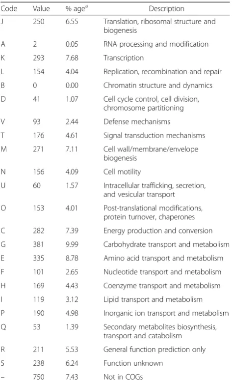
The genome ofE. coliBG1 consists of 4,782,107 bp with no discernible plasmid (no match retrieved with PlasmidFinder version 1.3 [29]), and a G + C content of 50.7%. The genome has been assembled into 84 contigs. Of the 4562 predicted genes, 4465 coded for protein and 97 were RNA-related (including eight 5S rRNA genes, suggesting the presence of 8 rRNA operons, and 86 tRNA genes). In addition, 22 pseudo genes were identified. Among the 4465 protein coding genes, 3831 (85.8%) had an assigned func-tion while the 634 remaining genes (14.2%) encoded pro-teins annotated as hypothetical or unknown. In addition, the BG1 genome contained 38 predicted insertion sequences (ISs), 4 intact and 1 questionable prophage regions, and 2 CRISPR elements suggesting possible genetic crosstalk, such as horizontal gene transfer among theE. coli population. The genome properties are presented in Table 3. The distribution of genes into COGs functional categories is summarized in Table 4.
Extended insights
Genome repertoire comparison
It is admitted that bacterial genome sequences show sig-nificant diversity due to horizontal gene transfers, gene loss and other genomic rearrangements [1]. In this report, characteristics of whole genome datasets of a selection of E. coli strains were compared with those of E. coli BG1 (Table 5). Our main objective was to com-pare the genome of BG1 with that of bovine (K71 and W26) and human (SE15 and Nissle) commensal E. coli strains, but we also included a bovine pathogenic strain (VL2732) and a human EHEC pathogen (Sakai), as the bovine intestine is the main reservoir of EHEC [10]. A human uropathogenic strain (NCTC9001T), which is also theE. colitype strain, was also included as reference. These strains were assigned to different phylogroups (Additional file 1: Table S1; Fig. 2). As expected, the Table 3Genome statistics
Attribute Value % of Totala
Genome size (bp) 4,782,107 100.00
DNA coding (bp) 4,218,785 88.22
DNA G + C (bp) 2,424,397 50.70
DNA scaffolds 84
Total genes 4562 100.00
Protein coding genes 4465 97.88
RNA genes 97 2.13
Pseudo genes 22 0.48
Genes in internal clusters 1171 25.67
Genes with function prediction 3831 83.98
Genes assigned to COGs 3814 83.60
Genes with Pfam domains 275 6.03
Genes with signal peptides 174 3.81
Genes with transmembrane helices 1080 23.67
CRISPR repeats 2
a
The total is based on either the size of the genome in base pairs or the total number of proteins coding genes in the annotated genome
All the information has been obtained from Prokka annotation
Table 4Number of genes associated with general COGs functional categories
Code Value % agea Description
J 250 6.55 Translation, ribosomal structure and biogenesis
A 2 0.05 RNA processing and modification
K 293 7.68 Transcription
L 154 4.04 Replication, recombination and repair
B 0 0.00 Chromatin structure and dynamics
D 41 1.07 Cell cycle control, cell division, chromosome partitioning
V 93 2.44 Defense mechanisms
T 176 4.61 Signal transduction mechanisms
M 271 7.11 Cell wall/membrane/envelope
biogenesis
N 156 4.09 Cell motility
U 60 1.57 Intracellular trafficking, secretion, and vesicular transport
O 153 4.01 Post-translational modifications, protein turnover, chaperones
C 282 7.39 Energy production and conversion
G 381 9.99 Carbohydrate transport and metabolism
E 335 8.78 Amino acid transport and metabolism
F 101 2.65 Nucleotide transport and metabolism
H 169 4.43 Coenzyme transport and metabolism
I 119 3.12 Lipid transport and metabolism
P 190 4.98 Inorganic ion transport and metabolism
Q 53 1.39 Secondary metabolites biosynthesis, transport and catabolism
R 211 5.53 General function prediction only
S 238 6.24 Function unknown
– 750 7.43 Not in COGs
a
greatest difference in genome size was observed between BG1 and the EHEC strain Sakai (the genome size of BG1 is 812,370 bp smaller than the Sakai genome [17.0% of the BG1 genome]). This difference could be explained by the number of mobile genetic elements: the Sakai genome contains 18 prophage regions (at most 5 in the BG1 genome) and 80 insertion sequences (38 in the BG1 genome) [30]. About half of the Sakai-specific sequences are of bacteriophage origin and carry the genes involved in EHEC pathogenesis (bloody diarrhea, hemolytic uremic syndrome) [30]. More surprisingly, the chromosome length of the commensal E. coli Nissle 1917 is 659,093 bp larger than the BG1 genome (13.8% of the BG1 genome). E. coli Nissle 1917 is a human commensal strain known to be a successful colonizer of the human gut and used as a probiotic for the treatment of various intes-tinal disorders [31]. It is well documented that the Nissle genome carries at least three genomic islands (GEIs) inserted at different tRNA sites (serX, argW and pheV) probably acquired by horizontal gene transfer [32, 33]. These GEIs contained genes encod-ing proteins considered as fitness factors (microcins, iron uptake systems, proteases …) contributing to sur-vival of E. coli Nissle and successful colonization of the human body [32, 33]. These GEIs were found in
non-pathogenic E. coli strains but were also
frequently distributed among ExPEC strains [32]. Sequence comparison showed that the genes carried by Nissle 1917 GEIs (mch, mcm, iro, iuc, sat, iha, ybt) are absent in the BG1 genome, suggesting the absence of these GEIs in BG1.
In accordance with the differences in genome size, the highest number of tRNA genes, described as common sites for integration of foreign DNA elements (bacterio-phages, genomic islands), were detected in the genome
of E. coli strains Nissle and Sakai (121 and 103 tRNA genes, respectively while only 86 were identified in the BG1 draft genome (Table 5). The genome of the remaining strains carried 62 (in the type strain NCTC 9001T) to 85 tRNA-encoding genes (Table 5). These numbers may be slightly different depending on the an-notation pipeline used for the draft genome sequences.
Virulence factors
The genes encoding virulence factors in theE. coli BG1 genome were analyzed using blastn against the Virulence Factors Database genomic dataset [34]. A total of 164 genes encoding virulence factors were identified in BG1 (Additional file 2: Table S2), while 181 and 202 genes encoding virulence factors were found in the reference strains NCTC86 and NCTC9001T, respectively. In-depth analysis of the BG1 genome showed that most of these genes are involved in bacterial adherence to the host epi-thelium, iron acquisition systems (siderophores) and flagella synthesis. As expected, genes coding for toxins produced by pathogenic E. coli strains responsible for diarrhea or intestinal damage in mammals (Shiga-toxin, heat stable [ST] toxin,heat-labile [LT] toxin, heat-stable enterotoxin 1 [EAST1], cytotoxic necrotizing factor 1 [CNF1]) are absent in the BG1 genome. TheE. coliBG1 genome also lacks the genes encodingα-hemolysin and enterohemolysin which are involved in the virulence of pathogenicE. colistrains.
Adherence systems
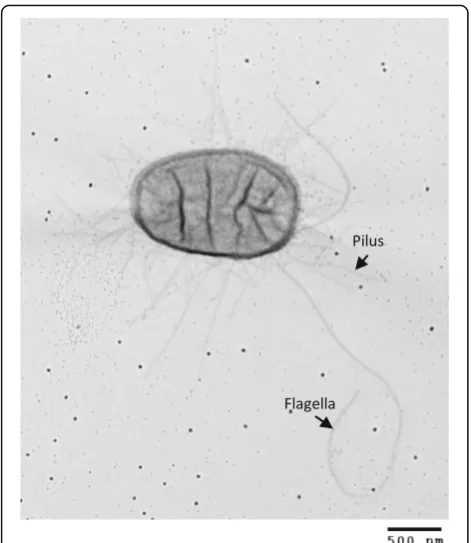
A total of 49 genes coded for the synthesis of organelles involved in adherence ofE. colito host intestinal epithe-lium (Additional file 3: Table S3). Accordingly, the trans-mission electron micrograph of E. coli BG1 showed numerous fimbriae surrounding the bacteria (Fig. 1). Removal of partial genes and incomplete gene clusters Table 5Characteristics of whole genome datasets of differentE. colistrains
Strain name Phylo group Origina Plasmid(s) Genome size (bp) [chromosome + plasmid(s)]
G + C ratio (%) CDS (nb) Protein coding regions (nb)
rRNA operons
(nb)b tRNA genes(nb)
BG1 B1 Bc 0 4,782,107 50.7 4562 4465 8 86
K71 B1 Bc 0 5,115,070 50.7 5178 4872 4 65
W26 B1 Bc 0 5,118,532 50.6 4925 4852 4 66
Nissle 1917 B2 Hc 0 5,441,200 50.6 5417 4970 10 121
SE15 B2 Hc 1 4,839,683
[4,717,338 + 122,345]
50.7 4763 4572 7 85
NCTC86 A Hc 0 5,111,920 50.6 5243 4934 7 87
VL2732 A Bp 0 4,664,032 50.6 4615 4363 4 71
Sakai E Hp 2 5,594,477
[5,498,450 + 92,721 + 3306]
50.5 5447 5324 7 103
NCTC9001T B2 Hp 0 5,038,133 50.6 5154 4859 6 62
a
B: bovine; H: human; c: commensal; p: pathogen
b
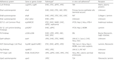
revealed that BG1 possessed the genetic information re-quired to encode 12 potentially functional full adherence systems (Table 6). All these systems are known to be produced by pathogenicE. coliand to adhere in vitro to different cells lines (Table 6) (for reviews see [35–37]).
These adherence systems reflect the ability of
commensalE. coli to colonize distinct niches during its transit through the different compartments of the bovine gastro-intestinal tract. It is also of interest to note that some of these adherence systems possess characteristics corre-sponding to physiological conditions encountered in the bovine gastro-intestinal tract: i)eaeHexpression is induced at 39 °C, the internal bovine temperature, but not at 37 °C [38] ii) the pili HCP is involved in adherence of E. colito bovine gut explants [39] and iii) the F9 fimbriae are essen-tial for in vivo colonization of calves [40]. Furthermore, the stgandF9gene clusters are strongly associated withE. coli belonging to phylogenetic group B1 [41, 42]. To broaden these results, in silico analysis of adherence systems carried by additionalE. colistrains (human and bovine commensal and pathogenic isolates) (Additional file 1: Table S1; Add-itional file 4: Figure S1) was also performed. A hierarchical clustering based on the presence/absence of 78 distinct ad-herence systems encoding genes was built using R (version 3.3.1) [43]. As shown in Additional file 4: Figure S1, bovine
and human E. coli strains were not separately distributed (the closest relative strains to BG1 were the humanE. coli strains S11 and IAI1 [Additional file 4: Figure S1]) suggest-ing that the adherence systems are associated with the adaptation ofE. colito a specific habitat (i.e. the digestive tract) rather than host specificity. As expected, the uro-pathogenic strain NCTC9001T possesses the pap ACDE-GHIK genes which are specific to UPEC strains [44].
Some of these adherence systems possess redundant properties: EhaB, ELF, HCP and UpaG are known to bind to laminin and curli, EhaA, EhaB, EhaC, ECP, F9, EaeH, HCP and UpaG are involved in biofilm formation (Table 6). This suggested an important role of both laminin binding and biofilm formation in sur-vival and/or multiplication of commensal E. coli. Laminin is an extracellular matrix commonly present in the mammalian intestine which act as an interlink-ing molecule in connective tissues that promote bac-terial adhesion and colonization to the host tissues [45]. Moreover, commensal E. coli strains can reside in mixed biofilms in the mucus layer covering the mouse intestine [4, 46]. Because the survival of E. coli depends on anaerobes that degrade polysaccharides included in the mucus layer, it has been hypothesized that the anaerobes in the mixed biofilms provide E. Table 6Adherence systems encoded by theE. coliBG1 genome
Adherence system Gene or genes cluster Pathotypea in vitro cell adherenceb Receptor
Curli fimbriae csgDEFG, csgBA EHEC, ETEC, aEPEC, APEC T84 Matrix, plasma
proteins
EhaA autotransporter ehaA EHEC, EAEC, ETEC, AIEC, EPEC Primary bovine epithelial cells (terminal rectum)
Unknown
EhaB autotransporter ehaB EHEC, UPEC, ETEC, EIEC, EAEC NAc Collagen I, laminin
EhaC autotransporter ehaC (yfaL) EHEC, UPEC Unknown Unknown
ECP (E. coliCommon Pilus) ecpRABCDE ETEC, EHEC, NMEC, EAEC, aEPEC, septicemia
HT29, Hep-2, HeLa, HTB-4 Arabinosyl residues
ELF (E. coliLaminin-binding Fimbriae)
ycbQRST EHEC, aEPEC HT29, Hep-2, MDBK Laminin
F9 Fimbriae z2200-z2206 EHEC, UPEC, APEC, AIEC,
EAEC, EPEC
EBL Bovine fibronectin,
Galβ1-3GlcNAc
EaeH adhesin eaeH UPEC, EHEC, ETEC, NMEC UM-UC-3, Caco-2, CHO,
HeLa, Vero
Unknown
HCP (Hemorrhagic Coli Pilus) hcpABC (ppdD-hofBC) ETEC, EHEC, aEPEC, APEC T84, Caco-2, HeLa, Hep-2, MDBK, cow colon explants
Laminin, fibronectin
Stg fimbriae stgABCD APEC, UPEC UM-UC-3, INT 407 Unknown
T1P (Type I pili) fimBE, fimAICDFGH UPEC, aEPEC, EAEC, APEC, STEC HeLa, REC, colonic and ileal enterocytes
Mannose
UpaG autotransporter upaG UPEC T24 Fibronectin, laminin
a
See the“Abbreviations”paragraph
b
Cell lines: T84 (human colonic adenocarcinoma), HT29 (human colorectal adenocarcinoma), Hep-2 (epithelial cells from epidermoid carcinoma of the human larynx), HeLa (human cervix epithelial carcinoma),HTB-4 (human bladder transitional carcinoma), MDBK (Madin-Darby bovine kidney), EBL (embryonic bovine lung), UM-UC-3 (human bladder carcinoma), Caco-2 (human colon carcinoma), CHO (Chinese hamster ovary), Vero (kidney epithelial cells from an African green monkey), INT 407 (HeLa derivative), REC (humen B cell lymphoma), T24 (human bladder transitional carcinoma)
c
coli with monosaccharide locally rather than from a mixed pool available to all species [4, 46]. Therefore, the mixed biofilm formation can results in a more efficient carbon source for commensal E. coli strains in the mammalian gut [4, 46].
As discussed above, the adhesion systems encoded by the BG1 genome were associated with E. coli strains mostly isolated from clinical cases (Table 6). However, it is important to note that the BG1 genome did not carry the genes encoding the F17, F5 and F41 fimbriae and the afimbrial adhesin CS31A mainly associated with bovine pathogenic E. coli strains involved in diarrhea [47]. For example, a recent epidemiological study showed that the F5/F41 fimbriae were prevalent among bovine diarrhea-genicE. coliisolated in France [48]. The genes encoding F17, F5 and F41 are not detected in the genome of the human and bovine E. coli strains included in this study suggesting that these adherence systems are specific to bovine intestinal pathogenicE. coli.
Flagella synthesis
A total of 47 genes encoding proteins required for flagella synthesis were present in the BG1 genome. Accordingly, the transmission electron micrograph of E. coli BG1 showed peritrichous flagella attached to the bacterial cell surface and clearly distinct from fimbriae (Fig. 1). Flagella are mainly locomotive or-ganelles allowing bacterial movements. However, it is well documented that the flagella (also known as H-antigen) of some pathogenic E. coli mediate the adhe-sion to or invaadhe-sion of epithelial cells (NMEC, aEPEC, ETEC, EAEC, EHEC, APEC) and contribute to bio-film formation (UPEC, ETEC) (for a review see Zhou et al. [49]). In particular, flagella of aEPEC, ETEC and EHEC strains specifically recognized a receptor located at the microvillus tips of human enterocytes [50]. Interestingly, E. coli BG1 possesses the genetic information required to encode the flagella H21, a H antigen type reported to be involved in the invasion of EHEC O113:H21 into HCT-8 colonic epithelial cells [49]. Also, it should be noted that STEC strains with serotype O159:H21 have been isolated from bo-vine as well as porcine feces [51, 52].
Iron acquisition systems
Complete genetic information required for enterobactin synthesis (entABCDEFS) and ferric-enterobactin uptake (fepABCDEFG) was present in the genome of E. coli BG1 (Additional file 2: Table S2). Siderophores, includ-ing enterobactin, are mechanisms secreted by E. colito scavenge iron in order to survive and multiply in hosts or external environments. Siderophores are usually de-scribed as crucial for the proliferation of pathogenic E. coli in the host and have been classified as virulence
factors. However, enterobactin is frequently produced by commensal E. coliisolated from healthy mammals (hu-man and animal isolates) [53]. ent and fep genes were also found in the genome of the reference strain NCTC86 (data not shown). Accordingly, Pi et al. have demonstrated that enterobactin plays a fundamental role in the colonization of healthy mouse gastro-intestinal tract by non-pathogenicE. coli[54].
Ethanolamine utilization
In a previous study, we demonstrated that ethanolamine present in the bovine gut is used by EHEC as a nitrogen source [11]. Furthermore, ethanolamine promotes expression of fimbrial genes and influenced EHEC ad-herence to epithelial cells [55]. Interestingly,E. coliBG1 is unable to degrade ethanolamine present in the bovine intestine, while the EHEC reference strain EDL933 gains a growth competitive advantage by assimilating ethanol-amine in bovine intestinal content [11]. Therefore, we performed in-depth analysis of the genes involved in ethanolamine utilization in order to understand the in-ability of the commensal strain BG1 to use ethanolamine as a nitrogen source.
The degradation and assimilation of ethanolamine by EHEC EDL933 requires exogenous adenosylcobalamin (Ado-Cbl) and are encoded by 17 genes included in the eut operon [11]. In this study, we used blastn and Sea-view (version 4.6.1) [56] to compare the eutgenes ofE. coli BG1 with those of EHEC EDL933. Sequence align-ment showed 317 SNPs between the two eut operons (97.82% identity) (Additional file 5: Table S4). In addition, no premature stop codon was detected and only 34 amino acid changes due to non-synonymous SNPs were identified among the 17 predicted polypep-tides encoded by the eutoperon of BG1 (Additional file 5: Table S4). Furthermore, the presence of a 72 bp inser-tion was also identified in the eutTgene coding for
co-balamin adenosyltransferase in the BG1 genome
compared with the EDL933 genome (Additional file 6: Figure S2). It is important to note that ethanolamine ammonia-lyase, the key enzyme in ethanolamine degrad-ation, required the Ado-Cbl cofactor produced by EutT to be active. The 72 bp insertion sequence at position 395 resulted in a modified translated polypeptide with 24 additional amino acids at position 132. The possible EutT conformation illustrated in Fig. 3 was predicted using Phyre (version 2.0) [57] and showed that 18 of the 24 amino acids encoded by the 72 bp sequence were predicted to form an additional alpha helix in the BG1 EutT protein.
of an additional alpha helix in BG1 EutT cobalamin ade-nosyltransferase, we suspected a reduced or abolished ethanolamine ammonialyase activity, which could ex-plain the inability of BG1 to assimilate ethanolamine in the bovine digestive tract.
Conclusion
The comparison of whole genomes provides information on gene content and organization, and gives an overview of how organisms are related. The draft genome se-quence ofE. coliBG1 isolated from the bovine intestine is now available and can provide valuable information at the genomic scale to explore the genetic and functional features adapted to the bovine gut. The genome of E. coliBG1 can be used as a reference for subsequent evo-lution and comparative studies (some examples of gen-ome comparative analysis have already been described in this report).
As expected, the BG1 genome does not carry the genetic information encoding toxins responsible for
enabling successful colonization of the host rather than strict markers of pathogenesis. In contrast, the factors responsible for disease establishment or intestinal dam-ages during infection (e.g. aqueous or hemorrhagic diar-rhea), such as toxins or the type III secretion system, appear to be true virulence factors.
Additional files
Additional file 1: Table S1.E. colistrains included in this study (XLSX 13 kb)
Additional file 2: Table S2.Genes encoding virulence factors in theE. coli BG1 genome (XLSX 31 kb)
Additional file 3: Table S3.Genes encoding adherence systems inE. coli BG1 genome (XLSX 11 kb)
Additional file 4: Figure S1.Hierarchical clustering ofE. colistrains according to adherence systems encoding genes. The dendrogram and associated heatmap are generated on the basis of gene presence/absence considering 78 genes involved in adherence, using binary distance and complete clustering method, R version 3.3.1. [43]. Blue color indicates gene presence, red gene absence. The origin of each strain is identified with B (Bovine) or H (Human). The color of the strain name corresponds to its phylogroup as in Fig. 2. (DOCX 67 kb)
Additional file 5: Table S4.Genes encoding the transport and assimilation of ethanolamine inE. coliBG1 genome. (XLSX 12 kb)
Additional file 6: Figure S2.Nucleotide sequence alignment of the eutTgene. The sequences of theeutTgene and the translated EutT polypeptide were aligned respectively fromE. colistrains BG1 and EDL933 using Seaview version 4.6.1 [56]. (DOCX 15 kb)
Abbreviations
AIEC:Adherent-invasiveE. coli; APEC: Avian pathogenicE. coli; EAggEC: EnteroaggregativeE. coli; EHEC: EnterohemorrhagicE. coli; EPEC: EnteropathogenicE. coli; ETEC: EnterotoxigenicE. coli;
ExPEC: Extraintestinal pathogenicE. coli; NMEC: Neonatal meningitisE. coli; STEC: Shiga-producingE. coli; UPEC: UropathogenicE. coli
Acknowledgements
The authors thank Frédérique Chaucheyras-Durand for critical reading of the manuscript, Alexandra Durand and Marine Bertoni for excellent technical assistance, Brigitte Gaillard-Martinie for the transmission electron microscopy and Olivier Bouchez for genome sequencing. The genome sequencing was performed at the GeT core facility, Toulouse, France (http://get.genotoul.fr), and was supported by France Génomique National infrastructure, funded as part of the“Investissement d’avenir”program managed by the Agence Nationale pour la Recherche (contract ANR-10-INBS-09). We are also grateful to the Genotoul bioinformatics platform Toulouse Midi-Pyrenées (Genotoul Bioinfo) for providing computing and storage resources.
Authors’contributions
EF and YB designed and coordinated the study; PA and CK performed the bioinformatic analysis; CK performed the genome assembly and annotation; AS performed the laboratory experiments; EF, YB, PA, AS and CK wrote the manuscript. All the authors read and approved the final manuscript.
Competing interests
The authors declare that they have no competing interests.
Publisher’s Note
Springer Nature remains neutral with regard to jurisdictional claims in published maps and institutional affiliations.
Author details
1Université Clermont Auvergne, INRA, MEDIS, F-63000 Clermont-Ferrand, France.2Plateforme Bioinformatique Toulouse, Midi-Pyrénées UBIA, INRA, Auzeville Castanet-Tolosan, France.
Received: 7 March 2017 Accepted: 21 September 2017
References
1. Welch RA. The genusEscherichia. In: Dworkin M, Falkow S, Rosenberg E, Schleifer K-H, Stackebrandt E, editors. The Prokaryotes, vol. 6. Third ed. Berlin: Springer; 2006. p. 60–71.
2. Blount ZD. The unexhausted potential ofE. coli. elife. 2015;4:e05826. 3. Jones SA, Gibson T, Maltby RC, Chowdhury FZ, Stewart V, Cohen PS,
Conway T. Anaerobic respiration ofEscherichia coliin the mouse intestine. Infect Immun. 2011;79:4218–26.
4. Conway T, Cohen PS. Commensal and pathogenicEscherichia coli metabolism in the gut. Microbiol Spectr. 2015; doi:10.1128/microbiolspec. MBP-0006-2014.
5. Kaper JB, Nataro JP, Mobley HL. PathogenicEscherichia coli. Nat Rev Microbiol. 2004;2:123–40.
6. Karmali MA, Gannon V, Sargeant JM. Verocytotoxin-producingEscherichia coli(VTEC). Vet Microbiol. 2010;140:360–70.
7. Oshima K, Toh H, Ogura Y, Sasamoto H, Morita H, Park SH, Ooka T, Iyoda S, Taylor TD, Hayashi T, et al. Complete genome sequence and comparative analysis of the wild-type commensalEscherichia colistrain SE11 isolated from a healthy adult. DNA Res. 2008;15:375–86.
8. Rasko DA, Rosovitz MJ, Myers GS, Mongodin EF, Fricke WF, Gajer P, Crabtree J, Sebaihia M, Thomson NR, Chaudhuri R, et al. The pangenome structure of Escherichia coli: comparative genomic analysis ofE. colicommensal and pathogenic isolates. J Bacteriol. 2008;190:6881–93.
9. Toh H, Oshima K, Toyoda A, Ogura Y, Ooka T, Sasamoto H, Park SH, Iyoda S, Kurokawa K, Morita H, et al. Complete genome sequence of the wild-type commensalEscherichia colistrain SE15, belonging to phylogenetic group B2. J Bacteriol. 2010;192:1165–6.
10. Bertin Y, Chaucheyras-Durand F, Robbe-Masselot C, Durand A, de la Foye A, Harel J, Cohen PS, Conway T, Forano E, Martin C. Carbohydrate utilization by enterohaemorrhagicEscherichia coliO157:H7 in bovine intestinal content. Environ Microbiol. 2013;15:610–22.
11. Bertin Y, Girardeau JP, Chaucheyras-Durand F, Lyan B, Pujos-Guillot E, Harel J, Martin C. EnterohaemorrhagicEscherichia coligains a competitive advantage by using ethanolamine as a nitrogen source in the bovine intestinal content. Environ Microbiol. 2011;13:365–77.
12. Joensen KG, Tetzschner AM, Iguchi A, Aarestrup FM, Scheutz F. Rapid and easy in silico serotyping ofEscherichia coliisolates by use of whole-genome sequencing data. J Clin Microbiol. 2015;53:2410–26.
13. Zankari E, Hasman H, Cosentino S, Vestergaard M, Rasmussen S, Lund O, Aarestrup FM, Larsen MV. Identification of acquired antimicrobial resistance genes. J Antimicrob Chemother. 2012;67:2640–4.
14. Clermont O, Christenson JK, Denamur E, Gordon DM. The Clermont Escherichia coliphylo-typing method revisited: improvement of specificity and detection of new phylo-groups. Environ Microbiol Rep. 2013;5:58–65.
15. Rice P, Longden I, Bleasby A. EMBOSS: the European Molecular Biology Open Software Suite. Trends Genet. 2000;16:276–7.
16. Askari Badouei M, Jajarmi M, Mirsalehian A. Virulence profiling and genetic relatedness of Shiga toxin-producingEscherichia coliisolated from humans and ruminants. Comp Immunol Microbiol Infect Dis. 2015;38:15–20. 17. Bok E, Mazurek J, Stosik M, Wojciech M, Baldy-Chudzik K. Prevalence of
virulence determinants and antimicrobial resistance among commensal Escherichia coliderived from dairy and beef cattle. Int J Environ Res Public Health. 2015;12:970–85.
18. Kaas RS, Leekitcharoenphon P, Aarestrup FM, Lund O. Solving the problem of comparing whole bacterial genomes across different sequencing platforms. PLoS One. 2014;9:e104984.
19. Mariette J, Escudie F, Allias N, Salin G, Noirot C, Thomas S, Klopp C. NG6: Integrated next generation sequencing storage and processing environment. BMC Genomics. 2012;13:462.
20. Andrews, S. FastQC: a quality control tool for high throughput sequence data. 2010. http://www.bioinformatics.babraham.ac.uk/projects/fastqc/. Accessed 10 Oct 2016.
22. Seemann T. Prokka: rapid prokaryotic genome annotation. Bioinformatics. 2014;30:2068–9.
23. Geer LY, Marchler-Bauer A, Geer RC, Han L, He J, He S, Liu C, Shi W, Bryant SH. The NCBI BioSystems database. Nucleic Acids Res. 2010;38:D492–6. 24. Krogh A, Larsson B, von Heijne G, Sonnhammer EL. Predicting
transmembrane protein topology with a hidden Markov model: application to complete genomes. J Mol Biol. 2001;305:567–80.
25. Petersen TN, Brunak S, von Heijne G, Nielsen H. SignalP 4.0: discriminating signal peptides from transmembrane regions. Nat Methods. 2011;8:785–6.
26. Grissa I, Vergnaud G, Pourcel C. CRISPRFinder: a web tool to identify clustered regularly interspaced short palindromic repeats. Nucleic Acids Res. 2007;35:W52–7.
27. Varani AM, Siguier P, Gourbeyre E, Charneau V, Chandler M. ISsaga is an ensemble of web-based methods for high throughput identification and semi-automatic annotation of insertion sequences in prokaryotic genomes. Genome Biol. 2011;12:R30.
28. Arndt D, Grant JR, Marcu A, Sajed T, Pon A, Liang Y, Wishart DS. PHASTER: a better, faster version of the PHAST phage search tool. Nucleic Acids Res. 2016;44:W16–21.
29. Carattoli A, Zankari E, Garcia-Fernandez A, Voldby Larsen M, Lund O, Villa L, Moller Aarestrup F, Hasman H. In silico detection and typing of plasmids using PlasmidFinder and plasmid multilocus sequence typing. Antimicrob Agents Chemother. 2014;58:3895–903.
30. Hayashi T, Makino K, Ohnishi M, Kurokawa K, Ishii K, Yokoyama K, Han CG, Ohtsubo E, Nakayama K, Murata T, et al. Complete genome sequence of enterohemorrhagicEscherichia coliO157:H7 and genomic comparison with a laboratory strain K-12. DNA Res. 2001;8:11–22.
31. Lodinova-Zadnikova R, Sonnenborn U. Effect of preventive administration of a nonpathogenicEscherichia colistrain on the colonization of the intestine with microbial pathogens in newborn infants. Biol Neonate. 1997;71:224–32. 32. Grozdanov L, Raasch C, Schulze J, Sonnenborn U, Gottschalk G, Hacker J,
Dobrindt U. Analysis of the genome structure of the nonpathogenic probioticEscherichia colistrain Nissle 1917. J Bacteriol. 2004;186:5432–41. 33. Sun J, Gunzer F, Westendorf AM, Buer J, Scharfe M, Jarek M, Gossling F,
Blocker H, Zeng AP. Genomic peculiarity of coding sequences and metabolic potential of probioticEscherichia colistrain Nissle 1917 inferred from raw genome data. J Biotechnol. 2005;117:147–61.
34. Chen L, Yang J, Yu J, Yao Z, Sun L, Shen Y, Jin Q. VFDB: a reference database for bacterial virulence factors. Nucleic Acids Res. 2005;33:D325–8. 35. Antao EM, Wieler LH, Ewers C. Adhesive threads of extraintestinal
pathogenicEscherichia coli. Gut Pathog. 2009;1:22.
36. Croxen MA, Law RJ, Scholz R, Keeney KM, Wlodarska M, Finlay BB. Recent advances in understanding enteric pathogenicEscherichia coli. Clin Microbiol Rev. 2013;26:822–80.
37. McWilliams BD, Torres AG. EnterohemorrhagicEscherichia coliadhesins. Microbiol Spectrum. 2014; doi: 10.1128/microbiolspec.EHEC-0003-2013.
38. Easton DM, Allsopp LP, Phan MD, Moriel DG, Goh GK, Beatson SA, Mahony TJ, Cobbold RN, Schembri MA. The intimin-like protein FdeC is regulated by H-NS and temperature in enterohemorrhagicEscherichia coli. Appl Environ Microbiol. 2014;80:7337–47.
39. Xicohtencatl-Cortes J, Monteiro-Neto V, Ledesma MA, Jordan DM, Francetic O, Kaper JB, Puente JL, Giron JA. Intestinal adherence associated with type IV pili of enterohemorrhagicEscherichia coliO157:H7. J Clin Invest. 2007;117:3519–29. 40. Dziva F, van Diemen PM, Stevens MP, Smith AJ, Wallis TS. Identification of
Escherichia coliO157 : H7 genes influencing colonization of the bovine gastrointestinal tract using signature-tagged mutagenesis. Microbiology. 2004;150:3631–45.
41. Lymberopoulos MH, Houle S, Daigle F, Leveille S, Bree A, Moulin-Schouleur M, Johnson JR, Dozois CM. Characterization of Stg fimbriae from an avian pathogenicEscherichia coliO78:K80 strain and assessment of their contribution to colonization of the chicken respiratory tract. J Bacteriol. 2006;188:6449–59. 42. Wurpel DJ, Totsika M, Allsopp LP, Hartley-Tassell LE, Day CJ, Peters KM,
Sarkar S, Ulett GC, Yang J, Tiralongo J, et al. F9 fimbriae of uropathogenic Escherichia coliare expressed at low temperature and recognise Galbeta1-3GlcNAc-containing glycans. PLoS One. 2014;9:e93177.
43. Team RC. R: A language and environment for statistical computing. Vienna, Austria: The R Project for Statistical Computing; 2016.
44. Arthur M, Campanelli C, Arbeit RD, Kim C, Steinbach S, Johnson CE, Rubin RH, Goldstein R. Structure and copy number of gene clusters related to thepap P-adhesin operon of uropathogenicEscherichia coli. Infect Immun. 1989;57:314–21.
45. Simon-Assmann P, Spenle C, Lefebvre O, Kedinger M. The role of the basement membrane as a modulator of intestinal epithelial-mesenchymal interactions. Prog Mol Biol Transl Sci. 2010;96:175–206.
46. Leatham-Jensen MP, Frimodt-Moller J, Adediran J, Mokszycki ME, Banner ME, Caughron JE, Krogfelt KA, Conway T, Cohen PS. The streptomycin-treated mouse intestine selectsEscherichia coli envZmissense mutants that interact with dense and diverse intestinal microbiota. Infect Immun. 2012;80:1716–27. 47. Nagy B, Fekete PZ. EnterotoxigenicEscherichia coliin veterinary medicine.
Int J Med Microbiol. 2005;295:443–54.
48. Valat C, Forest K, Auvray F, Metayer V, Meheut T, Polizzi C, Gay E, Haenni M, Oswald E, Madec JY. Assessment of adhesins as an indicator of pathovar-associated virulence factors in bovineEscherichia coli. Appl Environ Microbiol. 2014;80:7230–4.
49. Zhou M, Yang Y, Chen P, Hu H, Hardwidge PR, Zhu G. More than a locomotive organelle: flagella inEscherichia coli. Appl Microbiol Biotechnol. 2015;99:8883–90.
50. Sampaio SC, Luiz WB, Vieira MA, Ferreira RC, Garcia BG, Sinigaglia-Coimbra R, Sampaio JL, Ferreira LC, Gomes TA. Flagellar cap protein FliD mediates adherence of atypical enteropathogenicEscherichia colito enterocyte microvilli. Infect Immun. 2016;84:1112–22.
51. Baranzoni GM, Fratamico PM, Gangiredla J, Patel I, Bagi LK, Delannoy S, Fach P, Boccia F, Anastasio A, Pepe T. Characterization of shiga toxin subtypes and virulence genes in porcine shiga toxin-producingEscherichia coli. Front Microbiol. 2016;7:574.
52. Pigatto CP, Schocken-Iturrino RP, Souza EM, Pedrosa FO, Comarella L, Irino K, Kato MA, Farah SM, Warth JF, Fadel-Picheth CM. Virulence properties and antimicrobial susceptibility of Shiga toxin-producingEscherichia colistrains isolated from healthy cattle from Parana State. Brazil Can J Microbiol. 2008;54:588–93.
53. Searle LJ, Meric G, Porcelli I, Sheppard SK, Lucchini S. Variation in siderophore biosynthetic gene distribution and production across environmental and faecal populations ofEscherichia coli. PLoS One. 2015;10:e0117906. 54. Pi H, Jones SA, Mercer LE, Meador JP, Caughron JE, Jordan L, Newton SM,
Conway T, Klebba PE. Role of catecholate siderophores in gram-negative bacterial colonization of the mouse gut. PLoS One. 2012;7:e50020. 55. Garsin DA. Ethanolamine utilization in bacterial pathogens: roles and
regulation. Nat Rev Microbiol. 2010;8:290–5.
56. Gouy M, Guindon S, Gascuel O. SeaView version 4: A multiplatform graphical user interface for sequence alignment and phylogenetic tree building. Mol Biol Evol. 2010;27:221–4.
57. Kelley LA, Mezulis S, Yates CM, Wass MN, Sternberg MJ. The Phyre2 web portal for protein modeling, prediction and analysis. Nat Protoc. 2015;10:845–58. 58. Field D, Garrity G, Gray T, Morrison N, Selengut J, Sterk P, Tatusova T,
Thomson N, Allen MJ, Angiuoli SV, et al. The minimum information about a genome sequence (MIGS) specification. Nat Biotechnol. 2008;26:541–7. 59. Woese CR, Kandler O, Wheelis ML. Towards a natural system of organisms:
proposal for the domains Archaea, Bacteria, and Eucarya. Proc Natl Acad Sci U S A. 1990;87:4576–9.
60. Garrity GM, Bell JA, Lilburn T. Phylum XIV. Proteobacteriaphyl.nov. In: Garrity GM, Brenner DJ, Krieg NR, Staley JR, editors.Bergey's Manual of Systematic Bacteriology. Second edition,Volume2,Part B. New York: Springer; 2005: p. 1. 61. Garrity GM, Bell JA, Lilburn T. Class III. Gammaproteobacteriaclass. nov. In:
Garrity GM, Brenner DJ, Krieg NR, Staley JR, editors. Bergey's Manual of Systematic Bacteriology. Second edition, Volume 2, Part B. New York: Springer; 2005. p. 1.
62. Euzeby J. Validation list no. 106. Validation of publication of new names and new combinations previously effectively published outside the IJSEM. Int J Syst Evol Microbiol. 2005;55:2235–8.
63. Garrity GM, Holt JG. Taxonomic outline of the archaea and bacteria. In: Garrity GM, Boone DR, Castenholz RW, editors. Bergey's Manual of Systematic Bacteriology, vol. 1. Second ed. New York: Springer; 2001. p. 155–66. 64. Hill LR, Skerman VBD, Sneath PHA. Corrigenda to the approved lists of
bacterial names: edited for the international committee on systematic bacteriology. Int J Syst Bacteriol. 1984;34:508–11.
65. Rahn O. New principles for the classification of bacteria. Zentralblatt für Backteriologie, Parasitenkunde, Infektionskrankheiten und Hygiene. 1937;96:273–86. 66. Skerman VBD, McGowan V, Sneath PHA. Approved lists of bacterial names.
Int J Syst Bacteriol. 1980:225–420.
68. Ashburner M, Ball CA, Blake JA, Botstein D, Butler H, Cherry JM, Davis AP, Dolinski K, Dwight SS, Eppig JT, et al. Gene ontology: tool for the unification of biology. The Gene Ontology Consortium. Nat Genet. 2000;25:25–9. 69. Stoddard SF, Smith BJ, Hein R, Roller BR, Schmidt TM. rrnDB: improved tools
for interpreting rRNA gene abundance in bacteria and archaea and a new foundation for future development. Nucleic Acids Res. 2015;43:D593–8.
• We accept pre-submission inquiries
• Our selector tool helps you to find the most relevant journal
• We provide round the clock customer support
• Convenient online submission
• Thorough peer review
• Inclusion in PubMed and all major indexing services
• Maximum visibility for your research
Submit your manuscript at www.biomedcentral.com/submit
![Table 1 Classification and general features of E. coli BG1 [58]](https://thumb-us.123doks.com/thumbv2/123dok_us/650124.2064686/2.595.305.535.263.692/table-classification-general-features-e-coli-bg.webp)
![Fig. 2 Phylogenetic tree highlighting the position ofestablished with CSI phylogeny 1.4 [28] using the genome of K71 as a reference and standard input parameters](https://thumb-us.123doks.com/thumbv2/123dok_us/650124.2064686/4.595.56.291.526.732/phylogenetic-highlighting-position-ofestablished-phylogeny-reference-standard-parameters.webp)
![Fig. 3 Predicted secondary structure modeling of the EutT protein of E. coli BG1 obtained with Phyre version 2.0 [57]](https://thumb-us.123doks.com/thumbv2/123dok_us/650124.2064686/9.595.56.540.87.451/predicted-secondary-structure-modeling-protein-obtained-phyre-version.webp)