di
ff
erent geometries as a
template for physiological callus
formation: evaluation of
collagen 3D assembly
1
3.1 Abstract
Bone tissue regeneration involves different healing stages and the resulting final hard tissue is formed from natural templates such as fibrous collagen, soft and hard callus and capillary bed. This work aims to evaluate the efficiency of different scaf-fold geometries with a novel approach: exploring the relationships among scaffold morphologies, cell activity and collagen 3D organization, which serves as a natural template for subsequent mineralization. Among the possible systems to fabricate scaffolds, solvent casting with particulate leaching and microfabrication were used to produce random versus ordered structures from poly-d,l-lactic acid. In vitro biological testing was carried out by culturing a human osteosarcoma derived os-teoblast cell line (MG63) and measuring material cytotoxicity, cell proliferation and migration. Assemblage of collagen fibers was evaluated. A preliminary study of col-lagen distribution over the two different matrices was performed by confocal laser microscopy after Direct Red 80 staining. Both of the scaffolds were seen to be a good substrate for cell attachment, growth and proliferation. However, it seems that
1The work presented in this chapter was published in Journal of Tissue Engineering and
3.2 Introduction
Bone fracture healing can be considered as a four-step process. The initial inflam-matory cellular response due to a fracture leads to the formation of soft callus (fibrocartilage), serving as a guide for the subsequent hard callus production, i.e., the first osteogenetic phase[62, 1]. Cortical and trabecular bone are developed and remodeled in the final stage. Each newly-formed tissue starts to grow from the pre-vious tissue, which represents the appropriate template. The whole mechanism of bone healing comprises these overlapping stages [64, 65, 66].
The repair process involves different cells and molecules which contribute at different levels, playing precise biological roles. These include inflammatory cells, vascular cells, osteoblasts, osteoclasts, pro-inflammatory cytokines, pro-osteogenic factors and angiogenic factors [133].
Collagen represents the most important and the richest protein of connective tissue in mammals and it is the main constituent of extracellular matrix (ECM) and soft callus. It is a fibrous protein and several different types have been identified. Col-lagen type I is predominant in developing bone tissue and it is principally produced by osteoblasts.
The main focus of bone tissue engineering is to support and guide the healing process and bone reconstruction, in our case, by using scaffolds designed as temporary ECMs able to direct normal cell function [3, 134]. Recent studies have shown that cells are sensitive to chemistry, stiffness and surface properties of the scaffold [3, 92, 135]. Therefore, successful bone repair depends on the three-dimensional (3D) scaffold design which works as a bioactive support for host cells as they are organized into a functional tissue [136, 137]. Important matrix parameters are pore size, distribution and interconnectivity, mechanical integrity and surface characteristics [41, 75, 138, 139, 140, 141].
to evaluate how different scaffold morphologies influence osteoblast activity as well as collagen assembly. Collagen organization and callus formation play fundamental roles as gap filler and blueprint for subsequent ECM maturation and mineralization [145]. The ECM of cancellous bone is characterized by irregularly arranged collagen fibers and prior callus formation is fundamental for angiogenesis and final tissue formation [146]. Identifying the geometric structure of matrix (pore size and shape) and its role in collagen fiber organization represents a good starting point to address a strategy for directing a more biomimetic and controlled mineralization [3].
In this work poly-d,l-lactic acid (PdlLA) was used. This polymer is widely employed in scaffold production for bone tissue regeneration due to its mechanical properties and processability into a variety of shapes and sizes [123, 147, 148, 149, 150]. Two different models of scaffolds were produced: ordered, structured microfabri-cated scaffolds with a spatially well defined porosity and salt-leached sponges with interconnected, randomly distributed pores.
3.3 Materials and Methods
Materials
Poly-d,l-lactic acid (PdlLA, type RESOMER®207, MW= 252 kDa) was purchased from Boehringer Ingelheim, Germany (polymer was used without further purifica-tion) and dichloromethane (DCM) and dimethylformamide (DMF) were purchased from BDH Chemicals (UK) and J.T.Baker (Holland), respectively.
Sca
ff
olds
Scaffolds with regular geometry were produced with a home-made microfabrica-tion apparatus [144]. PdlLA was dissolved in dichloromethane: dimethylformamide (70:30 v/v) to prepare a 20% (w/v) solution. This concentration was selected as being the optimal one after performing multiple microfabrication tests. Scaffolds were made with equidistant rows on 20 sequential orthogonal layers produced by extrusion of solution filaments through a syringe needle (33 gauge). The distance between rows was set to 375 µm.
Sponges with randomly distributed pores were produced by solvent casting into cylinders and particulate leaching [156, 157, 158]. PdlLA was dissolved in dichloromethane: dimethylformamide (70:30 v/v) to prepare a 6.5% (w/v) solution. Sieved NaCl par-ticulates with a dimensional range between 240 and 315µm were mixed in, obtaining a final concentration polymer/porogen of 1:40 w/w. The mixture was air dried (24-36 hours) and immersed in deionized water (dH2O) for 3 days with change every 6 hours for the removal of the porogen.
All the scaffolds, which were prepared by casting into cylinders were cut into discs with a diameter of 10 mm sterilized with aqueous ethanol solution 70% (v/v) at 4°C for 24 hours and dried under a hood at room temperature.
Sca
ff
old characterization
Scanning Electron Microscopy (SEM) imaging
samples were sputter-coated with a thin gold layer under argon atmosphere (SEM Coating Unit PS3, Assing S.p.A., Rome, Italy).
Scaffold porosity evaluation
Considering the different geometrical features of scaffolds, two different methods were chosen to calculate scaffold porosity. Microfabricated scaffold porosity was evaluated from SEM images by ImageJ software [159, 160] while salt leached scaffold porosity by a liquid displacement method similar to a previously described technique [161, 162, 163].
Microfabricated scaffold pore analysis was performed using the public domain Im-ageJ, program developed at the US National Institutes of Health (NIH). It was used to measure the area of pores selected on SEM images. To obtain percent porosity the sum of the areas of the pores was divided by the total exposed sample area. In liquid displacement method ethanol 100% was used since it can simply infiltrate into the pores without dissolving the polymer. Briefly, the sponge was dipped into a 5 ml graduated cylinder containing a known volume of ethanol (V1). A series
of rapid evacuation– depressurization cycles were assessed to allow the ethanol to penetrate and fill the pores and completely evacuate entrapped air. The volume of ethanol after immersing the scaffold was measured asV2. The ethanol-impregnated
scaffold was taken away from the cylinder and the remaining ethanol volume was recorded asV3. The porosity of the scaffold was given by the formula
p= V1−V3 V2−V3.
In both cases the measures of porosities were the result of a triplicate analysis.
Lactate Dehydrogenase (LDH) Assay
After incubation, PBS was collected and added to the seeded wells in an amount equal to 1/3 of the medium for 24 hours. LDH in vitro test was carried out following the manufacturer’s instructions. Wells seeded with the same concentration of cells and standard culture medium were used as control. The absorbance of the assay mixture was measured at 492 nm ad 600 nm with a photometric microplate reader (Multiskan EX, ThermoLabsystems, Finland). For each type of scaffold four reading of three parallel samples were performed: average value and standard deviation are reported.
Biological characterization
After sterilization with ethanol, 70%, scaffolds were placed in 48-well plates and washed several times with sterile, distilled water. Samples were seeded with 1·104 cells, human osteosarcoma derived osteoblasts (MG63). After 24 hours, scaffolds were moved from the original well plate to a new one to avoid considering cells attached on the tissue culture plate (TCP) and incubated for a total of 28 days. Results were evaluated at five experimental time points: 4, 7, 14, 21 and 28 days. 48-well plates, seeded with the same cell density, were utilized as controls. Minimum essential medium (MEM) supplemented with 10% Fetal Bovine Serum (FBS), 1% Penicillin, 1% Glutamax, 1% Vitamin, 1% non-essential amino acids comprised the culture medium. Cells were incubated at 37ºC in a 5% CO2 atmosphere incubator, with medium changes every 2-3 days.
AlamarBlue Assay
a photometric microplate reader (Multiskan EX, ThermoLabsystems, Finland) and the percentage of reduced alamarBlue was calculated [164].
Confocal Laser Microscopy (CLM) imaging
Evaluation of cell attachment, growth, distribution and migration on the seeded scaf-folds was performed by confocal laser microscopy (Nikon Eclipse, Ti-E) after Phal-loidin Rhodamine (Biosource International, Invitrogen) and DAPI (Sigma Aldrich) staining according to the manufacturer’s protocol. Fixation with a formaldehyde so-lution (4% formaldehyde in PBS soso-lution) and permeabilization with TritonX (0.2% TritonX in PBS solution) were performed before staining.
CLM was also used to evaluate collagen production and distribution after Direct Red 80 (Sigma Aldrich) staining. A published protocol modified specifically for our samples was used [154]. Direct Red 80 staining, more frequently used for collagen detection in tissue histological sections[165, 166, 167], has also been tested to mark collagen for CLM imaging [152]. The staining is not specific for the different types of collagen. Samples were washed in dH20, pre-treated for 40 minutes with a solution of 0.2% phosphomolybdic acid (PMA), and incubated for 30 minutes in a solution consisted of 0.1% Direct Red 80 diluted in saturated aqueous picric acid. At the end each sample was washed in 0.01 M HCl for 2 min. PMA was used to make colorless cytoplasm and HCl was used to remove non specific staining.
Representative images are shown.
Scanning Electron Microscopy (SEM) imaging
Statistical analysis
3.4 Results and Discussion
Collagen behaves as a support structure for tissues, controlling cell shape, diff er-entiation and regeneration. In the intermediate stage of the bone healing process, collagen is the main constituent of callus, which is fundamental because it stabilizes the fracture site, behaves as a support structure for tissue ingrowth and guides the final angiogenesis and tissue mineralization [62]. In particular, the orientation of collagen fibrils leads to the formation of woven, lamellar or primary parallel fibered bone [168]. Soft and hard callus can be considered physiological models for de-signing biomimetic scaffolds for a tissue engineering approach. A bioresorbable and osteoconductive scaffolding support should trigger and address cell attachment, mi-gration, ECM production, assembling, mineralization and angiogenesis.
In this work the functional role of scaffold geometry as template for physiological callus formation and proper collagen spatial organization was evaluated. After pre-vious scaffold characterization by imaging analyses (SEM) and material cytotoxicity assessment, preliminary biological tests were performed. Cell proliferation, distri-bution and growth were considered and collagen distridistri-bution and arrangement were qualitatively analyzed.
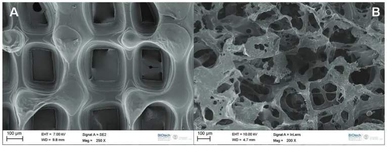
Figure 3.1: Scanning electron microscopy images of PdlLA scaffolds: (A) micro-fabricated scaffold, (B) salt-leached sponge.
contrast, in the microfabricated scaffold (Fig. 3.1B) the pore distribution was quite regular with square shapes, with a side length of around 250 µm. In the micro-fabricated constructs, the surface appeared smoother and the pore walls appeared thicker than in the salt-leached sponges.
Salt leached sponges porosity was calculated by liquid displacement method and re-sulted 86%±2%. Conversely, microfabricated scaffold porosity, analyzed by ImageJ software, was 61% ±4%
Cytotoxicity testing on the hypothesis that traces of solvent may have a negative effect on biocompatibility was performed.
Figure 3.2: LDH assay of PdlLA scaffolds: salt-leached sponges and microfabri-cated scaffolds are not cytotoxic.
Figure 3.3: AlamarBlue assay for MG63 cell culture on PdlLA scaffolds and 48-well culture plate used as control.
The proliferation trend was further confirmed by CLM imaging analyses. Images in Fig. 3.4 show attachment and proliferation of MG63 cells on PdlLA scaffolds. Increase in cell number could be observed with incubation time. Cells started in-vading scaffold pores and adhering to the matrix. Furthermore, at longer culture time, a deeper migration of cells inside and between the pores was visible. Pore sizes were large enough to permit ingress of cells, with concurrent bridging of cells between pore walls. Fig. 3.4, G and H, show that cells almost completely covered the scaffolds after 21 days of culture.
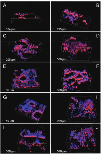
Cell migration is evident from Fig. 3.5, where confocal depth projection images show the outer and inner part of the seeded scaffolds. The figures result from stacking multiple horizontal images corresponding to different focused planes. The profiling depth is reported on the images.
Microfabricated scaffolds and salt-leached sponges, at the first analyzed time point (Fig. 3.5A and B), showed a cell distribution mainly on the outer surface, where cells were previously seeded. Afterward, the internal regions started to be invaded (Fig. 3.5 C, D, E, F, G, H, I, J). In general, a homogeneous cell distribution was shown within the scaffold thickness indicated.
SEM analyses were in concordance with the confocal microscopy results in terms of cell adhesion and distribution, and also gave a closer view of cell and cell-material interactions.
Figure 3.5: 3D Confocal Laser Scanning Microscopy images of MG63 cells stained with Rhodamine Phalloidin and DAPI adhered to PdlLA scaffolds after A, B) 4 days, C, D) 7 days, E, F) 14 days, G, H) 21 days, and J, K) 28 days of cell culture. On the left the microfabricated scaffolds, on the right the salt-leached sponges. Stack depths are specified.
was further characterized by a qualitative imaging analyses to check if different scaf-fold architectures can influence ECM production and assembly. Cells were fixed and stained with Direct Red 80 to visualize the collagen with the confocal microscope.
Figure 3.6: SEM images of the PdlLA scaffolds after A, B) 21 days, C, D) 28 days of cell culture. On the left the microfabricated scaffolds, on the right the salt-leached sponges. Arrows indicate cell bridging mechanism and spreading behavior. Different magnification are reported.
CLM images show an increase in collagen amount and a more complex organiza-tion with incubaorganiza-tion time (Fig. 3.7). Until day 7, collagen structure appeared quite similar on the two different scaffolds: it did not seem organized and the acquired emission signal describes a modest overall amount. Differences were visible at the last cultured time points: in salt-leached sponges a more complex collagen organiza-tion could be distinguished. In fact in microfabricated scaffolds (Fig. 3.7J) collagen showed an inhomogeneous planar organization (2D), whereas osteoblasts on salt-leached sponges (Fig. 3.7K) seemed to guide collagen with a fiber-like organized structure arranged more in 3D.
3.5 Conclusions
The functional role of scaffolds with different geometries as templates for physiolog-ical collagen mesh formation was studied. Two different 3D porous PdlLA scaffolds provided an appropriate environment for cells growing and good proliferation, as well as effective migration. Furthermore, collagen production and its organization on the scaffolds with a porosity randomly distributed seemed to be more suitable for a bone tissue regeneration application with osteoblasts producing a collagen architecture similar to the natural callus matrix.