Mohammed Al .; et al… - 512- MINIMALLY INVASIVE TECHNIQUES IN MANAGEMENT OF INTRA-ARTICULAR
CALCANEAL FRACTURES
Mohammed Al Ahmady Abd EL Reheem,Mohsen Mohamed Abdou,Adel Mohamed Abd El Azim,Hossam Fathy Mahmoud,
Orthopedic Surgery department; Faculty of Medicine-Zagazig University-Egypt
ABSTRACT
Background: Intra-articular calcaneus fractures are commonly occured after high-energy trauma. Avariety of techniques exists for anatomic reduction and surgical fixation .The optimal management of displaced intra-articular calcaneus fractures is controversial and represents a topic of sustained interest and research for the past two decades .
Open reduction and internal fixation (ORIF) via an extensile L-shaped approach has gained many soft tissue complications.
These complications include deep and superficial infections and wound sloughs, which reportedly occur in 1.8% to 27% of patients. This high frequency of infection is likely attributed to thin soft-tissue envelope around the calcaneus especially the lateral wall, which is exposed for surgery . Recently, less invasive surgical techniques for treating displaced intra-articular calcaneus fractures have been undertaken in an attempt to reduce complication rates and promising clinical and
radiographic outcomes.These recent techniques include limited-incision sinus tarsi
ORIF,percutaneous stabilization with pins and /or screws, and minimally invasive plate osteosynthesis (MIPO(
Subjects:This study was conducted on 12 patients (10 males 83.3% & 2 females 16.7 %) with displaced intra-articular calcaneal fractures treated with less invasive techniques in the Orthopedics and Traumatology Department of Zagazig University Hospitals from April 2017 to October 2017 with a follow up period of six months. .
This study was done in Zagazig University Hospitals,Egypt on 12 patiens with displaced intraarticular calcaneal fractures including displaced Essex-Lopresti fractures, Sanders type II fractures, Sanders type III fractures in patients with multiple co morbidities.
Results & Discussion Results
Collected data will be presented in tables and suitable graphs and analyzed by computer software (SPSS version 19) using appropriate statistical methods.
Discussion done on results compared to related relevant literatures and specific researches to explain the reasons for getting such results.
Conclusion
less invasive surgical techniques for treating displaced calcaneus fractures are very effective and smart procedurs to reduce complications and improve recovery when surgery is indicated.
Keywords: Less invasive, Calcaneus, Fractures, Fixation Corresponding Authors:
Mohammed Al Ahmady Abd EL Reheem,M.B.B.C.H, Mohsen Mohamed Abdou,MD, Adel Mohamed Abd El Azim,MD, Hossam Fathy Mahmoud,MD.
Tel:01063433225-01281218950 Email:Dr_ma2020@yahoo.com
INTRODUCTION
isplaced intra-articular calcaneal
fractures are considered a therapeutic challenge due to fracture complexity and multiple treatment options.
Although fractures of the tarsus are relatively uncommon, the calcaneus is the most
frequently injured bone in this area
(1).Calcaneus fractures accounts for about 2% of all fractures and the proportion of intra-articular calcaneal fractures with involvement of the posterior subtalar joint is approximately 75% of calcaneal fractures. Intra-articular
fractures carry a high morbidity;
Mohammed Al .; et al… - 513- approximately 20% are not able to return to
work within a year, rendering intra-articular calcaneal fractures costly on a socio-economic aspect (2)
The primary goal of operative treatment of intra-articular calcaneal fractures is to restore the congruence of the subtalar joint and the secondary goal is to restore height, width, and alignment of the calcaneus. The aim of treatment is pain free ambulation, the ability to return to work and the ability to wear regular shoes (3)
Operative treatment has been divided into closed, percutaneous, and open techniques. Open reduction and internal fixation (ORIF) has been the standard method of treatment in last two decades. Although, ORIF provides good to excellent results to anatomically
reduce the subtalar joint, its main
complications are wound dehiscence and infection which may occur in up to 30% of patients (1)
There are also, some relative
contraindications for open operative treatment including severe co-morbidities, peripheral vascular disease and skin blisters. All of these increase risk of wound complications.
To avoid these complications, various less invasive techniques were introduced to reduce and fix the displaced fractures .
One of the earliest less invasive techniques was devised by the German surgeon Westhues in 1934 (4). It was later modified and popularized by Gissane and Essex Lopresti..
In the last ten years less invasive techniques has been used and gained more popularity in treatment of intra-articular calcaneal fractures
PATIENTS & METHODS . Patients
This study was conducted on 12 patients (10 males 83.3% & 2 females 16.7 %) with displaced intra-articular calcaneal fractures treated with less invasive techniques in the Orthopedics and Traumatology Department of Zagazig University Hospitals from April 2017 to October 2017 with a follow up period of six months .
I- Patient Selection: :
1- Type of fracture: Displaced intra-articular calcaneal fractures, ¬¬Sanders classification
type II and III and fracture variants with
minimal posterior facet fragment
comminution.
2- Age : 18-60 years . The mean age was 36 years
3- Timing of surgery: all fractures were treated within 14 days
Exclusion criteria:
1 Neglected fracture ( more than 14 day)
2 Non-displaced fractures ( Sanders type
I)
3 Open fractures, Infection and bad skin
conditions.
4 Non compliant patients ( dementia,
drug abusers)
5 Crushed calcaneus (Sanders type IV(
II- Age incidence:
This study was performed on patients with age range from 18 years to 60 years with mean age 36 years.
III- Sex:
10 patients were males(83.3%) , 2 were females(16.7%)
IV-Mechanism of trauma:
Most injuries (9 cases) occurred following falling from height (75%) while 2 cases following direct blow to the foot(16.6%) and one case following RTA (8.3%).
Methods
On admission, all patients were subjected to: I- History taking:
Personal history:
Name, Age, Sex, Residency, special habits.
Occupation and the level of physical activity Present history:
Mechanism of trauma.
Duration of trauma. Past history:
-Medical history: for pre-existing significant co-morbid conditions
II-Clinical Examination: General ex:.
Blood pressure, pulse, temperature,
respiration and other body systems. Local ex:.
Tenderness: at the hind foot
Swelling at the hind foot
Soft tissue & skin condition and peripheral circulation
Mohammed Al .; et al… - 514- III- Elevation ,rest, ice and posterior slap
. IV- Radiological evaluation:
A)Plain X-ray calcaneus that includes lateral, axial and Broden’s views. Broden’s views are special calcaneal radiographic projections to show the congruence of the subtalar joint. They are taken at 10°, 20°,30 and 40° to the horizontal.
The protocol of radiological evaluation in this study involved measuring 3 angles and 3 distances.
The angles measured are: (Fig.1)
1 .Bohler’s tuber joint angle by was measured using the highest points of the calcaneal tuberosity, the subtalar joint and the anterior process. It is taken as a relative measurement
of the degree of compression and deformity in calcaneal fractures. Normally, this angle ranges from 25° to 40°. (12(
2. The crucial angle, as described by Gissane, is the angle formed by the posterior facet and the line from the calcaneal sulcus to the tip of the anterior process of the calcaneus. This angle is normally between 120° and 145°.(13(
3. The posterior facet inclination angle, as described by Sarrafian (14) , is the angle formed by the two intersecting lines drawn along the surface of the posterior facet and along the upper surface of the calcaneal tuberosity. Normally, this angle measures an average of 65°, with a range of 55–75° .
(Fig.1) Calcaneal angles.
The distances measured are: (Fig.2)
1. The length of the calcaneus is measured on the lateral view from the most posterior point of the tuberosity to the calcaneocuboid joint. 2. .The height of the posterior facet was
measured by a line perpendicular on the
calcaneal axis to the highest point of the posterior facet.
Mohammed Al .; et al… - 515- (Fig.2)Calcaneal distances.
B)CT scan of ankle and subtalar joints: for assessment and classification of intra-articular fractures as Sander’s classification is based on coronal CT.
V- Laboratory evaluation:
Routine preoperative laboratory investigations were done (complete blood picture, liver
function tests, kidney function tests,
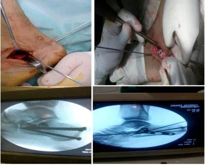
coagulation profile) VI-Operative Procedures: a) Percutaneous Fixation:
1- One or two 4.5 or 6.5 mm cannulated screws are directed from posterior to anterior to hold the primary fracture fragments. In cases of joint depression the higher one
should be inserted through the subchondral bone (Fig.3(
2- One or two screws were inserted from inferior to superior direction to hold the sustentaculum and or the posterior facet fragments to prevent late collapse of fragments.
Oblique image views were taken to be sure that the guide pin of cannulated screw is not penetrating the joint
3. Intrafocal screw fixation
:Following fixation of primary and secondary fractures any unstable large fragment was fixed with one screw
Mohammed Al .; et al… - 516- b) Limited-incision Sinus Tarsi Technique:
A small 2- to 4-cm sinus tarsi incision permits direct visualization of the posterior facet fragment for reduction, as well as of the anterolateral fragment and the lateral wall. This approach allows for the insertion of a small, low-profile plate if needed and decreases dissection and elevation of the peroneal tendons, thus theoretically lowering the risk of tendon irritation and subluxation.
Because the sural nerve is largely avoided, the risk of postoperative neuralgia or neuroma formation is minimized. along a line from the tip of the fibula to the base of the fourth metatarsal (Figure 4). The extensor digitorum brevis is retracted cephalad to permit exposure of the sinus tarsi and direct visualization of the posterior facet of the calcaneus.
Fig. (4): Sinus tarsi approach .
A Schanz pin is then placed through a stab incision in the posteroinferior calcaneal tuberosity from lateral to medial to allow for distraction, provide control of the tuberosity fragment, and aid reduction. After removing any fibrous debris and fat from the sinus tarsi, the lateral wall and the posterior facet fragment are mobilized using a knife or small elevator. Care is taken to avoid significant dissection of the peroneal tendons that are retracted posteriorly as needed. Using fluoroscopy to check alignment and length, two guide pins from a large cannulated screw set are placed from the calcaneal tuberosity
into the anterior distal portion of the calcaneus.
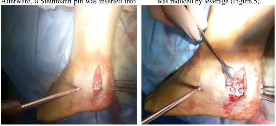
c) Minimally Invasive Plate Osteosynthesis (MIPO):
In the MIPO technique, the patient was placed in a lateral position on a radiolucent operating table and a tourniquet was applied .
Mohammed Al .; et al… - 517- After dividing the subcutaneous tissue
directly to the bone, a periosteum elevator was used to create a lateral calcaneal subfascial channel to fit the calcaneal plate . Afterward, a Steinmann pin was inserted into
the calcaneal tuberosity from the lateral side and traction was applied along the long axis of the foot to correct the varus and length of calcaneus while the posterior articular surface was reduced by leverage (Figure.5).
Fig. (5): Minimally Invasive Plate Osteosynthesis (MIPO).
RESULTS
In this study, 12 patients are followed up and constituted the basis of this prospective study with a follow up period of six months.
3 cases were operated by percutaneous fixation while 8 cases were operated by limited-incision sinus tarsi technique and one
case by minimally invasive plate
osteosynthesis (MIPO). Functional Results: A) Pain :
The pain was assessed according to AOFAS
questionnaire. 9 patients had mild
(occasional) pain, two patients had no pain, and one patient had moderate (daily) pain. B) AOFAS score
According to AOFAS score and clinical evaluation, the functional results were graded as excellent in five patients (41.7%), good in 6 patients (50%), fair in one patient (8.3%), with no poor results. The excellent and good results were considered as satisfactory ones while the unsatisfactory included the fair results. Thus, satisfactory results were found
in 11 patients (95.91%), and the
unsatisfactory ones were found in 1 patient (8.3%).
Radiological Results: 1-Bohler's angle:
There is significant improvement of the
Bohler's angle at the final follow up (P <0.001). The mean preoperative Bohler's
angle was 12.67° and the mean postoperative angle was 18.48°, and in final follow up was 18.14°.
2-Angle of Gissane:
There is significant improvement of the Gissane angle at the final follow up (P <0.001). The mean preoperative angle of Gissane was 117.24°, and the mean immediate postoperative angle was 107.57, and in final follow up was 107.57°
3-Subtalar joint arthritis
Inspite of the short term follow up period, all
patients were evaluated for subtalar
osteoarthritic changes according to Paley and Hall system. Grade 0: 7 cases, Grade 1: 4 cases, Grade 2: 1 case (and no patients with grade 3).
DISCUSSION
Calcaneus is the most frequently fractured tarsal bone, accounting for about 2 % of all fractures, often derived from high-energy trauma in young patients.
The ideal treatment for displaced
intra-articular calcaneal fractures remains
Mohammed Al .; et al… - 518- fragment reduction, with anatomical Bohler’s
angle restoration, and subtalar joint congruity predict higher functional scores as well as a lower incidence of post-traumatic arthritis. (1, 2(
Open reduction and internal fixation (ORIF) via an extensile L-shaped approach has gained wide popularity because this procedure can provide a good anatomic alignment of the calcaneus and congruity of the posterior subtalar joint and stable fixation which allow good mobilization. ORIF with an extensile exposure is often plagued by soft tissue complications(3(
These complications include deep and superficial infections and wound sloughs, which reportedly occur in 1.8% to 27% of patients. This high frequency of infection is likely attributed to thin soft-tissue envelope around the calcaneus especially the lateral wall, which is exposed for surgery(3(
In our study, less invasive surgical techniques for treating displaced intra-articular calcaneus fractures have been undertaken in an attempt to reduce complications and improve recovery when surgery is indicated. These early results reported reduce complication rates and promising clinical and radiographic outcomes in certain fracture patterns and patient populations (4).These recent techniques include limited-incision sinus tarsi ORIF, percutaneous stabilization with pins and /or
screws, and minimal invasive plate
osteosynthesis .
In our study, most patients (91.7%) had a satisfactory functional end results. By using the AOFAS score, the functional results were graded as excellent in five patients (41.7%), good in 6 patients (50%), fair in one patient (8.3%), with no poor results. The excellent and good results were considered as satisfactory ones while the unsatisfactory included the fair results. Thus, satisfactory results were found in 11 patients (91.7%), and the unsatisfactory ones were found in 1 patient(8.3%)
The numbers of good or excellent clinical results were comparable to the results in a series of calcaneal fractures using similar percutaneous fixation technique in the literature (21). The satisfactory results in this study are higher than those achieved by
Schepers et al (1); they treated 59 patients by percutaneous fixation. AOFAS Hind foot scores were good to excellent in 72%.
Comparing with open reduction and internal fixation in os calcis fractures, the satisfactory results in this study are higher than those achieved by Asik et al (20), They treated 19 fractures in patients with open reduction and internal fixation, Their results after two year follow up period were excellent in five feet (26%), good in eight feet, fair in four feet (21%); and poor in two feet (11%)
The results in this study are higher than those achieved by Wang et al (21) .In their study, 47 cases (50 feet) treated with calcaneal plates. After a follow-up of 12–34 months, the rate of excellent-good results was 80.0% . Also, the results are higher than those achieved by Makki et al (22); they operated 45 patients by open reduction and internal fixation. According to AOFAS scale, excellent and good results were achieved in 35 patients 77.7%. In this study, the mean AOFAS ankle-hind foot score is higher than that achieved by Potter and Nunley (23) , they operated 81 fractures in 73 patients with mean AOFAS ankle-hind foot score 65.4.
The higher results in this study can be explained by its short follow up period, as longer follow up periods could reveal more complications regarding subtalar arthritis and flattening of calcaneal angles.
The results are nearly matched with that achieved by Rammelt et al (25). They
operated patients with percutaneous
arthroscopically assisted reduction and screw fixation with mean AOFAS score 92.1 points. The results were also matched with those achieved by Walde et al (24), who used closed reduction and percutaneous K-wires, reported thirty-four (50.7%) patients had a restricted range of motion of up to 15° and more than half (58.2%) of the patients had achieved more than 75% of their total range of motion in the subtalar joint.
Mohammed Al .; et al… - 519- (80%). There was severe limitation of
movements in one case(1.6%),while
moderate limitation of movements was recorded in the remaining 10 cases (17%). In the present study, The pain was assessed according to AOFAS questionnaire. 9 patients
(75%) had mild(occasional) pain,two
patients(16.7%) had no pain, and one patient(8.3%) had moderate (daily) pain. The results are nearly matched with that achieved by Yeung et al (26); they reported that four patients (16.6%) were pain free on the latest follow-up. There were 16 (patients (66.6%) with mild pain, four patients (16.6%) with moderate pain, and none with severe pain.
Those results were lower than those achieved by Walde et al (24),who showed that Thirty-seven (55.3%) of the 67 patients had no pain while full weight bearing or could walk at least 4 hours without pain at last follow-up evaluation. Nine (13.4%) patients had constant pain.
In this study, the lower results regarding the pain could be due to early weight bearing in some patients, development of subtalar joint arthritis, peroneal tendons irritation at the lateral aspect of calcaneus either due to bulging of the lateral wall after trauma or from the fixation technique using transverse screw, or due to calcaneal broadening after trauma.
Complications:
Complications of calcaneal surgery include
subtalar joint arthritis ,Malreduction
nonunion, heel pain and numbness, wound infection , skin flap necrosis and implant problems (27)
Inspite of the short term follow up period, all
patients were evaluated for subtalar
osteoarthritic changes at the end of follow up period according to Paley and Hall system. In the present study, The result of subtalar joint arthritis was Grade 0: 7 cases, Grade 1: 4 cases, Grade 2: 1 case (and no patients with grade 3)according to Paley and Hall system. There was no significant correlation between the results of subtalar joint arthritis and the functional outcome, probably due to short follow up period of the cases.
The results were lower in comparison with those achieved by Wang et al (28), in their
study using less invasive fixation techniques, 15 patients with 16 displaced intra-articular fractures of the calcaneus were treated by percutaneous reduction and cannulated screw fixation, no subtalar joint arthritis was encountered in any patient at 6 months.
In the present study, Wound infection occurred in one case(8.3%) , The patient was 44years old with bilateral intra-articular calcaneal fracture Sanders type III operated by bilateral sinus tarsi approach .The right side infected after 15 days. Debridment was done and hardware was removed .Below knee cast for 3 weeks and the patient final AOFAS score was 77 with fair result.
SUMMARY & CONCLUSION Less invasive surgical techniques for treating displaced intra-articular calcaneus fractures have been undertaken in an attempt to reduce complications and improve recovery when surgery is indicated. These early results reported reduce complication rates and promising clinical and radiographic outcomes in certain fracture patterns and patient populations.These recent techniques include
limited-incision sinus tarsi ORIF,
percutaneous stabilization with pins and /or screws, and MIPO.
A thorough understanding of the clinical and radiographic anatomy of the calcaneus and its articulations is crucial when attempting less
invasive procedures for intra-articular
calcaneus fractures. These emerging
techniques may be beneficial in patients with
soft-tissue compromise, multiple
comorbidities, and displaced intraarticular fractures with minimal comminution . Limitations&Recommendations:
1- Small size sample (12 cases(
it is recommended to enlarge sample size 2 -follow up for 6 monthes
it is recommended to widen period for follow up for more results evaluation and data
collection. REFERENCES
1- Schepers T, van Lieshout EMM, van Ginhoven TM. Current concepts in the treatment of intra-articular calcaneal fractures: results of a nationwide survey. Int Orthop 2009; 32:711–5. 2- Rockwood Ca Jr, Greene DP. Rockwood and
Mohammed Al .; et al… - 520-
3- Gougoulias N, Khanna A, McBride DJ, Maffulli N. Management of calcaneal fractures: systematic review of randomized trials. Br Med Bull 2011; 97.
4- Westhues H. A new treatment method for the calcaneus fracture. Arch Orthop Unfallchir 1943; 35:121
5- Kimberly S, Belte Ann Harris. Interrater reliability of subtalar neutral, calcaneal inversion and eversion. JOSPT 2007; 12:1
6- Song KS, Kang CH, Min BW. Preoperative and postoperative evaluation of intra-articular fractures of the calcaneus based on computed tomography scanning. J Orthop Trauma 2015; 11:435–40.
7- Schwall R, Junge RH, Zenker W. Treatment of intra-articular calcaneus fractures with a para-articular external fixator. Unfallchirurg 2000; 103:1065–72 .
8- Maskill JD, Bohay DR, Anderson JG. Calcaneus fractures: a review article. Foot Ankle Clin 2005; 3:463–89
9- McMinn RMH. Last’s anatomy: Regional and applied, 8th edition: Churchill Livingstone 2003; pp 230-5
10- Standring S. Gray’s Anatomy: The Anatomical Basis of Clinical Practice. Elsevier Health Sciences UK; 2009; 6:838-63
11- Stehlík J, Štulík J. Calcaneal fracture. In: Jindrova MJ, editor. Calcaneal fracture. First. Prague: Galen Publishing house; 2010; 2: 11–23. 12- Rammelt S, Zwipp H. Calcaneus fractures: facts,
controversies and recent developments. 2004; 443–61.
13-Arvind S, Subhash Joshi SS. Internal architecture of calcaneus : correlations with mechanics and pathoanatomy of calcaneal fractures. 2016; 115– 22.
14- Rick Buckley, Andrew Sands. Midffot-cuboid crush fracture [Internet]. AO foundation, AO Surgery Reference. 2008 [cited 2010 Oct 14]. p. 11–8. Available from:https://www2.aofoundation.org/wps/portal/s urgery/CuboidDorsolateral.
15-Frank H. Netter. Netter orth anatomy. Saunders; 5 edition; 2010. p. 624.
16- William Pl, Warwick R.Osteology. In Grey’s anatomy. 36th edition. Osteology. London. Churchill Livingstone 1986; pp 409-11.
17-Rockar P a. The subtalar joint: anatomy and joint motion. J Orthop Sports Phys Ther. 1995; 6:361– 72 .
18-Ava Kinesiology, NTUPT [Internet]. [Cited 2015 May 7]. Available from:http://www.pt.ntu.edu.tw/hmchai/Kinesiolog y/KINlower/Foot.files/FootKinematics.htm#top 19-Macey L, Benirschke S, Sangeorzan B, Hansen S.
Acute Calcaneal Fractures: Treatment Options and Results. J Am Acad Orthop Surg]. 2011 ; 1:36–43.
20-Asik M,bSen C, Bilen FE,Hamzaoglu A. Surgical management of intra-articular calcaneus fractures Acta Orthop Traumatol Turc 2002; 36:35–41. 21-Wang J-H, Wu Y, Yang M-H. Internal fixation
with plates: the best method to cure intra-articular calcaneal fractures? Chin J Orthop Trauma 2006; p8.
22-Makki D, Alnajjar HM, Walkay S, Ramkumar U, Watson AJ, Allen PW. Osteosynthesis of displaced intra-articular fractures of the calcaneus: a long-term review of 47 cases. J Bone Joint Surg Br 2010; 92:693–700.
23-Potter MQ, Nutley JA. Long-term functional outcomes after operative treatment for intra-articular fractures of the calcaneus. J Bone Joint Surg Am 2016 ;( 91):1854–60
24-Walde TA, Sauer B, Degreif J, Walde HJ. Closed reduction and percutaneous Kirschner wire fixation for the treatment of dislocated calcaneal fractures: surgical technique, complications, clinical and radiological results after 2–10 years. Arch Orthop Trauma Surg. 2012; 128:585–591. 25-Rammelt S, Gavlik JM, Barthel S, et al. The value
of subtalar arthroscopy in the management of intra-articular calcaneus fractures. Foot Ankle Int 2002; 23:906–16
26-Yeung Y-K, Ho Y-F. Percutaneous Fixation of Displaced Calcaneal Fracture. J Orthop Trauma Rehabil. 2017; 15(1):5–9.
27-Thordarson, DB, Krieger, LE. Operative vs. nonoperative treatment of intra-articular fractures of the calcaneus: a prospective randomized trial. Foot Ankle Int 2012; 17: 2–9.