Available Online at www.ijpret.com
644
INTERNATIONAL JOURNAL OF PURE AND
APPLIED RESEARCH IN ENGINEERING AND
TECHNOLOGY
A PATH FOR HORIZING YOUR INNOVATIVE WORK
WEB BASED JAW PATHOLOGY USING IMAGE PROCESSING
Y. P. LAKADE1, S. R. JADHAO2
1. M.E, Department of Computer Science & Engineering, Sant Gadge Baba Amaravati University 2. Assistant Professor, Department Of CSE, B.N.C.O.E., Pusad, India.
Accepted Date: 15/02/2014 ; Published Date: 01/04/2014
\
Abstract:Development of web based technology in medical field has emerged as one of the major research domain in recent decade to extract knowledge. Information technology offers the promise of more structured, accessible, secure, and clinically rich information on populations of patients. Radiography plays a key role in the diagnosis. Dentists trained in radiographic interpretation can determine the presence of tumors or growths. Dentists are less confident about their ability to interpret radiographic images accurately. This lack of confidence could potentially lead to a failure to recognize or misdiagnosis significant pathology. In past 50 years, the world population is growing rapidly that lead to the shortage of health care. In order to utilize the limited medical resources and provide more efficient health care services we use web based system to empowering the patients and saving the cost while maintaining the better quality of care. In this paper our concern is to presents an approach which will be useful for detection of jaw tumor using image processing.
Keywords: Ameloblastoma, Image Processing, Client-Server
Corresponding Author: MR. Y. P. LAKADE
Access Online On:
www.ijpret.com
How to Cite This Article:
YP Lakade, IJPRET, 2014; Volume 2 (8): 644-649
Available Online at www.ijpret.com
645 INTRODUCTION
Ameloblastoma is a benign odontogenic tumor usually located in the jaw bone[1]. The tumor is thought to originate from sources that include residual epithelium from tooth germ; epithelium of odontogenic cysts; stratified squamous epithelium; and epithelium of the enamel organ. In this ameloblastoma comes under epithelial odontological benign tumor. Ameloblastoma is divided into 3 clinic radiologic groups.
Classic solid/multicystic ameloblastoma (SMA): SMA is a slow growing, locally invasive, epithelial odontogenic tumor of the jaws with a high rate of recurrence if not removed adequately, but with virtually no tendency to metastasize.
Unicystic ameloblastoma (UA):The unicystic ameloblastoma (UA) represents an ameloblastoma variant, presenting as a cyst. Patient suffering from this tumor at the age 20–30 years & no sex predilection, location in posterior region of mandible.
Peripheral ameloblastoma(PA): Also known as soft tissue ameloblastoma of the gingival. The extraosseous/peripheral ameloblastoma (PA) is the extraosseous counterpart of the intraosseous solid/multicystic ameloblastoma.
Literature review/survey
Hazem m. El-bakry & nikos mastorakis [2], used a new fast algorithm for dental diseases detection. This algorithm relies on performing cross correlation in the frequency domain between input image and the input weights of fast neural networks (fnns).
Wang Yingli Zhao Xueli Wang Ming[3] uses top-hat algorithm which is applied to medical image for gray image processing, according to different image, take different gray value, in order to achieve the different disease case image processing.
Banumathi [4], used Radial Basis Function(RBF) Neural Network for automated diagnosis of cysts in dental X-ray Images. The severity of the cysts is then evaluated based on gray level properties, circularity and area. The results obtained provide the complete information about severity of the cysts by increasing the diagnostic ease of the dental surgeon.
Ingrid Nurtanio [5], used active contour models that’s assist the clinical expert in locating potentially cyst or tumor cases for further analysis.
Available Online at www.ijpret.com
646
Ştefan Oprea et al [7], used image processing techniques to show how image processing
techniques will help check the x-ray and examine the extent to which the caries lesion is present and then classify the type of caries present in the dental radiograph.
PROPOSED SYSTEM
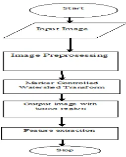
Fig 1 Block Diagram of Proposed System Architecture
Input to the system is medical images & for proper diagnosis of disease images require to be proper, accurate so here we using medical image processing so that proper image should be take for processing. Intra-oral and extra-oral images are an indispensable diagnostic tool for pathologic conditions of the oral and maxillofacial structures. There are very few instances when a diagnosis is actually made from a radiograph. Most pathologic conditions in the oral and maxillofacial region share some radiographic characteristics, so use of radiographs should always be viewed as only a part of the diagnostic procedure.
Methodology
Available Online at www.ijpret.com
647
A. Image Preprocessing
In the Image Preprocessing stage[8], Image enhancement is done to improve the interpretability or perception of information in images for human viewers, or to provide better input for other automated image processing techniques. The input image is preprocessed by increasing the gray levels in the image using linear contrast stretching.
B. Segmentation by Thresholding Method
Threshold segmentation is one of the simplest segmentation methods. The input gray scale image is converted into a binary format. The method is based on a threshold value which will convert gray scale image into a binary image format.
C. Marker Controlled Watershed Transform Algorithm
The Marker – Controlled watershed segmentation[10] solves the problems of lack of smoothness in the image and the over segmentation, faces by the previous segmentation algorithm. The Marker based watershed segmentation can segment unique boundaries from an image or stack of images.
1. Compute a segmentation function. This is an image whose dark regions are the objects to be segmented.
2. Compute foreground Markers. These are connected blobs of pixels within each of the objects.
3. Compute background Markers. These are pixels that are not part of any object.
4. Modify the segmentation function so that it only has minima at the foreground
and background Marker locations.
5. Compute the watershed transform of the modified segmentation function.
D. Morphological Operations
Available Online at www.ijpret.com
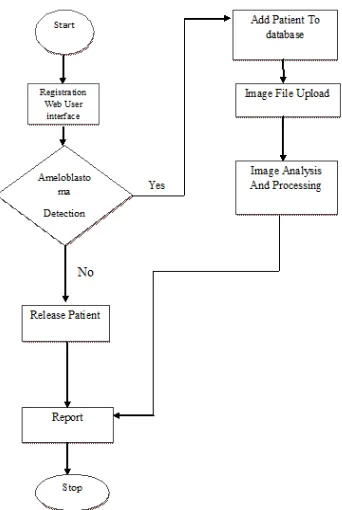
648 FLOWCHART OF PROPOSED SYSTEM
Fig 3. Shows flowchart of proposed system
CONCLUSIONS
In this paper we have proposed a system that will provide rapid remote expertise to diagnosis to patient based on communication technology. Medical field require appropriate health infrastructure and expertise for effective delivery and output. Here we use image processing technique to process x-ray of patient to detect tumor in jaw and give status of the tumor. That helps doctor to diagnosis the tumor as early as possible.
REFERENCES
1. M. Anthony Pogrel, Brian L. Schmidt, Chad G. Robertson” Clinical Pathology: Odontogenic and Nonodontogenic Tumors of the Jaws”..
Available Online at www.ijpret.com
649 3. Wang Yingli Zhao Xueli Wang Ming “Based on the top-hat algorithm of oral medical image information recognition and data processing”,2012 International Conference on Measurement, Information and Control (MIC). 978-1-4577-1604-1/12/$26.00 ©2012 IEEE..
4. A. Banumathi, J. Praylin Mallika , S. Raju and V. Abhai Kumar, “Automated Diagnosis and Severity Measurement of Cyst in Dental X- ray Images using Neural Network”, Biomedical Soft Computing and Human Sciences, Vol.14,No.2,pp.103-108 (2009).
5. Ingrid Nurtanio, I Ketut Eddy Purnama, Mochamad Hariadi, and Mauridhi Hery Purnomo2 “Cyst and Tumor Lesion Segmentation on Dental Panoramic Images using Active Contour Models”, IPTEK, The Journal for Technology and Science, Vol. 22, No. 3, August 2011.
6. Mukesh Kumar, Kamal K. Mehta, “A Texture based Tumor detection and automatic Segmentation using Seeded Region Growing Method”, IJCTA JULY-AUGUST 2011.
7. Ştefan Oprea, Costin Marinescu , Ioan Liţă, Mariana Jurianu, Daniel Alexandru Vişan, Ion
Bogdan Cioc,” Image Processing Techniques used for Dental X-Ray Image Analysis” 978-1-4244-3974-4/08/$25.00 ©2008 IEEE
8. W. Gonzalez,’ Digital Image Processing, 2nd ed. Prentice Hall, Year of Publication 2008
9. Introduction To Radiologic Diagnosis Of Disease Of The Oral And Maxillofacial Region-Guidelines.
10.K. Anuradha, K. Sankaranarayanan “Detection of Oral Tumor based on Marker – Controlled Watershed Algorithm”, International Journal of Computer Applications (0975 – 8887) Volume 52– No.2, August 2012.