EVALUATION OF ROOT ANATOMY AND MORPHOLOGY OF
MANDIBULAR PREMOLARS WITH CBCT IN IRANIAN POPULATION
ROGHANIZAD NASRIN1, KHALILAKZOHREH2, PANJNOUSH MEHRDAD3 AND
GHOLAMSHAHI MAHBOUBEH4*
1
Assistant Professor of Endodontics, Dental School, Islamic Azad University, Tehran, Iran. 2
Assistant Professor of Endodontics, Dental School/Iranian Center for Endodontic Research, Islamic Azad University, Tehran, Iran.
3
Assistant Professor of orofacial radiology, Dental School, Tehran University of Medical science, Tehran, Iran.
4*
Post Graduated Student, EndodonticsDept, Islamic Azad University, Dental Branch, Tehran, Iran.
ABSTRACT
There are different methods for investigating anatomy and morphology of root canal. One useful clinical method is applying cone beam computed tomography which lessens the limitations of two dimensional radiography images and it has fewer rays with higher resolution. Considering the importance of anatomy and morphology of canal in cleaning and formation of root canal system, based on incompatible results from various studies, and due to insufficient researches in terms of planning and research methodology, this study aimed to investigate root anatomyand mandibular first and second canal by CBCT in Iranian population during 2014-2015. This study was performed cross-sectional. Studied population included mandibular premolarteeth that were healthy and recognizable crown with healthy and evolved apex without central or internal desorption and root crack and without restoration from CBCT images. 300 CBCT images were studied statistically. What we examined was teeth of mandibular first and secondpremolarin terms of gender, root length, root number, numbers of each root canals, canal types, presence of isthmus, intervalscanal orifice , and finally anatomy symmetry. Statistical analysis was conducted by SPSS software. 95% of mandibular first premolar teeth in left side were single rooted and 5% were double rooted, 76/5% were single canal and 23/5% was double canal. In right side, 96/3% were single rooted and 2/7% were double rooted, 78% single canal and 21/9% were dc. 96/6% of second premolar teeth in left side were single rooted and 4% were double rooted, 85/5% were single rooted and 14/5% were double in right side 99/3% were single rooted and 0/7% were double rooted and 92/5% were single canal and 7/5% were double canal which all indicated that there was no significant difference between first and second premolars. Based on results it was illustrated that first and second premolar teeth were almost single rooted (95%, 99/6%) with one canal (type I) and secondlytype V had the most prevalence rate. Second premolars in nearly all cases were single rooted and there was no difference in tooth type. Direction of root curvature was direct in most cases (54%) and then distal direction was in second priority (37%) and there was no difference in tooth type.
KEYWORDS: premolar canals, CBCT, root and canal anatomy, mandibularpremolar teeth
INTRODUCTION
Base tooth anatomy is main basis for science of root treatment. Today, root apex is not the only area in endodonticscience but the idea ofthree-dimensionalroot canal fillingimplies that although
working length and maintaining it is more important, access to all complications of canal inside is also crucial in order to facilitate root canal filling(1).Different methods are for investigating anatomy and morphology of root canal which all
includes dental pulpcoloration with clearing,
study of different sections by disc and providing dental models via clear resin which all are part of laboratory equipment's.(3,4). One useful clinical method is cone beam computed tomography (CBCT) which decreases limitations of two dimensional radiography and it has less ray and more resolution (1,11). Studies indicated that for assessing dental sections, CBCT is more useful in anatomy investigations than other methods of radiography (1, 2).In 2015 HakanArslan conducted cone-beam computed tomographic study of root canal systems in mandibular premolars in a Turkish
population. 287 CBCT images from
mandibularpremolar teeth were provided from patients and then they studied in terms of different formations of orifice, age and gender of patient, dental type, its site, canal form, presence of C form canal, and radicular groove. Circle form of canal and condition of being circle were studied in terms of distance from orifice. A theoretical model has been defined for determining orifice form. Orifice form is almost planar (37%) and it is like key holes (23%).prevalence of T form canal was 3/8% and C form was 2/1%. And also prevalence of circle form of canal in middle area and 1/3 epical was 95/1% and prevalence of radicular groove was 24% in first premolarand 4.5% in second premolar. Average length of distance to circle formation of canal form had relation with patient age (5). In 2015, Felsypremila et al studied Anatomic symmetry of root and root canal morphology of posterior teeth in Indian subpopulation using cone beam computed tomography. They selected 246 CBCTimages and they anterior teeth without dentalresorption were examined in each quadrant.Form 3015 examined teeth (811 maxilla premolar, 845 mandibular premolar, 738 maxillary molars, 621 mandibular molar), There was no significant difference in root number and symmetry of maxillary first and second premolar (81/5%). mandibular second premolar had more symmetry than second molar. The most prevalence rate of anatomy was observed in maxilla first premolar (double rooted, double canal), maxilla second premolar (single rooted and four canal), mandibular first and second premolar
(single rooted and single canal), maxillary first and second molar (three root and fourth canal), mandibular first and second molar (double rooted and three canal). In contrast, there was the most asymmetry between maxillary second molars (8).Considering the importance of anatomy and morphology of canal in cleaning and shaping of root canal system, based on incompatible results from various studies, and due to insufficient researches in terms of planning and research methodology, this study aimed to research effect of gender in root with mandibular first and second premolar morphology and teeth canals of CBCT in Iranian population.
RESEARCH METHODOLOGY
This study was performed cross-sectional. Studied population included mandibularpremolar teeth that were healthy and recognizable crown with healthy and developedapex without external or internal resorption and root crack and without restoration from CBCT images. 300 CBCT images were studied statistically. What we examined was teeth of mandibular first and secondpremolarin terms of gender, root length, root number, numbers of each root canals, canal types, presence of isthmusand finally anatomy symmetry. Root numbers, canal numbers in each root, canal type in each three dimension of axial, coronal and sagittal were studied. Canal types were reported based on categorization ofVertucci Root length were . measured in two view of coronal and sagittal. Root curvature and root curvatureangle were examined in both view of coronal and sagittal through Schnieder method and curvature angle were recorded by degree. Axial slice were in 0.2mm sections. It should be mentioned that all noted cases were studied by endodontic and radiologist's
professional assistance twice. And also
* Cross sectional CBCT images of the mandibular premolar a) axial plan, b) sagittal plan c) coronal plan
RESULTS
In this study, 300 CBCT images of 1133 teeth
including 593 firstpremolarteeth and 540
mandibular secondpremolarwere examined among which images belonged to 141 men and 159 women with average age of 42 (20-62 year old). First and secondpremolarteeth were single rooted in most
cases (95% and 99/6%) and single canal (type I) and then the most prevalence designed to type V and respectively to type III, II, IV. 3 cases did not belong to Vertucci’s categorization (table 1,2). Prevalence of more than two canal was greater in women than men while this difference was not statistically significant (p>0/1) (diagram 1).
Diagram 1
Table 1
Distribution of canal type in respect of tooth under study
type Canal I
II III
IV V
Other issues Total
type Tooth
left mandibular first premolar 226(75/6%)
5(1/7%) 16(5/4%)
1(0/3%) (15/7%)47
(0/3%) 296 (100%)
right mandibular first premolar 232(7/3%)
3(1%) 13(4/3%)
1(0/3%) 48(16%)
- 297(100%)
left mandibular second premolar 232(77/3%)
3(1%) 4(1/3%)
1(0/3%) 31(10/3%)
1(0/3%) 272(100%)
right mandibular second premolar 248(82/4%)
3(1%) 1(0/3%)
1(0/3%) 14(4/7%)
1(0/3%) 268(100%)
Table 2
Distribution of canal number in respect of tooth under study
number Canal 1
2 bigger than 2 total
Tooth type
left mandibular first premolar 226 (76/4%)
69 (23/3%) 1
(0/3%) 296
(100%)
right mandibular first premolar 232
(78/1%) 65(21/9)
- 297
(100%)
left mandibular second premolar 232
(78/1%) 39
(14/3%) 1
(0/4%) 272
(100%)
right mandibular second premolar 248
(92/5%) 19
(7/1%) 1(0/4%)
268 (100%)
In almost all double canal cases, isthmus was observed in 1/3 epical. Orifice site in nearly all cases was located in CEJ area. Orifice form was also observed in CEJ area ellipticalforms in most cases and in a few cases was circle form. Mandibular first premolarteeth in left side were 95% single rooted, 5% double rooted, 76/5% single canal and 23/5% double canal. In right side they were 96/3% single rooted and 2/7% double rooted,
Table 3
Distribution of root number in respect of tooth under study
Root number 1
2 Total
type Tooth
left mandibular first premolar 248
(95/9%) 12
(4/1%) 296
(100%)
right mandibular first premolar 289(97/3%
8(2/7%) 297
(100%)
left mandibular second premolar 271(99/6%)
1(0/3%) 272
(100%)
right mandibular second premolar 266(99/3%)
2(0/7%) 268
(100%)
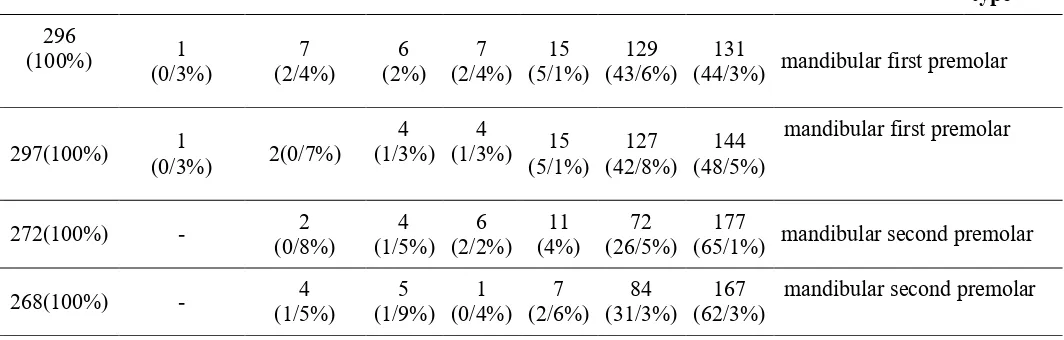
Other cases such as distribution of root curvature, separation location of canals, root length and angel of root curvature of premolarteeth are stated in table 4-7.
Table 4
Distribution of root curvature regarding tooth under study
root of Direction curvature
direct distal
masial lingula
buccal
distobuccal
distolingual total
Tooth type
mandibular first premolar 131
(44/3%) 129
(43/6%) 15
(5/1%) 7
(2/4%) 6
(2%) 7
(2/4%) 1
(0/3%) 296
(100%)
mandibular first premolar 144
(48/5%) 127
(42/8%) 15
(5/1%) 4
(1/3%) 4
(1/3%) 2(0/7%)
1 (0/3%) 297(100%)
mandibular second premolar 177
(65/1%) 72
(26/5%) 11
(4%) 6
(2/2%) 4
(1/5%) 2
(0/8%) -
272(100%)
mandibular second premolar 167
(62/3%) 84
(31/3%) 7
(2/6%) 1
(0/4%) 5
(1/9%) 4
(1/5%) -
Table 5
Distribution of detachment location of canals regarding under study tooth type
Location of canal detachment Middle third
Third epical Total
Tooth type
left mandibular first premolar 57
(82%) 12
(18%) 69
(100%)
right mandibular first premolar 58
(89%) 7
(11%) 65
(100%)
left mandibular second premolar 27
(69/2%) 12
(31/8%) 39
(100%)
right mandibular second premolar 15
(78%) 4
(22%) 19
(100%)
Table 6
Distribution of root length by consideration of under study tooth
Root length Tooth type Minimum
Maximum Mean±S.D
C.V
left mandibular first premolar 17
27 21/6±1/8
8/37
right mandibular first premolar 17/3
26/6 21/6±1/9
8/99
left mandibular second premolar 18
27/2 1/74±2/9
8/03
right mandibular second premolar 16/5
27/7 1/9±22
8/77
Table 7
Distribution of angel of root curvature by teeth considering under study
Angle of root curvature
type Tooth
Minimum
Maximum mean±S.D
C.V
Left mandibular first premolar 0
61/31 16/22±16/36
96/7
right mandibular first premolar 0
62/28 15/46±13/95
1/3409
left mandibular second premolar 0
52/51 13/47±9/37
1/14 47
right mandibular second premolar 0
61/55 13/53±9/94
14/960
DISCUSSION
Based on results of this study generally in Iranian race mandibular first premolar teeth in left side were 95% single rooted, 5% double rooted, 76/4% single canal and 23/3% double canal. In right side 96/3% they were single rooted and 2/7% double rooted, 78% single canal and 21/9% double canal. Second premolarteeth in left side were 99/6% single rooted, 4% double rooted, 85/3% single canal and 14/3% double canal. In right side they were 99/3% single rooted, and 7% double rooted, 92/5 single canal, and 7/1 were double canal which proved there was no significant difference between first and second premolarin both side. First results
right side they were 87% single root and 12/7% double rooted, 66/5% single canal and 30% double canal which indicated that there was no significant difference between first premolars in both side. In right side they were 78% single root, 66/5% single canal and 33/5% double canal which there was no significant difference first premolars in both side. In right side they were 95/% single rooted, 4/4% double rooted, 83/4% single canal and 16/6% double canal while there was not significant
difference in canal numbers between
secondpremolars of both side (7).In our study mandibular first premolar teeth in left side were 95% single rooted and 5% double rooted, 76% single canal and 23/3% double canal. In right side they were 96/3% single rooted and 2/7% double rooted, 78% single canal and 21/9% double canal. Second premolarteeth in left side were 9/6% single rooted and 0/4% double rooted, 85/3% were single canal and 14/3% double canal. In right side they were 99/3% single rooted and 0/7% double rooted, 92/5% single canal and 7/1% double canal which there is no significant difference between first and second premolars in both sides. First double rooted premolars numbers in left side was higher and second premolars were almost single rooted in all cases. Prevalence rate of double canal teeth in our study was less especially second premolar teeth.In other study performed by Felsypremila et al studied Anatomic symmetry of root and root canal
morphology of posterior teeth in Indian
subpopulation using cone beam computed
tomography. They selected 246 CBCT images and investigated about posterior teeth without dental restoration and restoration in each quadrant. Mandibular second premolar had more symmetry in root and canal number (98/3%) than first premolars (96/1%). Most prevalence of anatomy was observed in mandibular first and second premolar with single rooted and single canal. In our study firstpremolars showed more symmetry (95%) that secondpremolar s (90%) in root number. But in canal numbers of both first and second premolar it was illustrated that there is similar symmetry equals to 83% may be due to racial difference and sample volume (8).Bulult et al studied about evaluation of root morphology and root canal configuration of premolars in the Turkish individuals using cone beam computed tomography. Mostmandibular premolars were single rooted. Prevalent anatomy in mandibular first premolar was single canal and type I (94%) and type V (3/2%) which mandibular second premolars with single canal and type I (98%) were most prevalent. There was no difference between types and numbers of canal
among men and women (9). However, in our research prevalence of a canal and type I in mandibular first premolars was (77/7%) and type V (15/6%). And it was (90%) of canal type I in second premolars and (7/7%) of canal type V. In Iranian population double canal premolars was more prevalent maybe due to racial difference or sample volume. AditiaShetty performed similar research in Indian population by CBCT. They studied 1186 mandibular first and second premolar teeth investigated by CBCT and indicated that most 83/8% first premolar teeth and second premolar 93/4% were type I and then type V had more prevalence rate (10).Jun-beam park performed similar research in Korean population by CBCT. Most first premolar s 99/3% and second premolars 99/9% were single rooted (11). Our research had similar results. Xuan Yu et al conducted Cone-beam computed tomography study of root and canal morphology of mandibular premolars in a western Chinese population. 149 CBCT images including 178 mandibular first premolar and 178 mandibular second premolar were studied. Results indicated that 98% of mandibular first premolar were single rooted and 2 percent were double rooted. 87% were single canal and 11/2% double canal and 0/% were with three canal. Prevalence of Cshape canal was 1/1 percent. All second premolars were single rooted and 97/2 percent were single canal and 2/2 % were double canal. And also prevalence of C-shape canal was 0/6%. First premolar type V had the most prevalence of canal while second premolar had low anatomic variation in Chine's population (1). About root curvature, Vertucci stated the most prevalence for first and second premolar teeth as buccal<distal<direct. As Sobhani et al stated the most prevalence of root is respectively as mesial<buccal<lingual<mesiolingual<mesiobuccal <distal<distolingual<distobuccal<direct. Inthis study most prevalence of root curvature was
respectively related to
distoligual<distobuccal<lingual=buccal< (CurveS) bayonet<mesial<distal< direct (12). The most brilliant achievement of this study is utilizing CBCT for investigation canal anatomy which is in vivo method and it is not like other methods as pulling tooth and it doesn’t have problems related to two dimensional images with common radiographies.premolarteeth studies which is the most difficult teeth problems in root therapy regarding to different anatomic aspects and high
sample volume by CBCT is brilliant
CONCLUSION
First and second premolarteeth were mostly single rooted (95%, 99/6%) and single canal (type I). Secondly, type V was mostly prevalent. Orifice site was in tooth center in CEJ area almost in all cases. Orifice formation in CEJ area and almost in all sites was elliptical and rarely was it circle form.Second premolars were single rooted in nearly all cases and there was no difference in tooth type. Direction of root curvature was direct in most cases (54%) and in other cases was distal (37%) and there was no difference in tooth type.Prevalence of S-curve
curvature had been observed in 2% of cases (22cases) which had more prevalence in first premolar (91%).
Limitations and problems
Study of more than 1000 CBCT in order to obtain appropriate samples.
Considering all CBCT samples took a lot time and it was so elaborating.
Recommendation
Study of other teeth by CBCT and compiling data as general reference in Iranian population.
REFERENCES
1. Amin Sobhani M, Razmi H, Sadegh M.
Evaluation of anatomy and morphology of human mandibular premolar teeth by cone-beam computed tomography in aIranian population.Journal of Dental Medicine- Tehran University of medical science. 2013 autumn; 26(3):203-10.
2. Sert S, Bayiril GS. Evaluation of root canal
configuration of the mandibular and
maxillary permanent teeth by gender in the Turkish population. J Endod 2004;30(6):391-398
3. Pope O, Sathorn C, Parashos P. A
comparative investigation of cone-beam
computed tomography and periapical
radiography in the diagnosis of a healthy periapex. J Endod. 2014 Mar; 40(3):360-5
4. Xuan Yu, Guo B, Ke Zeng Li K. Cone-beam
computed tomography study of root and canal morphology of mandibular premolars in a western Chinese population. BMC Medical Imaging. 2012 July; 12:18
5. Arslan H, Davut Capar I. A cone-beam
computed tomographic study of root canal systems in mandibular premolars in a Turkish
population: Theoretical model for
determining orifice shape. European Journal of Dentistry. 2015 Jan-Mar;9 (1) :11-19 6. Hess W. The anatomy of the root-canals of
the teeth of the permanent dentition, part 1. New York:William Wood and Co, .1925
7. Kazemipoor M, Poorkheradmand M,
Rezaeian M, Safi Y. Evaluation by CBCT of
root and kanal Morphology in Mandibulars Premolars in an Iranian population. Chin J Dent Res. 2015 Sep; 18(3):191-6
8. FelsypremilaG, Vinothkuma T, Kandaswamy
D. Anatomic symmetry of root and root canal morphology of posterior teeth in Indian subpopulation using cone beam computed tomography: A retrospective study. Eur J Dent.2015 October-December;9(4):500-507
9. Bulut D, Kose D, Ozcan G, Sekerci A.E,
Canger E, Sisman Y. Evaluation of root morphology and root canal configuration of premolars in the Turkish individuals using cone beam computed tomography. Eur J Dent.2015 October-December;9(4):551-554
10. Shetty A, Hegde M, Shetty S.A three
dimensional study of variation in root canal morphology using cone-beam computed tomography of mandibular premolars in south
Indian population.JCDR.2014 Aug;
8(8):ZC22-ZC24
11. Park J, Kim N, Ko Y. Evaluation ofroot anatomy and morphology of mandibular
premolars & molars with cone-beam
computed tomography in a koreanpopulation. Eur J Dent.2013;7(1):94-101
12. Amin Sobhani M, Razmi H, Sadegh M.