Available Online at www.ijpret.com 685
INTERNATIONAL JOURNAL OF PURE AND
APPLIED RESEARCH IN ENGINEERING
AND TECHNOLOGY
A PATH FOR HORIZING YOUR INNOVATIVE WORK
THE REVIEW OF CLASSIFICATION OF BRAIN TUMOR IN MRI USING
PROBABILISTIC NEURAL NETWORK
GAURAO P. MATE, PROF. M. A. KHAN
Dept. of E & TC, BNCOE, Pusad.
Accepted Date: 15/03/2016; Published Date: 01/05/2016
\
Abstract- Brain Tumor is one of the serious problems in the humans. The main cause of the brain tumor is the uncontrolled growth of the cells in brain. If it detects at an early stages it will cure rapidly but if not it can cause a very painful death. Also it can spread to the various part of the body such as lungs, etc. The Magnetic Resonance Imaging (MRI) plays a very vital role in studying the various diseases related to the brain. The classification of such MRI is necessary to distinguish the between the healthy person and the person having a tumor .The proposed work consist of three parts namely segmentation, feature extraction and Probabilistic Neural Network to classify the image whether it contains a tumor or not.
Keywords: Magnetic Resonance Imaging, segmentation, Feature Extraction, Probabilistic Neural Network.
Corresponding Author: GAURAO P. MATE
Access Online On:
www.ijpret.com
How to Cite This Article:
Gaurao P. Mate, IJPRET, 2016; Volume 4 (9): 685-690
Available Online at www.ijpret.com 686
INTRODUCTION
The accurate detection of brain tumor is necessary to avoid unusual death due to brain tumor. Normally the brain tumor arises from various parts in the brain such as brain cells, blood vessels or nerves that are present in the brain.
Segmentation plays a very vital role in determining the Region of Interest (ROI) in the brain. Segmentation can be semiautomatic or fully automatic. The fully automatic methods can be applied in order to avoid the intervention of the human operator to being involved. Also with the help of an image processing we can achieve accurate result with the reduction in the time to detect and classify the brain tumor.
METHODS
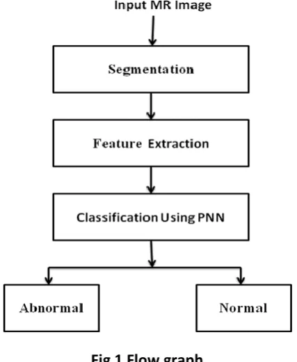
In the proposed work Saliency Map Algorithm is used to segment the Magnetic Resonance Image (MRI). The features are using the extended histogram and extended edge descriptors are used to extract the desired features from an image. Finally the Probabilistic Neural Network used to classify the image whether it contains the tumor or not.
Fig 1 Flow graph
1. SEGMENTATION
Available Online at www.ijpret.com 687
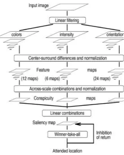
Fig 2 Saliency Map Model
This type of framework provides a massively parallel method for the fast selection of a small number of interesting image locations to be analyzed by more complex and time consuming object-recognition processes [1].
During the first phase the image is converted to nine different scales ranging from one to eight reduction factor.
At second step, the image is decomposed into a series of 42 feature maps. The first set of feature maps includes contrast, intensity and color double opponency maps which are generated from color channels. Firstly the intensity I is calculated as
𝐼 = (𝑟 + 𝑔 + 𝑏
3 )
Then for each pixel in the pyramid generate color channels 𝑅 =𝑟−(𝑏+𝑔)
2 for red, G=
𝑔−(𝑟+𝑏) 2
for green, B=𝑏−(𝑟+𝑔)
2 for blue, and Y=(𝑟 + 𝑔) 2 − |𝑟 −⁄ 𝑔| 2 − 𝑏⁄ for yellow and negative
values are set to zero. The detection of local orientation at each point in the given image is achieved using over complete steerable filters.
Available Online at www.ijpret.com 688
operation across spatial scales is done by interpolation to the fine scale and then point-by-point subtraction. Next there are twelve chromatic double opponency maps for the centre-surround differences between red-green and blue-yellow pairs of opposing channels. The final map consists of local orientation map generated from the intensity maps. There are such twenty four maps.
Finally normalization is carried out by normalizing the values in the map to a fixed range [0…M] in order to eliminate modality dependent amplitude difference.
The feature maps are combined into three conspicuity maps at the scale 4. This is obtained through across-scale addition by reducing each map to the lowest resolution (scale 4) and then doing point-by-point addition.
The algorithm identifies the maximum value of the saliency map as a centre of attention and then uses the winner takes-all neural network to identify secondary maximum in order to generate an approximation of switching the visual attention of the human eye.
2. FEATURE EXTRACTION
Features are extracted from the image so that it can be suitable for the further classification. There are two features are to be extracted
I. Extended Histogram Descriptors
II. Extended Edge Descriptors
I. extended histogram descriptors:
Normally the range of the gray level image between [0-255] but the image provided to us may or may not be between this ranges so by means of the extension we are just try to utilize the full range of an image, which results in better appearance of the image. So we can easily extract the desired feature. This will plots the number of pixels of a particular intensity on Y axis and the pixel intensity on X axis.
II. extended edge descriptors:
The Canny Edge detector is used here for detecting the edges. It continuous to search for an ideal step edge in the presence of Gaussian noise and defined a matched filter that could be approximated by the difference of a Gaussian.
Available Online at www.ijpret.com 689
3. PROBABILISTIC NEURAL NETWORK:
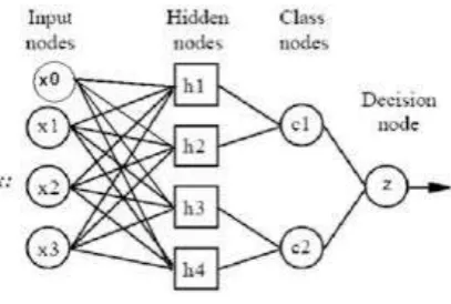
A probabilistic neural network (PNN) is a feed forward neural network, resulting from the Bayesian network and a statistical algorithm. In a PNN, the operations are organized into a multilayered feed forward network which includes four layers Input layer, Hidden layer, Pattern layer or Summation layer and an Output layer.
Fig 3 Probabilistic Neural Network
The probabilistic networks perform classification where target variable is available [9].
The input nodes are nothing but the set of measurements.
The second layer is consisting of the Gaussian functions which are formed using the given set of data points as a center.
The third layer performs the averaging operations on the outputs of the second layer for each class.
The fourth layer selects the largest value and after that associate class label is determine.
CONCLUSION:
Proposed System presents an efficient method of classifying MR brain Images whether it contains a tumor or not. In the proposed method with the help of features extracted from extended histogram and Canny edge detector as edge detector can be used to classify the image with the help of Probabilistic Neural Network and the accuracy of the neural network is made up to 100%.
REFERENCES:
1. Itti, L., Koch, C., Niebur, E. ‘A model of saliency-based visual attention for rapid scene
Available Online at www.ijpret.com 690
2. B. Balakumar and P. Raviraj , “Abnormality Segmentation and Classification of Brain MR
Images using Combined Edge, Texture Region Features and Radial basics Function”, Research Journal of Applied Sciences, Engineering and Technology’, 6(21), pp. 4040-4045,2013.
3. Shrasthta Chauhan, Mrs. Neha Shrma, “Brain Tumor Detection and Segmentation Using
Histogram. Thresholding and Artificial Neural Network Techniques”, International Journal of Advanced Research in Computer Science and Software Engineering, Vol.4, Issue 8, pp. 104-109, August 2014.
4. Shishir Dube , Suzie El-Saden , Timothy F. Cloughesy , Usha Sinha, “Content Based Image
Retrieval for MR Image Studies of Brain Tumors”, Proceedings of the 28th IEEE EMBS Annual International Conference New York City, USA, pp. 3337-3340, Aug 30-Sept 3, 2006.
5. P. Hari Krishnan, Dr. P. Ramamoorthy, “Fuzzy Clustering Based Ant Colony Optimization
Algorithm For MR Brain Image Segmentation”, Journal of Theoretical and Applied
Information Technology, Vol. 65, No. 33, pp. 645-649, 1st July 2014.
6. Jose Anand, K. Sivachandar, “An Edge Vector and Edge Map Based Boundary Detection
in Medical Images”, International Journal of Innovative Research in Computer and Communication Engineering, Vol. 1, pp.1051-1055, June 2013.
7. Song-yun Xie, Rang Guo, Ning-fei Li, Ge Wang, and Hai-tao Zhao ‘Brain MRI Processing
and Classification Based on Combination of PCA and SVM’ Proceedings of International Joint Conference on Neural Networks, Atlanta, Georgia, USA,pp.3384-3389,June 14-19, 2009.
8. Shrasthta Chauhan, Mrs. Neha Shrma ’Brain Tumor Detection and
Segmentation Using Histogram Thresholding and Artificial Neural Network Techniques .International Journal of Advanced Research in Computer Science and Software Engineering Volume 4,pp. 104-109 August 2014.
9. Shreepad S. Sawant, Preeti S. Topannavar “Introduction to Probabilistic Neural Network