Original Research Article
Clinical profile of systemic lupus erythematosus among children
less than 12 years
Senthil Kumar Andy
1, Elayaraja Kandasamy
2*
INTRODUCTION
Systemic lupus erythematosus (SLE) is an episodic multisystem autoimmune disease characterised by widespread inflammation of blood vessels and connective tissues and by the presence of antinuclear antibodies especially antibodies to native double stranded DNA. Its clinical manifestations are extremely variable, and its natural history is unpredictable. Untreated SLE is often progressive and has a significant fatality rate.1
It is the second commonest pediatric rheumatic disorder next to juvenile idiopathic arthritis. The clinical course
can range from mild to severe and can be potentially life threatening.1-5 SLE is a relatively rare disease in
childhood with estimated incidence ranging from 10 to 20 per 1 lakh children depending on ethnic population.1-5
Hence the study was undertaken to know about the varied clinical presentation, immunological status, disease activity and damage to organs at follow up.
The authors aimed at the study of the clinical profile of SLE among children less than 12 years attending an urban referral hospital, SLEDAI scoring at onset and follow up at 1 year and SLICC/ACR damage index at 1 year.
1Department of Paediatrics, Institute of Child Health and Hospital for Children, Egmore, Madras Medical College,
Chennai, Tamil Nadu, India
2Department ofPaediatrics, Government Villupuram Medical College, Villupuram, Tamil Nadu, India
Received: 03 January 2018 Accepted: 06 February 2018
*Correspondence: Dr. Elayaraja Kandasamy, E-mail: drelayaraja.k@gmail.com.
Copyright: © the author(s), publisher and licensee Medip Academy. This is an open-access article distributed under the terms of the Creative Commons Attribution Non-Commercial License, which permits unrestricted non-commercial use, distribution, and reproduction in any medium, provided the original work is properly cited.
ABSTRACT
Background: Systemic lupus erythematosus (SLE) is an episodic multisystem autoimmune disease characterized by widespread inflammation of blood vessels and connective tissues and by the presence of antinuclear antibodies especially antibodies to native double stranded DNA. The aim was to study the clinical profile of SLE among children less than 12 years attending an urban referral hospital, SLEDAI scoring at onset and follow up at 1 year and SLICC/ACR damage index at 1 year.
Methods: A descriptive, prospective and observational study was conducted in Medical, Nephrology, Rheumatology OPD wards, in ICH and HC, from November 2007 to August 2009 among all children diagnosed to have SLE. Results: In this study there were 50 cases over the last 2 years. Majority of the children were diagnosed within a year of their initial manifestation. The mean age at the time of onset of symptoms was 7.94 years. Female to male ratio in our study is 2.5:1.
Conclusions: Efforts should be directed in diagnosing at earlier stage itself for better outcome. SLEDAI and SLICC/ACR DI can be incorporated in routine follow up to detect mild to moderate and severe flare and extent of organ damage.
Keywords: Children, SLEDAI score, SLICC/ACR damage index, Systemic lupus erythematosus
METHODS
Present study was a descriptive/prospective observational study conducted in Medical, Nephrology, Rheumatology and OPD wards, in ICH and HC from November 2007 to August 2009
Study population includedall children diagnosed to have SLE with a Sample size of 50.
Inclusion criteria
All children <12 years diagnosed to have SLE.
This study was conducted in medical ward, nephrology ward, rheumatology OPD, in ICH and HC. Clinical features, laboratory investigations, treatment were followed for all the children. SLE disease activity index and SLICC/ACR damage index was done at diagnosis and follow up at 1 year.
Children were divided into three groups based on the age of onset as less than 2 years, between 2-10 years and 10-12 years. Clinical features, laboratory investigations and treatment were compared between the groups.
Statistical analysis
The sample size of the study was 50. Frequency of occurrence of clinical, laboratory, and treatment parameters were derived for all the 50 children. SLEDAI and SLICC/ACR damage index scores had their mean values computed. SLEDAI scores were compared between onset and follow up by one-way ANOVA, Fischer test and p values derived. Analysis between three groups of disease occurrence was done by Chi square test and p values obtained. P values less than 0.05 is taken as significant.
RESULTS
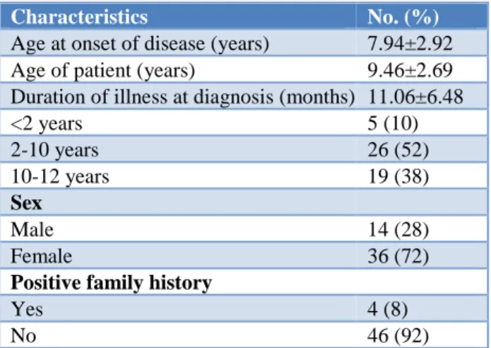
Age at onset of disease was 7.94±2.92 years (Table 1). Mean age of patients was 9.46±2.69 years. Duration of illness in patients prior to diagnosis was 11.06±6.48 months. Female to male ratio was 2.5:1. 14 (28%) of the children were male and 36 (72%) of the children were female. Family history of auto immunity in first and second-degree relatives is noted in 8% of patients.
Distribution patients according to the different organ involvement is presented in Table 2. In mucocutaneous involvement, 36 cases (72%) had malar rash followed by photosensitivity (52%) and oral ulcers (32%). Reticuloendothelial involvement in the form of lymphadenopathy was found in 16 cases (32%). Proteinuria more than 0.5 g/day was found in 20 cases (40%). 30% had nephrotic syndrome.
Renal biopsy was done in 14 patients. In GIT involvement 35 cases (70%) had hepatosplenomegaly.
Serositis was noted in 19 cases (38%). CNS involvement in the form of seizures was noted in 13 cases (26%). Cardiac involvement in the form of valvular lesions was found in 4 patients (8%).
Majority of patients had fever, 47 cases (94%). Haemolytic anemia was found in 10 patients (20%). Thrombocytopenia was found in 15 cases (30%). Leukopenia was found in 3 patients (6%).
Table 1: Demographic characteristics.
Characteristics No. (%)
Age at onset of disease (years) 7.94±2.92
Age of patient (years) 9.46±2.69
Duration of illness at diagnosis (months) 11.06±6.48
<2 years 5 (10)
2-10 years 26 (52)
10-12 years 19 (38)
Sex
Male 14 (28)
Female 36 (72)
Positive family history
Yes 4 (8)
No 46 (92)
Table 2: Distribution patients according to the different organ involvement.
Organs involvement N = 50 %
Mucocutaneous involvement
Malar rash 36 72
Photosensitivity 26 52
Oral ulcers 16 32
Alopecia 13 26
Discoid rash 2 4
Raynauds 0 0
Musculoskeletal involvement
Arthritis 30 60
Lymphadenopathy 16 32
Renal involvement
Proteinuria >0.5 g/day 20 40
Nephrotic syndrome 15 30
Hypertension 8 16
CVS, RS, GIT and CNS involvement
Hepatosplenomegaly 35 70
Serositis 19 38
Seizures 13 26
Cardiovascular 4 8
Organs (hematological and fever) involvement
Fever 47 94
Hemolytic anemia 10 20
Thrombocytopenia 15 30
Leukopenia 3 6
Table 3: Distribution of renal lesions.
WHO class N =14 % Outcome
Class 1 1 7.1 Improved
Class 2 1 7.1 Improved
Class 3 3 21.4 1-died
Class 4 6 42.8 Improved
Class 5 3 21.4 1-died
Table 4: Laboratory findings and treatment.
N =50 %
Lab findings
ANA 46 92
Anti DS DNA 26 52
C3/C4 40 80
ACL/LAC 3 (9) 34
Treatment
Prednisolone 50 100
Methyl prednisolone 15 30
Azathioprine 12 24
Cyclophosphamide 11 22
Mycophonelate mofetil 7 14
Out of the 50 cases with SLE, 46 cases (92%) had ANA positivity followed by 80% had low c3/c4 and 52% had Anti DS DNA (Table 4). All the 50 patients (100%) received prednisolone as treatment followed by 15 cases received methyl prednisolone and 12 patients had azathioprine. Cyclophosphamide was used as induction therapy in 8 cases (16%), maintenance therapy in nil (0%), and used in relapse 3 cases (6%). MMF was used as induction therapy in 5 cases (10%), maintenance therapy in 2 cases (4%) and MMF was not used for any of the relapse.
SLEDAI score at onset had mean value of 12.54±4.94 (Table 5). P value was 0.29. SLEDAI score at follow up of one year was 10.02±4.47. P value was 0.32. SLICC/ACR damage index at the end of 1 year had mean value of 0.68±1.63. P value was 0.3.
Table 5: SLEDAI score and SLICC/ACR damage index.
Mean SD P value
SLEDAI at onset 12.54 4.94 0.29 SLEDAI at 1 year 10.02 4.47 0.32
SLICC/ACR 0.68 1.63 0.38
Table 6: Sex distribution.
Sex <2 years (n = 5) 2-10 years (n = 26) 10-12 years (n = 19) Total
Distribution No % No % No % No %
Male 1 20 6 23.1 7 36.8 14 28
Female 4 80 20 76.9 12 63.2 36 72
F:M 5:0 3.3:1 1.7:1 P value (NS)
Mean duration at diagnosis 3.8 9.2 15.15 P<0.01
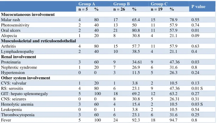
Patients were divided into three groups based on age at disease onset. Group A constituted 5 cases (10%), Group B constituted 26 cases (52%), and Group C 19 cases (38%). 14 (28%) of the children were male and 36 (72%) of the children were female. Female to male ratio was 2.5:1. Female to male ratio was 4:1 in Group A, 3.3:1 in Group B, and 1.7:1 in Group C. F: M ratio was 5:0 in Group A, 3.3:1 in Group B, and 1.7:1 in Group C (Table 6). P value was not significant. Mean duration of illness at diagnosis in months was 3.8 in Group A, 9.2 in Group B and 15.15 in Group C.
Malar rash was noted in 4 cases (80%) in Group A, 17 cases (65.4%) in Group B and 15 cases (78.9%) in Group C (Table 7). Photosensitivity was noted in 2 cases (40%) in Group A, 13 cases (50%) in Group B and 11 cases (57.9%) in Group C. Oral ulcers was found in 2 cases (40%) in Group A, 21 cases (80.8%) in Group B and 11 cases (57.9%) in Group C. P value was 0.01 which is significant. Alopecia was found in 1 case (20%) in Group A, 8 cases (30.8%) in Group B and 4 cases (21.1%) in group C. Arthritis was found in 4 cases (80%) in Group
A, 15 cases (57.7%) in Group B and 11 cases (57.9%) in Group C. Lymphadenopathy was noted in 2 cases (40%) in Group A, 10 cases (38.5%) in Group B and 4 cases (21.1%) in Group C. Proteinuria was found in 3 cases (60%) in Group A, 9 cases (34.61%) in Group B and 9 cases (47.36 %) in Group C. Nephrotic syndrome was found in 1 case (20%) in Group A, 7 cases (26.9%) in Group B and 6 cases (31.6%) in Group C. GIT involvement in the form of hepatosplenomegaly was found in 5 cases (100%) in Group A, 18 cases (69.2%) in Group B, and 12 cases (63.2%) in Group C. p value was 0.27. CNS involvement in the form of seizures was found only in 8 cases (30.8%) in Group B and 5 cases (26.31%) in Group C. p value was 0.31. Thrombocytopenia was noticed in 3 cases (60%) in Group A, 6 cases (23.1%) in Group B and 6 cases (31.6%) in Group C. Fever was noticed in all cases (100%) in Group A, 24 (92.3%) cases in Group B and 18 cases (94.7%) in Group C.
and 10 cases (52.6%) in Group C. Prednisolone was used in all cases (100%). Methyl prednisolone was used in 8 cases (30.8%) in Group B and 6cases (31.6%) in Group
C. Hydroxy chloroquine was used in 1 case (20%) in Group A, 7 cases (26.9%) in Group B and 7 cases (36.8%) in Group C.
Table 7: Distribution patients according to the different organ involvement.
Group A Group B Group C
P value
n = 5 % n = 26 % n = 19 %
Mucocutaneous involvement
Malar rash 4 80 17 65.4 15 78.9 0.55
Photosensitivity 2 40 13 50 11 57.9 0.74
Oral ulcers 2 40 21 80.8 11 57.9 0.01
Alopecia 1 20 8 30.8 4 21.1 0.09
Musculoskeletal and reticuloendothelial
Arthritis 4 80 15 57.7 11 57.9 0.63
Lymphadenopathy 2 40 10 38.5 4 21.1 0.4
Renal involvement
Proteinuria 3 60 9 34.61 9 47.36 0.03
Nephrotic syndrome 1 20 7 26.9 6 31.6 0.8
Hypertension 0 0 3 11.5 5 26.3 0.24
Other system involvement
CVS: valvular 1 20 1 3.8 2 10.5 0.13
RS: serositis 4 80 6 23.1 9 47.36 0.01 S
GIT: hepato splenomegaly 5 100 18 69.2 12 63.2 0.27
CNS: seizures 0 0 8 30.8 5 26.31 0.31
Hemolytic anemia 3 60 4 15.4 2 10.5 0.03 S
Leukopenia 0 0 1 3.8 2 10.5 0.54
Thrombocytopenia 3 60 6 23.1 6 31.6 0.25
Fever 5 100 24 92.3 18 94.7 0.8
Table 8: Laboratory findings and treatment.
Group A Group B Group C
P value
N % N % N %
Laboratory findings
ANA 5 100 24 92.3 17 89.5 0.7
Anti DS DNA 2 40 14 53.8 10 52.6 0.6
Low C3/C4 3 60 23 88.5 14 73.7 0.23
Treatment
Prednisolone 5 100 26 100 19 100 -
Methyl prednisolone 0 0 8 30.8 6 31.6 0.84
Azathioprine 1 20 5 19.2 6 31.6 0.51
Cyclophosphamide 2 40 4 15.3 5 26.3 0.3
MMF 0 0 3 11.5 4 21 0.14
Hydroxy chloroquine 1 20 7 26.9 7 36.8 0.62
Table 9: SLEDAI score and SLICC/ACR damage index.
Group A Group B Group C
One-way ANOVA
Mean S.D Mean S.D Mean S.D
SLEDAI at onset 9.6 1.34 13.31 5.69 12.26 4.18 F=1.2 P=0.29
SLEDAI at 1 year 7.2 1.09 10.5 4.69 10.11 4.58 F=1.16 P=0.32
SLEDAI 1 at onset was 9.6 (S.D 1.34) in Group A, 13.31 (S.D5.69) in Group B and 12.26 (S.D 4.18) in Group C. P value was 0.29 (Table 9). SLEDAI 2 at 1 year was 7.2 (S.D 1.09) in Group A, 10.5 (S.D 4.69) in Group B and 10.11 (S.D 4.58) in Group C. P value was 0.32. SLICC/ACR damage index was 0.8 (S.D. 0.83) in Group A, 0.68 (S.D. 1.02) in Group B and 0.61 (S.D 0.97) in Group C.
Out of the 3 cases (6%) who died SLEDAI 1 score had a mean of 22 (S.D 8.8). P value was 0.01 which is significant (Table 10). SLEDAI 2 at 1 year had a mean of 15.67 (S.D 9.86). p value was 0.01, which is significant. Out of the 3 cases who died SLICC/ACR damage index had a mean of 2.33 (S.D. 2.082). P value was 0.01 which is significant.
Table 10: SLEDAI score and SLICC/ACR damage index of died patients.
Death N Mean S.D T-test
SLEDAI 1 3 22.00 8.8 T=3.88 P=0.01 SLEDAI 2 3 15.67 9.86 T=2.36 P=0.01 SLICC/ACR 3 2.33 2.082 T=3.40 P=0.01
DISCUSSION
In this study there were 50 cases over the last 2 years. Majority of the children were diagnosed within a year of their initial manifestation. The mean duration of illness prior to diagnosis was 11.06 months in this study. The mean age at the time of onset of symptoms was 7.94 years which is lowest among other pediatric SLE studies from India and abroad.6,7 Female to male ratio in the
present study is 2.5:1 which is comparable with other studies.
SLE is a multisystem disorder and the manifestations can be variable. In atypical cases the diagnosis may be missed if the suspicion is not high. Such children may continue receiving treatment without diagnosis as exemplified by some cases. Antituberculosis treatment was received by 3 cases before diagnosis. One child being treated as pulmonary tuberculosis and dilated cardiomyopathy and another child with CNS involvement with MRI evidence of demyelination was found to be SLE. Both of them lacked the typical mucocutaneous features. The most common clinical manifestations were mucocutaneous involvement in the form of malar rash, photosensitivity, oral ulcers, and alopecia. Discoid rash was rare. Raynauds phenomenon was also not noticed. Musculoskeletal involvement in the form of arthritis and reticuloendothelial involvement in the form of lymphadenopathy was also commonly encountered. These results can be compared to other studies from India and abroad.6-8
Renal involvement was noticed in the form of proteinuria and nephrotic syndrome. Among the 8 patients with hypertension, 6 of them had renal involvement. Renal
biopsy was done in 14 cases out of 20 (70%). Majority of patients with lupus nephritis had pathological changes consistent with class 3, 4, 5 lesion. One patient with class 3 lesion and one patient with class 5 lesion died. 3 patients had peritoneal dialysis and 1 patient was on CAPD.
GIT involvement in the form of hepatosplenomegaly was found in majority of cases (70%). Serositis in the form of pleural or pericardial effusion was noticed in 38% of cases. Cardiovascular manifestations in the form of valvular involvement were found in 4 patients (8%). Dilated cardiomyopathy, pulmonary hypertension, mitral regurgitation and cardiac tamponade were the manifestations seen. The patient with pulmonary hypertension had anticardiolipin antibody positive which seems to be a manifestation of antiphospholipid antibody syndrome.
CNS involvement in the form of seizures is found in 13 cases (26%). Neuropsychiatric manifestations were found in 2 of these patients. CT scan was done in all of these patients. Cerebral atrophy (2 cases), CVA with multiple infarct (2 cases), Intracerebral bleed (2 cases) and demyelination (1 cases) were found from the study. The patient with demyelination had Hodgkins lymphoma. CT scan was normal in rest of the patients.
Fever was noticed in 47 patients. Majority of them presented with pyrexia of unknown origin and later found to be SLE. Hemolytic anemia was found in 10 cases. Thrombocytopenia was found in 30% of the total cases. Leukopenia was found in less no of patients (6%). Similar findings were reported by the other studies.6-8
ANA positivity was seen in 46 cases (92%). Anti-ds DNA antibody were positive in 52%. Though the values are lower when compared with other pediatric series from India, but Tan et al reported similar reports.9 The reason
was most of them were already started on steroids. Hypocomplementemia was noticed in 80%.
Prednisolone was used in all cases. Methyl prednisolone was used in 15 cases (30%) as induction therapy. Azathioprine was used as induction therapy in 8%, maintenance therapy in 10% and 6% cases of relapse. Cyclophosphamide was used in 22% of cases, majority as induction therapy. Children with lupus nephritis received methyl prednisolone and intravenous cyclophosphamide 6 monthly pulse doses to induce remission.
SLEDAI i.e. SLE disease activity index was determined at diagnosis and at follow up after 1 year.10 SLEDAI 1
had a mean of 12.54 and SLEDAI 2 had a mean of 10.02. This higher score of SLEDAI as compared with other studies suggests higher disease activity, diagnosis at later stages and poor prognosis.11 SLE associated injury was
measured by the systemic lupus international collaborating clinics/ACR Damage index. SLICC/ACR-DI.11 It was evaluated at the end of 1 year. It had a mean
suggests that it should be done frequently and requires follow up on long term basis.
Several studies on pediatric SLE suggest that age at onset modifies the expression of the disease in terms of clinical presentation, pattern of organ involvement and serological findings.12-14 In our study we analysed if SLE
has different clinical features in three specific age classes. Age less than 2 years constituted 10%, while 2-10years constituted 52% and 10-12 years 38%. Female to male ratio was 5:0 in group A, 3.3:1 and 1.7:1 in group B and group C respectively. Age at onset seems to affect clinical manifestations and prognosis of SLE. Those who had earlier onset had severe disease and worse prognosis. In group A disease duration at diagnosis was significantly shorter than the other two groups. In older patients mean disease duration at diagnosis was higher.
No significant difference between the groups was observed in the family history of autoimmune disorders, mucocutaneous involvement, musculoskeletal and reticuloendothelial involvement. Oral ulcers were significantly found in group B.
Renal involvement was significantly found in infantile SLE. Nephrotic syndrome had no difference. Hypertension was not found in infantile SLE, but other groups had no difference. Respiratory system involvement occurred significantly in infantile SLE than in other groups. GIT involvement was found in all infantile SLE patients. CNS involvement was not noticed in infantile SLE but found in other groups. Hemolytic anemia and thrombocytopenia was significantly found in infantile SLE. Fever had no difference between the groups. Laboratory findings had no difference.
Treatment with prednisolone, azathioprine, cyclophosphamide, MMF and hydroxychloroquine had no difference between the groups. Methyl prednisolone was not used in infantile SLE. Cases with lupus nephritis received methylprednisolone, cyclophosphamide, MMF to induce remission.
SLEDAI as index of disease activity had no difference within the groups. But the score was high. SLICC/ACR Damage index had no difference in the three age classes. Death occurred in 3 cases (6%). Cause of death being infection in all. One died due to sepsis, nocardiosis, lupus nephritis, another due to drug induced hepatitis and third due to infection. SLEDAI score in died patients had significant difference. SLICC/ACR DI had significant values with mean of 2.33.
CONCLUSION
SLE could present with varied clinical manifestations, some could be atypical and hence this diagnosis should be considered in case of multisystem involvement. Efforts should be directed in diagnosing at earlier stage itself for better outcome. SLEDAI and SLICC/ACR DI
can be incorporated in routine follow up to detect mild to moderate and severe flare and extent of organ damage. Renal biopsy can be done in all patients to detect earlier silent involvement, of kidney.
Lupus nephritis requires usage of methylprednisolone, pulse doses of cyclophosphamide at monthly intervals and mycophenolate mofetil. Adolescent group can be taken for future prospective studies.
Funding: No funding sources Conflict of interest: None declared
Ethical approval: The study was approved by the Institutional Ethics Committee
REFERENCES
1. Petty R, Laxer R. Systemic lupus erythematosus. In Cassidy JT, Petty RE, Laxer RM, Lindsley CB eds. Textbook of pediatric rheumatology, fifth edition. Elsevier Saunders, Philadelphia; 2005:342-391. 2. Mok CC, Mak A, Chu WP, To CH, Wong SN. Long
term survival of southern Chinese patients with systemic lupus erythematosus: a prospective study of all age groups. Medicine (Baltimore). 2005;84:218-24.
3. Lacks S, White P. Morbidity associated with childhood systemic lupus erythematosus. J Rheumatol. 1990;17:941-5.
4. Rood MJ, ten Cate R, van Suijlekom-smit LW, den Ouden EJ, Ouwerk-erk FE, Breedveld FC, et al. Childhood onset systemic lupus erythematosus: clinical presentation and prognosis in 31 patients. Scand J Rheumatol. 1999; 28: 222-6.
5. Lehman TJA, McCurdy DK, Bernstein BH, KING KK, Hanson V. Systemic lupus erythematosus in the first decade of life. J Pediatric. 1989;83:235-9. 6. Singh S, Kumar L, Khetarpal R, Aggarwal P,
Marwaha RK, Minz RW, et al. Clinical and Immunological Profile of SLE: Some unusual features. Indian Pediatr. 1997;34:979-86.
7. Chandrasekaran AN, Rajendran CP, Ramakrishnan S, Madhavan R, Pratibhan M. Childhood systemic lupus erythematosus. Indian J Pediatr. 1994;61:223-9.
8. Font J, Cervera R, Espinosa G, Pallares L, Ramos-casals M, Jimenez S, et al. Systemic lupus erythematosus (SLE) in childhood: analysis of clinical and immunological findings in 34 patients and comparison with SLE characteristics in adults. Ann Rheum Dis. 1998;57:456-9.
9. Tan EM, Cohen AS, Fries JF, Masi AT, McShane DJ, Rothfield NF, et al. The 1982 revised criteria for the classification of systemic lupus erythematosus. Arthritis Rhem. 1982;25(11):1271-7.
11. Brunner HI, Silverman ED, To T, Bombardier C, Feldman BM. Factors for damage in childhood-onset systemic lupus erythematosus: cumulative disease activity and medication use predict disease damage. Arthritis Rheum. 2002;46:436-44.
12. Costallat LTL, Coimbra AMV. Systemic lupus erythematosus: clinical and laboratory aspects to age at disease onset. Clin Exp Rheumatol. 1994;12:603-7.
13. Hashimoto H, Tsuda H, Hirano T, Takasaki Y, Matsumoto T, Hirose S. Differences in clinical and
immunological findings of systemic lupus erythematosus related to age. J Rheumatol. 1987;14:497-501.
14. Ting CK, Hsieh KH. A long-term immunological study of childhood onset systemic lupus erythematosus. Ann Rheum Dis. 1992;51:45-51.