Induction of Endometriosis by implantation of endometrial fragments in
female rats
Afsaneh Mohammadzadeh1M.D., Mahnaz Heidari1M.Sc., Haleh Soltan Ghoraii2M.D., Amir Hassan Zarnani3Ph.D., Marefat Ghaffari Novin1M.D.,Ph.D., Mohammad Mahdi Akhondi1Ph.D., Alireza Mossavie Jarahi1Ph.D., Farzaneh Mohammadzadeh4M.Sc.
1 Department of Reproductive Endocrinology and Embryology, Reproductive Biotechnology Research Center (RBRC), Avesina Research Institute (AIC), ACECR, Tehran, Iran.
2 Department of Genetic, Reproductive Biotechnology Research Center (RBRC), Avesina Research Institute (AIC), ACECR, Tehran, Iran.
3 Department of Immunology and Monoclonal Antibody, Monoclonal Antibody Research Center (MARC), Avesina Research Institute (AIC), ACERCR, Tehran, Iran.
4 Anatomy Department, Iran Medical Sciences University, Tehran, Iran. Received: 21, March, 2005; accepted: 20, April, 2006
Abstract
Background: Endometriosis is defined as the growth of endometrial tissues in ectopic places outside the uterus. This disease has an important effect on the health and fertility of affected women. It’s etiology is not clearly known. For better understanding the pathophysiology of this disease, many researchers study on several aspects of the disease on animals.
Objective: In this experimental study endometriosis was induced in female rats surgically and then its side effects were investigated with special focus on adhesion formation that is a major problem in women with this disease.
Materials and methods: In Protestrous phase, female rats were randomly divided into two groups. In both groups, under intra peritoneal anesthesia, laparotomy was done and left horn and associated fat were removed. In experimented group (A), the removed endometrium was cut to six square pieces (2mm each) and they were sutured to the peritoneum, near ovaries and subcutaneous. In sham group (B), the same procedure was done for the fat tissues around the removed horn and the pieces were sutured to the same places. After 8 weeks, in Protestrous phase, clinical adhesion and size of implants were evaluated.
Results: The total mean size of implants was calculated in each group, and this was significantly larger in experimented group (25.4 mm versus 2 mm p=0.000). The mean diameter of implants that calculated for each site of implantation in experimented group were significantly larger in left peritoneum (p=0.002), followed by right (p=0.000) and left (p=0.000) ovaries. The endometrial tissues grew in 100% of implants in subcutaneous area. Clinical adhesions (Score ≥ 2) were detected in 7 out of 10 in experimented group and in 2 out 10 in control group. The number of Esterous cycle were similar in both groups.
Conclusion: Our study showed that after inducing endometriosis by surgical approach, only endometrial implants grew as a cystic structures and this is a unique aspect of endometrial cells. Our results showed that endometriosis had a direct effect on adhesion formation, not surgery alone and induction of this disease didn't have any adverse effect on ovarian function in female rats.
Key words: Rat, Endometriosis, Endometrial implants, Fat implants, Adhesion formation
Introduction
Endometriosis is defined as the growth of endometrial stromal and glandular tissues in ectopic places outside the uterus (1). This disease involves
Correspondence Author:
Dr. Afsaneh Mohammadzadeh, Reproductive Endocrinology
and Embryology Department, Reproductive Biology and Biotechnology Research Centre, Avesina Research Institute (ARI), Tehran, Iran.
E-mail: af23mohammadzadeh@yahoo.com
women in reproductive age. Endometriosis is a major cause of infertility and gynecological problems such as dysmenorrhea, chronic pelvic pain and dysparonia in young women (1-3). Endometriosis is a progressive disease and one of the important aspects of this disease is adhesion formation. Firm and thick adhesion in pelvic area can destroy normal anatomy of female genital tract. This adhesion can be treated by laparoscopy or open laparotomy. In sever cases, total abdominal hysterectomy and bilateral oophorectomy may be considered.
Although nearly three quarters of a century have passed since the initial description of endometriosis, the current understanding of the etiology and pathophysiology of the disease still remains unclear (1-3). For better understanding the pathophysiology of this disease, many authors study on several aspect of the disease. Endometriosis spontaneously induces in human and primates who have menstrual cycles. Induction of this disease in human is not allowed by law and ethics. For this reason, finding a good model for inducing of endometriosis and evaluation of this disease is very important (1,4). One of the major reasons for this lack of information is the occurrence of this disease only in human and subhuman primates that is a situation which limits the design of controlled experiments (1,4). Because of this, there has been an obvious need for the development of techniques for induction of endometriosis in animals for research purposes (1,4,5).
The growth of autotransplanted uterine tissues in various ectopic location is not a new observation. In 1903, Stilling (6) found that pieces of uterine and vaginal tissues collected from immature rabbits would continue to grow when transplanted to the spleen of the same animal. The use of rabbit model for the study of endometriosis was first proposed by Jacobson (7) in 1922. When, the disease was induced surgically by autotranplanting uterine tissue into the peritoneal cavity. In addition to the rabbit model, endometrial tissue has grown successfully in the peritoneal cavity of the rhesus and cynomolgus monkey (8). Experiment on primates has a lot of problems including their large body size, and preparing suitable places for feeding and care for them. Therefore, female rat is a good animal model for study on endometriosis. Care and feeding of them are easier than primates. In addition, they have a suitable body size for working on them. While, they don't have menstrual cycles and endometriosis isn't induced in them spontaneously. In 1985, Vernon and Wilson suggested rats as a model for endometriosis (1). They divided rats to four groups. In first group, the distal of the right uterine horn was ablated and by a single tie of 4-0 nylon suture implanted on the peritoneum. In second group, after clamping of cervix, the uterus was instilled with 0.6 ml of sterile saline and then this suspension was aspirated into a sterile needle and syringe and was irrigated into the peritoneal cavity. In third group, they scraped the inner surface of the right uterine horn and flushed it into the peritoneal cavity with 1 ml of saline. And finally, the fourth group was the sham-operated control group. In this group, 4-0 nylon sutures, with or without fat tissues, were attached to the peritoneum. Only the autotransplantation of uterine pieces to the peritoneal
cavity (group 1) yielded endometriotic implants. Their plants looked like cystic and nodular structures and in histological examination; they were similar to the endometrium which contained stromal and epithelium. In Vernon and Wilson study, implantation was done only in peritoneal cavity (near to mesentery and utero-ovarian ligament) and the mean length of fragments was evaluated, but there was no report of adhesion in pelvic area. In 2001, Barkley et al (5) implanted
endometrial fragments or fat tissues to vagina and compared them and noticed that in rats with endometrial implantation, deformity and narrowing in vagina were seen. In 2002, Sharp-Thimms (9), studied on pathology of rat endometriosis and reported that the pathology in female rats was similar to human’s and rats are a good research model for the study of endometriosis.
We decided to induce endometriosis in female rats surgically and then investigate its side effects with special focus on adhesion formation that is a major problem in women with endometriosis and was not evaluated in other studies. In order to exclude the effect of surgery on adhesion formation, we compared experimented group with control group.
Materials and methods
Animal care
This clinical trial study was done on 20 female rats sparauge-dwley (2 months old). The rats were housed individually in hanging cages, and were maintained in accordance with the National Institute of Health Guide line for the care and use of laboratory animals. The facility was maintained on a 14-h light, 10-h dark lighting schedule and ambient temperature was kept between 23ºC and 2ºC (1,10,11). Female rats have a 4-5day cycle including: Proestrous (12h)(Follicular growth and peak Estrogen phase), Estrous (12h)(Ovulation phase), Metestrous (12h)(Corpus Luteum secrets Progesterone) and Diestrous (48h)(Corpus Luteum regression phase)(10). Without male rats, female rats are always in Diestrous phase and after mating with male rat, their phases will progress (10). In Estrous phase pregnancy will occur and after that, the vaginal plague will be observed. In this study for prevention of pregnancy, we had to do vasectomy for male rats. For evaluation of phases in female rats, vaginal pap smear was done daily. Only those rats exhibiting normal 4-5 days estrous cycle were subjected to surgical induction of endometriosis.
Surgery
In Proestrous phase or the phase of follicular growth and peak estrogen secretion, rats were divided randomly in two groups: Group A or Experimented
group (n-10) and group B or sham or control group (n=10). All rats were anesthetized intra peritoneally (i.p) with 20-30 mg/kg ketamine hydrochloride (Rotex, Germany). After perp and drep of the skin, a midline incision, was made to enter to abdominal cavity. Rats have bicorn uterus. The left uterine horn and associated fat tissue were removed while the left ovary was saved. The removed uterine horn was put in a sterile dish with culture media and with loop microscope (Olympus-SZX9, Japan), it was divided to 6 pieces by cutting it longitudely and dividing it to 6 pieces (2×2mm). Then endometrium from underling myometrium was separated. In experimented group (n=10), these 6 pieces were sutured to the different parts of peritoneal cavity (near kidneys, near mesenteric arteries and near both ovaries) with 4-0 round nylon (Murlticom, Turkey). In control group (n=10) the fat tissues around the removed uterine horn, were divided to 6 pieces and sutured in similar places. Then, the abdominal cavity was washed with warm and sterile physiological serum. The endometrial (fat) implants were sutured in subcutaneous of the rat’s skin.
Recovery
Rats were closely observed at least twice daily during the entire post surgical period. One hour after surgery, the animals could move and after 4 hours, they fed regularly. Two days later, they were put near the male rat for starting their Estrous cycles.
The rats were observed for 2 months. During this period, 10-12 Esterous cycles were recorded for each of them. Vaginal cytology was evaluated daily as an indirect index of ovarian activity. All of the rats had this normal Estrous cycle.
In second laparotomy, which was done in Proestrous phase too, under i.p anesthesia and with sterility method, at first the skin was cut and the endometrial tissues or fat were removed. In experimented group, cystic structures were observed but in sham group, the fat tissues showed no growth. Then the sutures were removed for microscopic evaluations and all observations about adhesions were scored according to Blauer’s scoring system (12): 0= no adhesion; 1= thin, easily separable adhesions; 2= thick adhesions limited to one area; 3= thick and widespread adhesions; 4=thick and widespread adhesions plus adhesions of viscera to the anterior/or posterior of abdominal wall. Any cystic formation and size and site of implantation were recorded carefully. The six endometriotic implants in each animal were measured (length x width) and an average volume (mm) for each animal was calculated. The tissues were put in 10% formalin and sent to laboratory for
microscopic evaluation. The pathologist didn’t know the group of rats, so, this study was double blind. Endometriosis was confirmed by detecting gland and stroma of endometrium in implants, by pathologist.
Statistical analysis
The data were analyzed by SPSS software and p<0.05 was considered to be significant.
Results
In both groups, age and weight of the rats and size and number of implanted tissues (Endometrium or fat) were similar. The total mean size of implants that calculated in each group was 25.4 mm but in control group there wasn't any sign of cystic formation. In all of endometrial implants in subcutaneous area, endometriosis was reported.
The mean diameter of implants that calculated for each site of implantation in experimented group after second laparotomy were significantly larger in left peritoneum (p=0.002), right (p=0.000) and left (p=0.000) ovaries. In right peritoneum, the finding wasn’t significant (p=0.006) (Table I).
Table I. Comparison of the size of endometrial implants in different places in experimented group before (the first size) and after (the second size) the second surgery.
Place 1st size1 2nd size2 P
Right peritoneum 2 1.5±1.363 0.006 Left peritoneum 2 2.6±1.944 0.002 Right Ovary 2 9.4±5.211 0.000
Left ovary 2 8.7±3.16 0.000
Subcutaneous 2 3.2±2.529 0.001
1 Mean diameter of The first size (mm) 2 Mean diameter of The second size (mm)
Clinical adhesions score ≥2 were observed in 7 rats in experimented group and in 2 rats in control group (table II).
Table II. Comparison of Clinical adhesions (score ≥2) between two groups.
Group A= surgically induced endometriosis or experimented group.
Group B= Sham control group.
Clinical adhesion score Group No. of Rats
0 1 2 3 4
Score ≥ 2
A 10 0 1 2 5 2 7 B 10 2 6 2 0 0 2
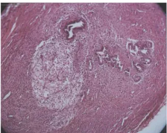
Figure 1. Right Ovary: Gland and Stroma of Endometrium.
Figure 2. Left Peritoneum: gland and stroma of endometrium.
Discussion
Vernon and Wilson (1) reported that, the auto transplantation of uterine tissues to the peritoneal cavity was the only way that yielded healthy endometriotic implants. These implants were seen as an ellipsoidal cystic structures that were composed of both endometrial gland and stroma of the uterus. Similarly, in our study, after inducing endometriosis by surgical approach, endometrial implants grew as a cystic structure. In microscopic examination, endometrial gland and stroma were seen. The total mean diameter of implants in experimented group was significantly larger than fat implants. In our study, the fat tissues didn't grow in control group. It showed well that only endometrial squares were able to grow. Brankly et al (5) in a study on vaginal
endometriosis in rats, showed that only endometrial implants were able to grow and induced cystic formation and adhesion and deformity in vagina. While fat tissues weren't able to grow. Our finding about fat tissues was similar to this study. In our study, we also reported the different places of implantation and found that in left peritoneum (p=0.002) right (p=0.000) and left (p=0.000) ovaries, the mean diameter of implants size after second laparotomy were significantly larger than before, but in right peritoneum, the finding wasn’t significant (p=0.006). This data analysis was not done before our study and we tried to find why it wasn't significant in right peritoneum. It might be due to cascade of mesenteric arteries that are dominant in left side. We sutured endometrial pieces in another place in subcutaneous and observed that endometrial tissues grew in all of these implants. It was similar to human that endometriosis has been reported in cesarian section scars. We suggest that subcutaneous is another good site for experimental study in rats. Our study showed that growth of endometrial implants made sever and firm adhesion. Clinical adhesions (score ≥2) were detected in 7 rats in experimented group and only in 2 rats in control group. Our study showed that firm and thick adhesion formation is an important aspect of endometriosis itself, and not the surgery alone.
In our study, we didn't carry out hormone analysis. We focused on normal Esterous cycles and if the rats had this normal pattern, they were selected for the study. The number of Esterous cycles were similar in both groups. It showed that induction of endometriosis didn't have any adverse effect on ovarian function in rats.
Conclusion
Our study showed that after inducing endometriosis by surgical approach, only endometrial implants grew as a cystic structures and this is a unique aspect of endometrial cells. The best site for implantation was subcutaneous area. Clinical adhesion (score ≥2) was detected in 7 rats in experimented group and 2 rats in control group. It showed that endometriosis had a direct effect on adhesion formation, not surgery alone. In addition, induction of this disease didn't have any adverse effect on ovarian function in female rats.
We didn’t study on the fertility status or abortion after induction of endometriosis in rats. We suggest this field to be studied in future study in rat model.
References
1. Denny E, Khan KS. Systematic reviews of qualitative evidence. What are the experiences of women with endometriosis. J Obstet Gynaecol 2006;26:501-506.
2. Tomassetti C, Meuleman C, Pexster SA, Mihalyi A, Kyama C, Sima P, et al. Endometriosis, recurrent miscarriage and
implantation failure: is there an immunological link? Reprod Biomed Online 2006;13:58-64.
3. Nothnick WB. Treating endometriosis as an autoimmune disease. Fert Steril 2001;76:223-231.
4. Sharpe-Timms KL. Using rats as a research model for the study of endometriosis. Ann Ny Acad sci 2002;955:318-327.
5. Berkley KJ, Cason A, Jacobs H, Bradshow H, Wood E. Vaginal hyperplasia in a rat model of endometriosis. Neurosci lett
2001;306:185-188.
6. Stilling D. Do endometrial fragments grow? Verch Dtsch Ges Pathol 1903:6:122.
7. Jacobson VC. The autotransplantation of endometrial tissue in rabbit. Arch Surg 1922;5:281.
8. Schenken RS, Asch RH, Williams RF, Hodgen GD. Etiology of infertility in monkeys with endometriosis: luteinized unruptured follicles, luteal phase defects, pelvic adhesions, and spontaneous abortions. Fertil Steril 1984;41:122.
9. Sharp-Thimms KL. Using rats as a reaserch module of the study of endometriosis. Ann N Y Acas sci 2002;:318-327.
10. Maeda KI, Kura SO, Tsukamura H. Physiology of reproduction, In the laboratory Rat: the handbook of experimental animal, Hrinke, C.J. Ed. Academic press. London. 2000;145-176.
11. Suckow MA, Danneman P, Brayton C. The laboratory animals, CRC press 2001;13-34.
12. Kocak I, Unlu C, Akcan Y, Yakin K. Reduction of adhesion formation with cross linked hyaloronic acid after peritoneal surgery in rats. Fertil Steril 1999;72:873-878.