ORIGINAL PAPERs
Mariusz Kochanowski
1, A–C, E, Paulina Łagodzińska
1, B–D, Jakub Marcinkowski
2, A,
Cezary Langot
1, E, Leszek Klimek
2, 3, C, E, FEstimation of Self-Etch Adhesive System and Resin
Cement Shear Bond Strength to Root Dentine with and
without Prior Etching – Pull-Out Test and SEM Analysis
Ocena wpływu dodatkowego trawienia na wytrzymałość połączenia
zębiny korzeniowej z wkładami koronowo-korzeniowymi
wzmacnianymi włóknami szklanymi, cementowanymi za pomocą
systemu samotrawiącego – test
pull-out
i analiza SEM
1 Department of Prosthetic Dentistry, Medical University of Lodz, Łódź, Poland
2 Department of Dental Technology of Chair of General Dentistry, Medical University of Lodz, Łódź, Poland 3 Department of Materials Research of Institute of Materials science and Engineering, Lodz Univeristy
of Technology, Łódź, Poland
A – research concept and design; B – collection and/or assembly of data; C – data analysis and interpretation;
D – writing the article; E – critical revision of the article; F – final approval of article
Abstract
Background. De-bonding is still considered to be the most common cause of failure concerning fiber posts. The proper application of adhesive is, therefore, crucial to improve the bond strength.
Objectives. This study evaluated whether the additional etching before the application of cross-generation self-etch adhesive and resin cement can increase the shear bond strength between the root dentine and fiber post’s surface.
Material and Methods. sixteen human one-canal teeth that had been extracted due to periodontal and orthodontic reasons were carefully selected for this study. They were divided into two groups of eight. Preparation of the speci-mens in both groups was equal except for the cementation procedure. The teeth were sectioned at the cementoe-namel junction. The post space was prepared to a depth of 8 mm and a diameter of 1.5 mm with a special machine ensuring the repeatability of post spaces’ shape. Posts were cemented using self-etch adhesive; in one group the root dentine was previously etched and the second group was without pretreatment. Posts were cemented with the use of metal rings. Then specimens were embedded in acrylic resin blocks. They were subjected to pull-out test using a Universal Testing Machine specially adapted to maintain the parallelism – regimen. Finally, the posts’ surface was evaluated in sEM observation. Obtained results were statistically analyzed.
Results. The mean shear bond strength values for group with prior etching of dentine were significantly lower (8.22 MPa) than for the group without prior etching (11.25 MPa). These findings were supported by sEM obser-vations, revealing that in the group with prior etching the resin-dentin-interface failure surpassed the resin-post-interface component.
Conclusions. Obtained results indicate that the self – etch adhesives’ bond strength to root dentine is decreased in case of prior etching (Dent. Med. Probl. 2014, 51, 2, 197–204).
Key words: fiber reinforced composite posts, self – etch adhesive, etching, root dentin, pull-out test.
Streszczenie
Wprowadzenie. Jednym z najczęściej spotykanych powikłań dotyczących wkładów wzmacnianych włóknami szklanymi (FRC) jest utrata retencji polegająca na ich odcementowaniu, dlatego właściwe zastosowanie systemów wiążących jest niezwykle ważne.
Cel pracy. Ocena wpływu dodatkowego wytrawienia zębiny na wytrzymałość połączenia między wkładami FRC a zębi-ną korzeniową po użyciu uniwersalnego systemu samotrawiącego typu cross-generation i cementu kompozytowego.
Dent. Med. Probl. 2014, 51, 2, 197–204
Fiber-reinforced composite (FRC) posts are nowadays commonly used in restorative dentistry. These posts are usually required in endodontical-ly treated teeth with coronal structure loss, espe-cially the anterior teeth. such teeth are already de-hydrated [1] and lack a great deal of their original structure, which makes them weaker and, there-fore, more prone to fractures [2]. However, such fractures of teeth with fixed posts are rather en-countered with metal or zircon posts, which have a very rigid nature [3–5]. The placement of a fi-ber post and core reduces the risk of such tooth fractures, even though the root stresses become higher than those reached with metal posts [4]. Because their elastic modulus is similar to that of dentine, the stresses along the fiber post’s surfaces decrease, and as a result, the risk of de-bonding is lowered [4]. However, retrospec-tive studies on the clinical performance of fi-ber posts have previously shown that de-bond-ing is considered to be the primary cause of FRC posts’ failure [6–8].
De-bonding usually occurs at the luting inter- face between root dentine and the resin cement [9, 10]. The most appropriate and efficient adhe-sives, and their procedural steps, should therefore be defined as a way to determine a clinically rele-vant and acceptable bonding strength.
In order to cement FRC posts, either the etch-and-rinse adhesives or self-etch adhesives can be used. Conventional etch-and-rinse adhesives are supposed to provide higher bond strength [11, 12]. However, the self-etch adhesives’ application tech-nique is simpler, more repeatable, and less sensi-tive (the issue of dentine wetness does not exist). self-etch adhesives do not require a separate etch-ing and rinsetch-ing step. Etchetch-ing and primetch-ing are car-ried out simultaneously because the acidic mono-mers are combined with the primer into one solu-tion. Currently used self-etch adhesives have the ability to penetrate beyond the smear layer and
modify it, and then ultimately incorporate it into the adhesive resin [13].
In this study, a cross-generation self-etch ad-hesive was used to cement FRC posts. The man-ufacturer mentions that, while working with these adhesives, the user has the option of etch-ing the prepared surface prior to the application of the self-etch adhesive. some researchers can be found incorporating this supposed advantage into their methods with the expectation of im-proving the performance of this self-etching ad-hesive [14]. However, there are several research-ers who have also proved that this additional etching does not actually improve the bonding strength capacity of self-etch adhesives at all, and may even decrease its bonding strength capaci-ty [15–17]. This phenomenon can be explained by the fact that the mere presence and micro-morphology of the prepared smear layer ulti-mately determines the micromechanical prop-erties and chemical interactions between the self-etch adhesives’ and root dentine’s compo-nents, as well as the structure of the hybrid lay-er [15–20]. Following such assumptions, any surface treatment of root dentine can influence the bonding outcomes, whether it will be etch-ing or the application of any chemical agents [14, 18–22].
In this study, the pull-out test was used to tain a quantitative assessment. As a way to ob-tain more consistent and reliable results, the steps of preparing specimens were specially formulat-ed, and the pull-out test was eventually modi-fied. Qualitative assessment was performed dur-ing sEM analysis of extruded posts.
The aim of this study was to evaluate whether the additional etching before applying the cross- -generation self-etch adhesive and resin cement can increase the bond strength between the root dentine and fiber post’s surface. The null hypoth-esis was that there would be no significant
differ-Materiał i metody. Badaniu poddano szesnaście jednokanałowych zębów usuniętych ze względów periodontolo-gicznych lub ortodontycznych. Podzielono je na dwie grupy. Próbki w obu grupach przygotowano w jednakowy sposób, poza procedurą cementowania. Na wysokości połączenia szkliwno-cementowego odcięto korony zębów. Łoża pod wkłady wypreparowano na głębokość 8 mm i o średnicy 1,5 mm. W tym celu użyto maszyny zapewniającej powtarzalność kształtu łoża. Wkłady zacementowano z użyciem systemu samotrawiącego. W jednej grupie dodat-kowo wytrawiono zębinę. Wkłady cementowano z użyciem metalowych pierścieni. Próbki zatopiono w akrylowych bloczkach i poddano je testowi wyciągania w uniwersalnej maszynie testującej, dbając o równoległość wszystkich elementów. Powierzchnię próbek poddano badaniu sEM.
Wyniki. Wykazano istotną statystycznie różnicę między wytrzymałością połączenia w grupie, w której dodatkowo wytrawiono zębinę (8,22 MPa) względem grupy, w której zębina nie była wytrawiona (11,25 MPa). Badania sEM wykazały, że w grupie z dodatkowym trawieniem połączenie było rozerwane głównie na granicy cement–zębina.
Wnioski. Uzyskane wyniki wskazują, że dodatkowe wytrawienie zębiny korzeniowej przed cementowaniem z uży-ciem systemów samotrawiących pogarsza wytrzymałość połączenia zębina korzeniowa–cement kompozytowy– –wkład FRC (Dent. Med. Probl. 2014, 51, 2, 197–204).
Słowa kluczowe: wkłady wzmacniane włóknami szklanymi, systemy samotrawiące, trawienie, zębina korzeniowa, test wyciągania.
ence between the shear bond strength of self-etch adhesive system to root dentine, with or without prior etching.
Material and Methods
sixteen human teeth with one canal (except lower incisors) that were previously extracted due to periodontal and orthodontic reasons were select-ed for this study. These teeth were storselect-ed in a 0.1% thymol solution during the course of the study and were used within a month after their extraction. Any teeth that had irregularly shaped or obliterat-ed canals and had a root length of less than 11 mm long were excluded from this study. Furthermore, any teeth with defects, fractures or previous endo-dontic treatment, were excluded as well.
The crowns were cut transversally at the ce-mentoenamel junction with a cylindrical dia-mond rotary bur (Dentsply Maillefer, Ballaigues, switzerland) at high speed under water coolant and discarded. The preparation of post space was performed on the specially constructed machine, which enabled us to standardize these spaces, and therefore eliminate any possible variations in the shape of the post space that might occur during typical manual preparation. The post space was enlarged under water coolant with Pesso®
Ream-ers No 2, 3 and 4 (Poldent, Warszawa, Poland). The final shaping of the post space was accomplished with a special drill, matched to the posts (FiberK-leer Parallel Post Kit™; Pentron Clinical
Technol-ogies Llc., Wallingford, CT) to a depth of 8 mm, with a diameter of 1.5 mm. It was then possible to obtain the same dimensions and proper adap-tation of the post, as well as the recommended 0.1–0.3 mm cement thickness [9]. This was fol-lowed by the preparation of the transverse micro-cuts on the surface of the root.
In this study, prefabricated FiberKleer® Posts
with a diameter of 1.5 mm (FiberKleer Parallel Posts™; Pentron Clinical Technologies Llc.) were
used. They are parallel and have a pre-silanized surface. The length of each post is 10.0 mm. These posts were chosen for this study because they are mounted with a retentive head that ensures consis-tent and repeatable action of grasping each speci-men during the pull-out test, and it also prevents any destruction of the post’s fibers induced by the holding jaws of the Universal Testing Machine.
These specimens were randomly assigned to two groups of eight teeth, each group being clas-sified according to the procedures that would be used to cement each post. In the first group (A), the additional steps of etching, rinsing and dry-ing of dentine preceded the application of the
self-etch adhesive system. In the second group (B), the same brand of self-etch adhesive was applied, but there was no prior etching of dentine. In the A-group, 37% phosphoric acid was applied into the post space for 15 s, activated, then thoroughly rinsed with a water-filled syringe. In the B-group, the post space was only rinsed with saline. In both groups, the post space was dried with ab-sorbent paper points (VDW GmbH, Munchen, Germany) so that the root dentine stayed moist, but not wet.
The following procedures were then per-formed equally in both groups. The self-etch ad-hesive was applied according to the manufactur-er’s instructions. The universal cross-generation adhesive (Iperbond®; Itena Clinical, Paris, France)
was mixed with its chemical bond activator (Bond Activator; Itena Clinical), and was then applied to the inner walls of the prepared post space with a microbrush, and rubbed in for 15 s [23]. Any ex-cess adhesive was then removed with sterile paper points. Following the adhesive application, the du-al-polymerizing luting resin cement (Dentocem®;
Itena Clinical) was then applied into the post space using a thin, self-mixing tip, and the post was then immediately placed to the full depth of 8 mm. Any excess cement was removed with a microbrush. The polymerizing light rays were transmitted par-allel to the long axis of the root and the tip of the polymerizing light was in direct contact with the coronal end of the post for 20 s. Due to the trans-lucency of these posts, and to the dual-polymeriz-ing action of the resin and cement, the most opti-mal bond strength in all regions of the post space could then be achieved [10, 20].
Cementation was performed with the use of specially fabricated metal rings that were fixed be-tween the retentive head of the post and the root. After the cementation procedures were finished, the specimens were then stored in a (0.9%) saline solution for 48 h to allow for the complete bonding between the adhesive system and resin cement.
The next step was to embed each specimen in-to a cylindrical block of auin-to-polymerizing acryl-ic resin (Duracryl®; spofa Dental, Jicin, Czech
Republic), while concomitantly maintaining the parallelism between all of the elements of the spec-imen. Previously made transverse cuts on the ex-ternal surface of the root ensured the complete re-tention of the specimen within the acrylic block. During the process of acrylic resin polymer-ization, the specimens were kept in a cold-wa-ter bath in order to eliminate the harmful in-fluence of heat. At this point the preparation of the specimens was completed. The parallelism between the post, canal and acrylic resin block was obtained.
This study was performed in cooperation with the Department of strength of Materials and structures, Technical University of Lodz. The pull-out test was conducted on the Universal Test-ing Machine Instron® 4485 (Instron 4485;
Nor-wood, MA, UsA) which was specifically adapted for this study. The pull-out test consisted of ex-truding the post from the canal along its long ax-is. The specimens were held in place in the ma-chine using two separate holding jaws. The acryl-ic resin block was secured to the lower jaw. The metal fixation ring was secured to the upper jaw. Both holding jaws were constructed in such a way that, while the specimen was being held firmly in place, the positioning of the jaws could be adjusted in any direction to assume the most optimal par-allel position. This enabled the placement of the specimen at the ideal angle of 0 degrees in relation to the long axis of the post.
The final step was performing the pull-out test (now modified) in the Universal Testing Machine. The extrusion load of 1 mm/min was applied to all the specimens under equal conditions.
The results were obtained in N (newtons) and converted to MPa (mega pascals), revealing the magnitude of the achieved shear bond strength.
The results were analyzed statistically (sta-tistica PL 6.0™; statsoft Polska, Kraków, Poland).
The verification of the normal distribution was made using the Kolmogorov-smirnov’s test. The comparative analysis of the strength obtained for specimens with and without previous etching was performed, using the student’s t-distribution test. statistical significance was set at p < 0.05.
The last approach was sEM (Hitachi® s-3000
N; Tokyo, Japan) examination of the extruded posts in the pursuit of visually identifying and verifying the surfaces at the de-bonding interface. Finally, cross-sections of two additional, previ-ously untested specimens representing each group (A and B) were analyzed microscopically to assess the quality of the bonding interface and to iden-tify the presence or absence of any gap that may have been created between the bonded surfaces. They were sliced and polished along their long axis with a metallographic grinder (Metapolan®;
Rathenower Optische Werke GmbH, Rathenow, Germany) under water coolant and then were ex-amined microscopically (MA-200; Nikon, Tokyo, Japan). To improve the quality of this microscopic image, the surfaces of both specimens’ cross-sec-tions were sprayed with a thermal layer of 99.99% Au (JEE-4X; Jeol, Tokyo, Japan) and were then al-so subjected to microscopic observation (Hitachi s-3000 N).
Results
The obtained results indicate the values of the pull-out bond strength in all specimens. In the group with pre-etched dentine, the mean shear
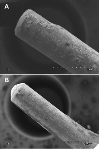
Fig. 1. Figure ‘A’ shows the sEM analysis for the post cemented with previous etching. The de-bonding failure had mixed characteristics with greater dentin resin inter-face component. The surinter-face of the post is almost entirely covered with the luting cement, as only a small area of the naked post can be seen. Figure ‘B’ shows the sEM analysis for a post that is cemented without previous etching. In this case the failure of bonding also had mixed charac-teristics, but the de-bonding occurred mainly at the post-cement interface. In addition, the bonding strength in the apical area appeared to be weaker, as in this region the resin remained on the post. Only small areas of remain-ing lutremain-ing cement can be seen on the surface of the posts.
Ryc. 1. Przykładowe obrazy uzyskane w skaningowym mikroskopie elektronowym po przeprowadzeniu ba-dawczej ekstruzji wkładów. „A” ukazuje wkład, który był zacementowany z uprzednim trawieniem zębiny. Zerwane połączenie ma charakter mieszany, głównie zerwanie połączenia nastąpiło na granicy cement/zębi-na korzeniowa. Powierzchnia wkładu jest prawie w ca-łości pokryta resztkami cementu. Rycina „B” ukazuje wkład, który był zacementowany bez dodatkowego tra-wienia zębiny. W tym przypadku zerwanie połączenia ma również charakter mieszany, ale przebiega głównie na granicy wkład/cement. Dodatkowo są widoczne po-zostałości cementu na wkładzie w jego wierzchołkowej części, co świadczy o słabszej wytrzymałości połączenia z zębiną korzeniową w części wierzchołkowej
bond strength value was 8.22 ± 0.83 MPa, with a range from 6.96 MPa to 9.40 MPa. In the group without previously etched dentine, the mean shear bond strength value was 11.25 ± 1.84 MPa, with a range from 8.69 MPa to 13.39 MPa. The distri-bution of strength values did not deviate from the normal distribution. The student’s t-distribution test revealed that there was a statistically signifi-cant difference between groups at p < 0.001.
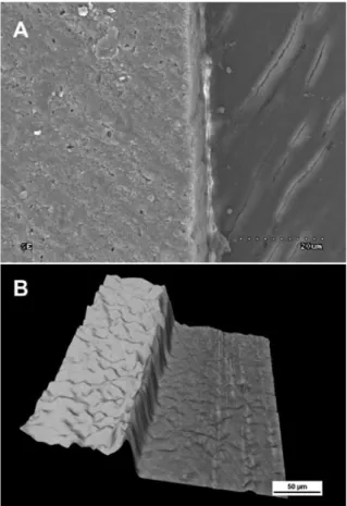
The recorded observations of the sEM analysis of the de-bonding-interface of extruded posts are presented in the included figures (Fig. 1). The im-ages of the bonding interface reveal the presence of a gap between the dentine and resin cement inter-face among the specimens that were treated with previous etching and the second additional micro-scopic examination supports this finding, and the included figure presents the transformation of the image showing the surface topography (Fig. 2). Fi-nally, the microscopic examination of the
speci-Fig. 2. Figure ‘A’ shows the sEM image of the cross-section of the specimen with etched dentine prior to application of the self-etch adhesive system. The gap between the resin cement and the dentine can be seen. The gap’s width amounts to several nanometers. since this gap is narrower than the diameter of any bacteria, it becomes irrelevant as a contamination risk factor. Figure ‘B’ shows the image obtained in the optical microscope made to confirm the presence of a gap. It was transformed so that the topography of the surface can be seen. such a projection shows the gap distinctly
Ryc. 2. Rycina „A” przedstawia obraz uzyskany w ska-ningowym mikroskopie elektronowym – przekrój przez próbkę, uprzednio wytrawioną. Widoczna jest szczelina między cementem a zębiną korzeniową, jej szerokość nie przekracza kilku nanometrów. W związ-ku z tym, że jest węższa od średnicy bakterii, nie sta-nowi wrót zakażenia. Rycina „B” przedstawia obraz uzyskany w mikroskopie optycznym wykonany w celu potwierdzenia obecności szczeliny. Obraz został wyko-nany w projekcji pokazującej topografię powierzchni przekroju i ukazuje wyraźnie istnienie szczeliny
Fig. 3. Figure ‘A’ shows the sEM image of the cross-section of the specimen without previous etching of dentine. There is no gap between any of the surfaces. The hybrid layer of the self-etching adhesive can be seen. ‘B’ shows the topography of the surface. No gap can be seen. The difference in height between the two layers can be seen; this can be caused by a difference in toughness between specific materials during polishing
Ryc. 3. Rycina „A” przedstawia obraz uzyskany w ska-ningowym mikroskopie elektronowym – przekrój przez próbkę zacementowaną bez dodatkowego tra-wienia. Nie zaobserwowano obecności szczeliny na granicy zębina/cement/wkład. Widoczna jest natomiast warstwa hybrydowa wytworzona przez system wiążący. Rycina „B” przedstawia obraz uzyskany w mikroskopie optycznym przedstawiający topografię powierzchni przekroju, który potwierdza brak szczeliny. Widoczna jest różnica w wysokości dwóch warstw, co może być spowodowane różną twardością materiałów podczas polerowania próbki
men without the additional prior etching found no gap (Fig. 3). The description for the images is in-cluded below each figure.
Discussion
The null hypothesis was that there would be no significant difference between the bond strength of self-etch adhesive systems to root den-tine, with or without prior etching, was rejected. The obtained results indicate that the shear bond strength is higher when self-etch adhesive is ap-plied according to the manufacturer’s directions without any prior modification of the root den-tine. The additional etching deteriorated the bond strength. This phenomenon can be explained by the fact that self-etch adhesives modify the smear layer and incorporate it into the hybrid layer. As the etching step results in complete removal of the smear layer and smear plugs, the micromechani-cal and chemimicromechani-cal interactions with them are inter-fered with [15, 16, 18]. Also, Van Landuyt et al. [15] reports that although the additional etching pre-ceding the application of self-etch adhesive im-proves the bond strength to enamel, it significant-ly impairs bonding effectiveness to dentine. Previ-ous etching results in an inferior quality of hybrid layer that contains submicron gaps, which is there-fore prone to nano-leakage [15]. This suggestion is supported by the sEM observation in our study (Fig. 1), because in specimens with prior etching, the failure of bonding occurred at the root dentine – luting cement interface. Additional microscopic examination of the bonding interface in a special-ly prepared specimen also showed the presence of the gap along the dentine-resin cement interface, which was further supported by the image show-ing the topography of this surface (Fig. 2). Even though the hybrid layer after additional etching
is thicker than the hybrid layer obtained without etching, its changed structure does not ensure suf-ficient anchorage for the resin [15]. Moreover, the bond strength is independent of the thickness of the hybrid layer [20, 24]. In addition, the chemical interaction between the functional monomers of the adhesive and the hydroxyapatite crystals that normally have a strong affinity for one anoth-er detanoth-eriorates, because this extra etching causes the hydroxyapatite crystals to be inaccessible [15]. They also suggest that after etching, the exposed dentine is too rich in collagen fibrils, and this dis-turbs the infiltration of the resin [25]. Other au-thors also claim that pre-etching causes deep den-tine demineralization leading to incomplete infil-tration of the resin. As a result, there is a ‘defective zone’ at the hybrid layer’s base, further decreas-ing the bond strength [15, 17, 26]. Alternatively, the acidic monomers contained in the self-etch solution condition the dentine in such a way that the collagen network is infiltrated properly and entirely [16, 17]. The other reason for decreased shear bond strength performance in specimens with pre-etching can be attributed to the presence of water while the polymerization process is tak-ing place. Besides the fact that while ustak-ing the self-etch adhesive systems there is always a risk of leav-ing too much water before polymerization, as they are water solutions, there is also an additional risk caused by the previous etching. After the removal of the smear layer, the dentinal tubules are opened and can cause dentinal fluid movement. This leads to additional water excess that impairs the bond-ing capacity [27, 28]. Over-wettbond-ing of the dentine combined with the highly water-permeable nature of self-etch adhesives [29] interferes with the po-lymerization and ultimately has negative effects on the performance of the adhesive resin, which results in a weaker bond strength. All of these hy-potheses can together serve to help explain the resulting weaker shear bond strength values ob-tained for specimens with dentine that was etched prior to the application of a self-etch adhesive.
Analysis of the data obtained from sEM im-ages indicated that there were notable differences found between the bond strength at the dentine/ /resin-interface and the fiber post/resin-inter-face, both in specimens with, and without, previ-ous etching. Our sEM observations and analyses were not only consistent with the data, but also re-vealed the specific contact areas that were respon-sible for the weaker bonding capacity. The sEM images clearly show the occurrence of de-bond-ing at either the dentine-resin-interface, the res-in-post-interface, or a combination of the two. In both groups the locations of bonding failure had mixed characteristics. However, in both cases there
Table 1. The table shows the shear bond strength values [MPa] obtained in both groups in the pull-out test in this study.
Tabela 1. Tabela ukazuje wartości wytrzymałości połą-czenia [MPa] uzyskane w badanych grupach w teście pull-out.
Group-A with previous etching
Group-B without previo-us etching
Max 9.40 13.39
Min 6.96 8.69
Mean 9.18 11.25
standard deviation 0.77 1.72
was adverse advantage of the de-bonding inter-face combination. The testing until failure of de- -bonding manifested better clinical performance in specimens without petching, because it re-garded the post-cement interface. Even though in such cases where it can be difficult to remove the cement debris from the inner surface of the post space, it has relevant clinical implications. The dentinal tubules are sealed off by the residual ce-ment and are more resistant to any possible con-tamination from the oral environment. On the other hand, in specimens with previous etching, the bond strength was better on the post-resin in-terface. This finding indicates that the adhesion to dentine is impaired. such observations only stand to further support the results obtained in our pull- -out test.
Due to the fact that de-bonding is the main cause of failure of FRC post and core treatments, the optimal adhesive procedural steps should be stated for the clinician to achieve maximum allow-able bond strengths. There are many tests aimed at assessing the bond strength value, and the rules of these tests should consist of the measurement of the force that is sufficient to dislodge the post, that ultimately depends on the effectiveness of the luting procedure to the root dentine [30]. In this study the pull-out test was used. The biggest chal-lenge was to maintain the axial alignment of all the elements throughout. In this study this was accomplished by yielding to the importance of
parallelism from the very beginning. Firstly, the specimens were prepared so that all the elements were set in one mutual axis. Then the specimen was fixed in the Universal Testing Machine with the aid of the moveable holding jaws, ensuring the proper axial alignment of the specimen during the pull-out test.
Any chemical root dentine surface treatment before the application of a cross-generation self-etch adhesive should be carefully considered as it may significantly influence their bond strength capacity. This refers not only to the 37% phosphor-ic acid used in this study, but also may apply to any other chemicals that have a similar effect on the smear layer [21].
Within the limitations of this study, it was concluded that the shear bond strength of self-etch adhesive system to root dentine is decreased when used in cases of additional prior etching. Moreover, in specimens without pre-etching of dentine, the failure of de-bonding was found to occur at the post-cement interface, while in spec-imens with previous etching of the root dentine, the failure of de-bonding was manifested at the dentine-cement interface. This indicates weak-er adhesion to dentine aftweak-er unnecessary remov-al of the smear layer. When etching and rinsing precede the application of a self-etch adhesive, this unfortunately results in the presence of a gap width of several nanometers along the dentine-resin cement interface.
References
[1] Papa J., Cain C., Messer H.H.: Moisture content of vital vs endodontically treated teeth. Endod. Dent. Trauma-tol. 1994, 10, 91–93.
[2] Reeh E.s.: Reduction in tooth stiffness as a result of endodontic restorative procedures. J. Endod. 1989, 15, 512– 516.
[3] Dilmener F.T., sipahi C., Dalkiz M.: Resistance of three new esthetic post-and-core systems to compressive loading. J. Prosthet. Dent. 2006, 95, 130–136.
[4] santos A.F.V., Meira J.B.C., Tanaka C.B., Xavier T.A., Ballester R.Y., Lima R.G., Pfeifer C.s.., Vesluis A.: Can fiber posts increase root stresses and reduce fracture? J. Dent. Res. 2010, 89, 587–591.
[5] Giovani A.R., Vansan L.P., de sousa Neto M.D., Paulino s.M.: In vitro fracture resistance of glass-fiber and cast metal posts with different lengths. J. Prosthet. Dent. 2009, 101, 183–188.
[6] Ferrari M., Cagidiaco M.C., Goracci C., Vichi A., Mason P.N., Radovic I., Tay F.: Long-term retrospective study of the clinical performance of fiber posts. Am. J. Dent. 2007, 20, 287–291.
[7] signore A., Benedicenti s., Kaitsas V., Barone M., Angiero F., Ravera G.: Long-term survival of endodon-tically treated, maxillary anterior teeth restored with either tapered or parallel-sided glass-fiber posts and full-ce-ramic crown coverage. J. Dent. 2009, 37, 115–121.
[8] Mannocci F., Bertelli E., sherriff M., Watson T.F.., Pitt Ford T.R.: Three-year clinical comparison of sur-vival of endodontically treated teeth restored with either full cast coverage or with direct composite restoration. J. Prosthet. Dent. 2002, 88, 297–301.
[9] D’Arcangelo C., Cinelli M., De Angelis F., D’Amario M.: The effect of resin cement film thickness on the pull-out strength of a fiber-reinforced post system. J. Prosthet. Dent. 2007, 98, 193–198.
[10] Giachetti L., Russo D.s., Bertini F., Giuliani V.: Translucent fiber post cementation using a light-curing ad-hesive/composite system: sEM analysis and pull-out test. J. Dent. 2004, 32, 629–634.
[11] Goracci C., sadek F.T., Fabianelli A., Tay F.R., Ferrari M.: Evaluation of the adhesion of fiber posts to intra-radicular dentin. Oper. Dent. 2005, 30, 627–635.
[12] Wang J., Chen Y.-M., Yip K., smales R.J., Meng Q.-F., Chen L.: Effect of two fiber post types and two luting ce-ment systems on regional post retention using the push-out test. Dental Mater. J. 2008, 24, 372–377.
[13] Eliades G., Watts D.C., Eliades T.: Dental hard tissues and bonding. Heidelberg: springer; 2005. p. 100–114. [14] Zhang L., Huang L., Xiong Y., Fang M., Chen J.H., Ferrari M.: Effect of post-space treatment on retention of
fiber posts in different root regions using two self-etching systems. Eur. J. Oral sci. 2008, 116, 280–286.
[15] Van Landuyt K.L., Kanumilli P., De Munck J., Peumans M., Lamberchts P., Van Meerbeek B.: Bond strength of mild self-etch adhesive with and without prior acid-etching. J. Dent. 2006, 34, 77–85.
[16] Torii Y., Itou K., Nishitani Y., Ishikawa K., suzuki K.: Effect of phosphoric acid etching prior to self-etching primer application on adhesion of resin composite to enamel and dentin. Am. J. Dent. 2002, 15, 305–308. [17] Walker M.P., Wang Y., swafford J., Evans A., spencer P.: Influence of additional acid etch treatment on
res-in cement dentres-in res-infiltration. J. Prosthodont. 2000, 9, 77–81.
[18] Hayashi M., Takahashi Y., Hirai M., Iwami Y., Imazato s., Ebisu s.: Effect of endodontic irrigation on bond-ing of resin cement to radicular dentin. Eur. J. Oral sci. 2005,113,70–76.
[19] Wu H., Hayashi M., Okamura K., Koytchev E.V., Imazato s., Tanaka s., sano H., Ebisu s.: Effects of light penetration and smear layer removal on adhesion of post-cores to root canal dentin by self-etching adhesives. Den-tal Mater. J. 2009, 25, 1484–1492.
[20] Akgungor G., Akkayan B.: Influence of dentin bonding agents and polymerization modes on the bond strength between translucent fiber posts and three dentin regions within a post space. J. Prosthet. Dent. 2006, 95, 368–378. [21] Demiryürek E.Ö., Külünk s., saraҁ D., Yüksel G., Bulucu B.: Effect of different surface treatments on the
push-out bond strength of fiber post to root canal dentin. Oral surg. Oral Med. Oral Pathol. Oral Radiol. Endod. 2009, 108, e74–e80.
[22] schwartz R.s.: Adhesive dentistry and endodontics. Part 2: Bonding in the root canal system – the promise and the problems: a review. J. Endod. 2006, 32, 1125–1134.
[23] Ferrari M., Vichi A., Grandini s., Geppi s.: Influence of microbrush on efficacy of bonding into root canals. Am. J. Dent. 2002, 15, 227–231.
[24] Hashimoto M., Ohno H., Endo K., Kaga M., sano H., Oguchi H.: The effect of hybrid layer thickness on bond strength: demineralized dentin zone of the hybrid layer. Dental Mater. J. 2000, 16, 406–411.
[25] Uno s., Finger W.J.: Function of the hybrid zone as a stress-absorbing layer in resin-dentin bonding. Quintes-sence Int. 1995, 26, 733–738.
[26] Jacques P., Hebling J.: Effect of dentin conditioners on the microtensile bond strength of a conventional and a self-etching primer adhesive system. Dental Mater. J. 2005, 21, 103–109.
[27] Pashley D.H., Carvalho R.M.: Dentine permeability and dentine adhesion. J. Adhes. Dent. 1997, 25, 355–372. [28] Zhang Z., Huang C., Zheng T., Wang s., Cheng X.: Effects of residual water on microtensile bond strength of
one-bottle dentin adhesive systems with different solvent basis. Chin. Med. J. 2005, 118, 1623–1628.
[29] Tay F.R., Pashley D.H., suh B.I., Carvalho R.M., Itthagarun A.: single-step adhesives are permeable mem-branes. J. Dent. 2002, 30, 371–382.
[30] Goracci C., Grandini s., Bossù M., Bertelli E., Ferrari M.: Laboratory assessment of the retentive potential of adhesive posts: A review. J. Dent. 2007, 35, 827–835.
Address for correspondence:
Paulina Łagodzińska
Department of Prosthetic Dentistry of Chair of General Dentistry Institute of Dentistry
Medical University of Lodz st. Pomorska 251
92-213 Lodz Poland
E-mail: paulina.lagodzinska@umed.lodz.pl Tel.: 42 675 74 50
Conflict of interest: None declared Received: 2.01.2014
Revised: 2.04.2014 Accepted: 14.05.2014
Praca wpłynęła do Redakcji: 2.01.2014 r. Po recenzji: 2.04.2014 r.
![Tabela 1. Tabela ukazuje wartości wytrzymałości połą- połą-czenia [MPa] uzyskane w badanych grupach w teście pull-out](https://thumb-us.123doks.com/thumbv2/123dok_us/8382587.2227125/6.892.107.426.205.381/tabela-tabela-ukazuje-wartości-wytrzymałości-uzyskane-badanych-grupach.webp)
