Address for correspondence
Jacek Matys
E-mail: [email protected]
Funding sources
none declared
Conflict of interest
none declared
Received on September 1, 2016 Revised on October 24, 2016 Accepted on October 26, 2016
Abstract
Dental impressions are mainly used to record the geometry of hard and soft tissue and the relations between teeth and the adjacent tissues. In implantology, precise implant impressions are necessary to obtain the best possible detailed reproduction of the implant site and the passive intraoral fit of the implant framework. However, ensuring those necessities in the interdisciplinary treatment of orthodontic patients requires re-vised impression techniques and materials. In this study a modified one-stage putty-wash pick-up implant impression procedure was used, incorporating the use of two additional silicone materials and modeling wax. Additionally, an Er:YAG laser was used to obtain abetter emergence profile for implant restorations and an immediate impression, which shortened the prosthodontic stage in patients undergoing orthodontic treatment. The technique described here for taking the impression and creating the emergence profile offers dental practitioners additional options in implantoprosthodontic treatment of orthodontic patients, as the method is characterized by simple preparation and satisfactory implant site reproduction.
Key words: dental implants, orthodontic treatment, Er:YAG laser, prosthetic impression, laser surgery Słowa kluczowe: implanty stomatologiczne, leczenie ortodontyczne, laser Er:YAG, wycisk protetyczny, chirurgia laserowa
DOI
10.17219/dmp/66363
Copyright
© 2017 by Wroclaw Medical University and Polish Dental Society
This is an article distributed under the terms of the Creative Commons Attribution Non-Commercial License (http://creativecommons.org/licenses/by-nc-nd/4.0/)
Laser instant implant impression method:
A case presentation
Metoda wycisku w implantacji natychmiastowej
z użyciem lasera (LIIIM) – prezentacja przypadku
Jacek Matys
1–3, A–D, Katarzyna Świder
3, D, Rafał Flieger
4, D, E1 Department of Dental Surgery, Wroclaw Medical University, Wrocław, Poland 2 Student of the Master in Laser Dentistry of Sapienza University of Rome, Italy 3 Private Practice, Wschowa, Poland
4 Private Dental Healthcare, Kościan, Poland
A – research concept and design; B – collection and/or assembly of data; C – data analysis and interpretation; D – writing the article; E – critical revision of the article; F – final approval of article
J. Matys, K. Świder, R. Flieger. Laser instant implant impression method 102
The use of endosseous titanium implants has been suc-cessfully implemented in the treatment of patients with edentulism of various extents.1–3 Nowadays, implants are
extensively used in several disciplines of dentistry. Pa-tients affected with malocclusion and partial edentulism frequently insist on being treated as quickly as possible. Therefore, an interdisciplinary approach and precise plan-ning are needed to obtain satisfactory results. Orthodon-tic treatment can contribute to the successful use of im-plants through vertical development of the peri-implant site (using tooth extrusion) and the creation of space for implant restorations.1 In addition, osseointegrated dental
implants have been used for a variety of purposes in or-thodontics. The placement of dental implants can provide a good quality of anchorage control, as well as a method to reposition the natural teeth.1–3
However, interdisciplinary treatment can entail some difficulties. If the implant placement takes place during orthodontic treatment and before its finalization, a sat-isfactory impression is hard to obtain. Elements of orth-odontic appliances (wires, brackets, bands) can trap and damage the impression material. Usually the wires can be pulled out without problems prior to taking the impres-sion, but removing brackets would be time-consuming and could disturb the treatment process. To overcome this problem, clinicians have come up with different methods to facilitate impressions of orthodontically banded teeth. The most widely used method is to cover the orthodon-tic brackets. Consequently, when the impression tray is removed the material tearing is minimized, or even elim-inated in some cases. Maeda et al. proposed the use of a tube as block-out material for orthodontic brackets and arch wire while taking the impression for the production of a mouthguard.4 Rilo et al. suggested using small
por-tions of utility wax; Croll and Castaldi proposed strips of utility wax; while Lorton recommended the use of strips of occlusal indicator wax compressed over bonded brackets prior to alginate impression.5–7 Sukotjo and Bocage
pro-posed the use of an implant surgical template.8 However,
all of these procedures involved the use of irreversible hy-drocoloid as impression material, which cannot yield sat-isfactory impression results for implant procedures.
Another challenge the clinician can encounter dur-ing implant-supported restoration treatment of an orth-odontic patient is the emergence profile. Multiple meth-ods have been described that utilize direct, indirect or combined techniques.9 Tarlow, as well as Macintosh and
Sutherland, formed the emergence profile on the master cast by trimming or burring the overabundance of the soft tissue substitute before the final crown framework was made.10,11 Reike suggested surgically repositioning a split
flap and overcontouring the soft tissue around the healing abutment.12 Other methods incorporate the use of interim
restorations made of an autopolymerizing resinor direct composite.13–17 Ntounis and Petropoulou proposed the
use of a screw-retained provisional restoration that was
adjusted regularly.15 Azercombined the method of using
a rotary instrument to reshape the stone cast with the use of a provisional autopolymerizing resin crown that was gradually built up each week by adding more resin to the external gingival contours.17
One technique that offers the possibility of executing the impression immediately after implant exposure is the use of the Er:YAG laser, widely employed in surgical pro-cedures. Identifying the patients’ periodontal biotype is fundamental to the optimal planning of therapeutic man-agement, especially in implantology.18 Matys et al.
pro-posed the use of erbium lasers only in cases with sufficient keratinized tissue thickness.19 Hence, when the attached
gingiva around the implants are insufficiently thick, the implants should be covered with subepithelial connective tissue grafts (SCTG) or free gingival grafts (FGG).20
To the best of the present authors’ knowledge, the laser instant implant impression method (LIIIM) developed by the authors has not previously been described in litera-ture. The method incorporates the use of two silicone im-pression materials and a modeling wax strip as block-out material for orthodontic brackets. An optimal emergence profile is obtained with the direct use of an Er:YAG laser.
Case presentation
A 20-year-old female patient was referred to one au-thor’s private practice (in Wschowa, Poland) from a gen-eral dentist, with the aim of restoring her missing teeth 12 and 22 (FDI notation system used), in which the germs had been missing from birth due to hereditary agenesis. It was decided that implantation would be the best course of action; however, as the patient wished to align her teeth in the process, she was referred to an orthodontist for assessment. Complete fixed orthodontic treatment was planned for the correction of crowding and a lack of space for future implants 12 and 22. The tooth align-ment process meant that implant treatalign-ment would have to be staged. Six months before the planned termination of the orthodontic treatment, two implants (Superline, Dentium, Suwon, Korea), 4.0 mm in diameter and 10 mm in length, were inserted under local infiltrative anes-thesia with articaine hydrochloride 4% plus epinephrine 1:100000 (Orablock
®
, Pierrel Group, Capua, Italy) and cover screws were fitted immediately.After half a year, during which the adjacent bone healed, the patient came back to continue her implanto-prosthodontic treatment. However, it is difficult to obtain accurate impressions before the completion of orthodon-tic treatment, since (as noted earlier) the brackets of orth-odontic devices trap and tear the impression material. To bypass these problems, a modified one-stage putty-wash pick-up impression technique was used, with open tray (direct) procedure and an application of modeling wax for undercut coverage.
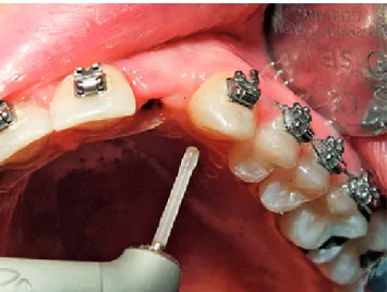
Prior to taking the impression, the orthodontic upper arch-wire was removed. To shorten the time of final crown cementation, the implants were exposed using an Er:YAG LiteTouch™ laser (Syneron™ Dental Lasers, Syneron Medi-cal Ltd., Yokneam, Israel) (Fig. 1) with the following settings: pulse energy 300 mJ, pulse frequency 18 Hz, energy den-sity per pulse 38.2 J/cm2, mode for soft tissue (ST), cooling
spray 5 mL/min, angle of the working tip 70o, size of the tip
1.0 × 17 mm, distance from the soft tissue 2 mm. The use of the laser allowed fast homeostasis of the wound. An imme-diate impression for the final prosthodontic restoration was possible thanks to the lack of postoperative bleeding. Addi-tionally, the use of the Er:YAG laser resulted in an emergence profile without any visible thermal damage (Fig. 2).
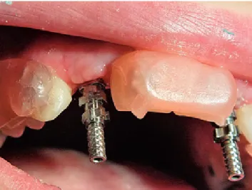
After this, direct transfer abutments were screwed onto the implants (Fig. 3). To facilitate access to the implant transfer, the open tray method was used; a custom im-pression tray was perforated (before the imim-pression was
taken, the hole was adjusted to line up with the position of the transfer) (Fig. 4). The undercuts around the brack-ets were blocked with a strip of Vertex™ Modelling wax (Vertex-Dental, Soesterberg, The Netherlands); the mate-rial was heated to obtain the necessary shape and then folded three times (Fig. 5). It is very important to leave the space near the implants and adjacent teeth free to al-low the impression material to enter. Light-bodied addi-tion silicone material (Variotime
®
Medium Flow, Heraeus Kulzer, Hanau, Germany) was placed around the implant with an injection tip. At the same time, putty-bodied addi-tion silicone material (Variotime®
Dynamix Monophase, Heraeus Kulzer, Hanau, Germany) was loaded onto the tray by a dental assistant, using the Variotime Dynamix®
Speed System (Heraeus Kulzer, Hanau, Germany). This allowed a full-arch impression with the transfer screws protruding through the tray (Fig. 6). Subsequently, with the tray still in place, the retaining screws were removed. In consequence, when the tray was taken out of the pa-tient’s mouth (using a swaying motion), the transfersFig. 1. Implants exposure and emergence profile was aquired using the Er:YAG LiteTouch™ laser
Fig. 2. Intraoral view of the emergence profile of the implant in position 22
Fig. 3. Placement of the implant transfers
J. Matys, K. Świder, R. Flieger. Laser instant implant impression method 104
were captured in the impression material. The presence of the wax prevented the impression material from tear-ing, and its thickness and ductility allowed easier tray re-moval. The implant analogs were then connected to the transfers in the impression material (Fig. 7–9). After the impressions were taken, temporary healing screws were placed onto the implants (Fig. 10). Healing screws with a diameter matching the implant emergence profile made with the laser were used, and after 7 hours the final screw-retained crowns were made by a dental laboratory and ce-mented onto the implants (Fig. 11).
Discussion
Implant impressions during fully-banded orthodontic treatment can cause many difficulties for dental practi-tioners, as elements of the orthodontic appliances
(brack-Fig. 5. The under cuts around the orthodontic brackets are blocket using the modeling wax strips – patient’s right side view
Fig. 6. View of the impression with the transfer screws protruded through the tray
Fig. 7. Close-up of the implant analogs in impression material
Fig. 8. Implant analog in position 22
ets, wires) can trap and tear the impression material. The easiest proven method to overcome these difficulties is to block the undercuts around the brackets with additional material. Maeda et al. proposed a use of a tube, while Su-kotjo and Bocagerecommend the use of an implant sur-gical template.4,8 However, those solutions are time
con-suming. The use of dental wax is a quick and inexpensive method that can accommodate orthodontically banded patients. The plasticity of the wax depends on the tem-perature. Due to its ductile nature when warmed, it al-lows easy shaping and molding. When cooled, however, it stays in the impression material and does not change its shape without extra applied force. The material has the added advantage of attaching well to brackets, in addition to peeling off easily. Consequently, it results in minimized material damage when the impression tray is removed. The first author to use this material was Lorton,who ad-vised the use of strips of occlusal indicator wax.7 Rilo et al.,
along with Croll and Castaldi, proposed a cheaper mate-rial: utility wax.5,6 Rilo et al. suggested using small portions
of the material, whereas Croll and Castaldi suggested us-ing strips.5,6 Forming small portions of wax takes a little
more time than the use of strips. The method proposed by the current authors incorporates the best of both of these techniques: folded strips of modeling wax are a faster and cheaper solution. Additionally, the thickness of the folded strip of wax allows simple tray removal even when using rigid A-silicone material.
The properties of the impression material (rigidity, ac-curacy) can influence the accuracy of the implant impres-sion, cast and framework.21 The most frequently applied
material for implant impressions is polyether; however, studies have shown that the use of A-silicon, as in the cur-rent study, yields comparable accuracy.21 The use of two
A-silicone materials allows improved dimensional accu-racy and less deformation22, compared to alginate
mate-rial that most cliniciansuse for orthodontic patients.2–8
It also permits better detail reproduction and adhesion between the impression materials, compared to irrevers-ible hydrocolloid material or an alginate and silicone com-bination.22
Studies assessing the prosthetic impression quality of an implant emergence profile using the Er:YAG are scarce in the scientific literature. Matys et al. used a 3-point prosthetic impression scale (PIS) to visually assess the accuracy of the prepared soft tissue using an erbium la-ser.19 They found an ideal projection of the soft tissue (no
bubbles or scratches: PIS1) in 4 cases; a satisfactory pro-jection of the soft tissue (small bubbles, scratches: PIS2) in 19 cases; and an inaccurate projection of the soft tissue (cavities, large cracks in the impression material: PIS3) in 7 cases. They concluded that in 70% of the cases (21/30) the quality of the implant emergence profiles prepared using an Er:YAG laser allowed a prosthetic impression to be taken immediately, without utilizing healing screws, which reduced the overall treatment time.19
The indirect techniques proposed by Tarlow or Macin-tosh and Sutherland require cast and/or soft tissue substi-tute modification, which prolongs the treatment and often requires excellent communication with the dental techni-cian.10,11 The result depends mainly on skills of the
techni-cian. Direct techniques are easier to carry out. However, the surgical intervention in soft tissue proposed by Reike entails extended impression waiting time due to the need for local anesthesia and the postoperative bleeding.12
So-lutions that incorporate the use of interim restorations very often do not ensure the ideal shape of the emergence profile (prefabricated abutments) or are dependent on the technician’s skills (custom abutments).13–17 Nevertheless,
the addition of an Er:YAG laser introduces a fast and easy method for obtaining an optimal emergence profile. The use of a laser excludes postoperative bleeding (optimal hemostasis), the need for suturing and local anesthesia.23
Laser intervention significantly reduces postoperative pain, discomfort and swelling,and causes only a minimal thermal rise in the bone around the implant.24–26 The
ab-Fig. 10. Intraoral view of interim healing screws
Fig. 11. View of the final screw retained crowns in position 12 and 22 – without the orthodontic wire
J. Matys, K. Świder, R. Flieger. Laser instant implant impression method 106
sence of bleeding permits immediate implementation of impressions for restorations, while other methods require healing time for the soft tissue.19
Conclusions
Regarding the new method presented here, the authors suggest that further comparison studies of the influence on implant collar height and crestal bone loss using LIIIM and traditional mucoperiosteal flap development are nec-essary. Furthermore, a long term randomized clinical trial should be performed to assess the emergence profile quality obtained using an Er:YAG laser.
Within the limitations of this single case study, the out-comes could indicate that the technique described here for taking the impression and creating the emergence profile offers dental practitioners additional options in the implantoprosthodontic treatment of orthodontic pa-tients, as the method is characterized by simple prepara-tion and satisfactory implant site reproducprepara-tion.
References
1. Rose TP, Jivraj S, Chee W. The role of orthodontics in implant den-tistry. Br Dent J. 2006;201:753–764.
2. Farret MM, Farret MM, Carlesso J, Carlesso O. Orthodontic treat-ment and implant-prosthetic rehabilitation of a partially edentu-lous patient. J Prosthodont. 2013;22:587–590.
3. Drago CJ. Use of osseointegrated implants in adult orthodontic treatment, a clinical report. J Prosthet Dent. 1999;82:504–509. 4. Maeda Y, Matsuda S, Tsugawa T, Maeda S. A modified method of
mouthguard fabrication for orthodontic patients. Dent Traumatol.
2008;24:475–478.
5. Rilo B, Lago L, Da Silva L, Fernández-Formoso N. Implant impression for full-banded orthodontic patient. J Oral Implantol. 2016;42:292–293. 6. Croll TP, Castaldi CR. Custom sports mouthguard modified for orth-odontic patients and children in the transitional dentition. Pediatr
Dent. 2004;26:417–420.
7. Lorton L. A method to facilitate impressions of orthodontically banded teeth. J Prosthet Dent. 1982;48:356.
8. Sukotjo C, Bocage V. Simplified fabrication of surgical template for orthodontic-implant treatment. J Prosthodont. 2006;15:59–61.
9. Alani A, Corson M. Soft tissue manipulation for single implant res-torations. Br Dent J. 2011;211:411–416.
10. Tarlow JL. Procedure for obtaining proper contour of an implant-supported crown, a clinical report. J Prosthet Dent. 2002;87:416–418. 11. Macintosh DC, Sutherland M. Method for developing an optimal
emergence profile using heat-polymerized provisional restora-tions for single-tooth implant-supported restorarestora-tions. J Prosthet
Dent. 2004;91:289–292.
12. Reikie DF. Restoring gingival harmony around single tooth implants. J Prosthet Dent. 1995;74:47–50.
13. Spyropoulou PE, Razzoog M, Sierraalta M. Restoring implants in the esthetic zone after sculpting and capturing the periimplant tissues in rest position, a clinical report. J Prosthet Dent. 2009;102:345–347. 14. Becker W, Doerr J, Becker BE. A novel method for creating an opti-mal emergence profile adjacent to dental implants. J Esthet Restor
Dent. 2012;24:395–400.
15. Ntounis A, Petropoulou A. A technique for managing and accu-rate registration of periimplant soft tissues. J Prosthet Dent.
2010;104:276–279.
16. Al-Harbi SA, Edgin WA. Preservation of soft tissue contours with immediate screw-retained provisional implant crown. J Prosthet
Dent. 2007;98:329–332.
17. Azer SS. A simplified technique for creating a customized gingival emergence profile for implant-supported crowns. J Prosthodont.
2010;19:497–501.
18. Bednarz W. The thickness of periodontal soft tissue ultrason-ic examination – current possibilities and perspectives. Dent Med
Probl. 2011;48:303–310.
19. Matys J, Dominiak M. Assessment of pain during uncovering implants with Er:YAG laser or scalpel for second stage surgery. Adv
Clin Exp Med. 2016, doi:10.17219/acem/62456.
20. Hsu YT, Shieh CH, Wang HL. Using soft tissue graft to prevent mid-facial mucosal recession following immediate implant placement.
J Int Acad Periodontol. 2012;14:76–82.
21. Wee AG. Comparison of impression materials for direct multi-implant impressions. J Prosthet Dent. 2000;83:323–331.
22. Peutzfeldt A, Asmussen E. Accuracy of alginate and elastomeric impression materials. Scand J Dent Res. 1989;97:375–379.
23. Walsh LJ. Erbium dental lasers and bone modification. Aust Dent
Pract. 2008;9:106–108.
24. Happe A, Körner G, Nolte A. The keyhole access expansion tech-nique for flapless implant stage-two surgery, technical note. Int
J Periodontics Restorative Dent. 2010;30:97–101.
25. Al-Khayatt AS, Eliyas S. Soft tissue handling during implant place-ment. Evid Based Dent. 2008;9:77.
26. Matys J, Botzenhart U, Gedrange T, Dominiak M. Thermodynam-ic effects after diode and Er:YAG laser irradiation of grade IV and V titanium implants placed in bone – an ex vivo study. Preliminary report. Biomed Engineering/Biomed Technik. 2016, doi: 10.1515/bmt-2015-0135.