SYNTHESIS OF HYBRID INORGANIC/ORGANIC NITRIC OXIDE-RELEASING SILICA NANOPARTICLES
FOR BIOMEDICAL APPLICATIONS
Alexis Wells Carpenter
A dissertation submitted to the faculty of the University of North Carolina at Chapel Hill in partial fulfillment of the requirements for the degree of Doctor of Philosophy in the
Department of Chemistry (Inorganic Chemistry). Chapel Hill
2012
Approved by:
ABSTRACT
ALEXIS WELLS CARPENTER: Synthesis of Hybrid Inorganic/Organic Nitric Oxide-Releasing Silica Nanoparticles for Biomedical Applications
(Under the direction of Professor Mark H. Schoenfisch)
Nitric oxide (NO) is an endogenously produced free radical involved in a number of physiological processes. Thus, much research has focused on developing scaffolds that store and deliver exogenous NO. Herein, the synthesis of N-diazeniumdiolate-modified silica nanoparticles of various physical and chemical properties for biomedical applications is presented.
To further develop NO-releasing silica particles for antimicrobial applications, a reverse microemulsion synthesis was designed to achieve nanoparticles of distinct sizes and similar NO release characteristics. Decreasing scaffold size resulted in improved bactericidal activity against Pseudomonas aeruginosa. Confocal microscopy revealed that the improved efficacy resulted from faster particle-bacterium association kinetics.
to <7 days.
An alternative strategy for increasing NO release duration involved directly stabilizing the N-diazeniumdiolate using O2-protecting groups. O2-Methoxymethyl 1-(4-(3-(trimethoxysilyl)propyl))piperazin-1-yl)diazen-1-ium-1,2-diolate (MOM-Pip/NO) was grafted onto mesoporous silica nanoparticles to yield scaffolds with an NO payload of 2.5 µmol NO/mg and an NO release half-life of 23 d. Doping the MOM-Pip/NO-modified particles into resin composites yielded antibacterial NO-releasing dental restorative materials. A 3-log reduction in viable adhered Streptococcus mutans was observed with the MOM-Pip/NO-doped composites compared to undoped controls.
To my parents, Mom and Pop,
whose love, support, and inspiration motivated me to pursue a PhD
and to my husband, Brandon,
ACKNOWLEDGEMENTS
The contents of this dissertation would be much less successful without the help and
support of many people. First, I would like to thank my advisor, Dr. Mark Schoenfisch, for
providing invaluable lessons about research, learning, teaching and life. I have truly enjoyed
being a part of the Schoenfisch lab, and I am grateful for all of the knowledge and
experiences that I have gained under his guidance. The multidisciplinary research conducted
in the Schoenfisch lab provided me with opportunities to think and learn about concepts
outside the area of synthetic chemistry, and for that I am incredibly appreciative. I am also
grateful for the opportunity to work alongside many great scientists from different disciplines
that provided refreshing perspectives, challenging questions, and an enjoyable work
environment. I thank Dr. Jae Ho Shin and Dr. Evan Hetrick for laying the ground work for
NO-releasing silica nanoparticles that shaped my graduate career. I thank Brittany Worley
for hitting the ground running and helping me round out the quaternary ammonium particle
project. I thank Justin Johnson for helping with the electrospinning portion of the
surface-modified particle project, and Ahyeon Koh for helping with imaging, experimental design,
and keeping us from electrocuting ourselves. I thank Danielle Slomberg for doing confocal
microscopy experiments that were such great contributions to the bacteria aspects of my
dissertation. I thank Katey Reighard for enthusiastically helping kick start the NO- releasing
and Laurel Miner for providing great discussions, guidance, motivation, and friendships. I am
indebted to Dr. Masaomi Matsumoto for invaluable discussions and advice regarding organic
chemistry. I am also thankful to our collaborator Dr. Joseph Saavedra for sharing his
extensive knowledge of diazeniumdiolate chemistry with me.
I am grateful for the mentoring opportunities I experienced as a graduate student. I
had the pleasure of working with exceptional undergraduates and high school students: Sarah
Newton, Steven Nutz, Abriana Johnson and David Gu. I loved seeing their enthusiasm for
learning and scientific research. I hope that they gained as much from our mentoring
relationship as I did.
I also thank Dr. Carrie Donley for instruction on XPS analysis, Dr. Wallace Ambrose
for assistance with TEM, and Dr. Amar Khumbar for help with SEM and EDS. I would like
to thank Dr. Marc ter Horst for discussions and assistance with NMR investigations, as well
as Dr. Habibi Sohrab for support and insight into ESI/MS analysis.
I thank my husband, Brandon Carpenter, for his unwavering support, endless patience
and constant love, which helped me to persevere the hard times and celebrate the good times.
I thank my parents, Jim and Nancy Wells, for being amazing role models and instilling in me
the importance of hard work, dedication, and the desire to succeed. The love, support and
TABLE OF CONTENTS
LIST OF TABLES ... xiii
LIST OF FIGURES ...xv
LIST OF SCHEMES...xx
LIST OF ABBREVIATIONS AND SYMBOLS ... xxi
CHAPTER 1. DESIGNING SILICA PARTICLES FOR THE DELIVERY OF THERAPEUTIC NITRIC OXIDE ...1
1.1 Overview of silica nanoparticles for drug delivery ...1
1.1.1 Sol–gel chemistry...2
1.1.2 Drug delivery ...10
1.1.3 Influence of nanoparticle properties on nanoparticle-cell interactions ...13
1.1.4 Targeting strategies ...19
1.2 Silica nanoparticles for the delivery of nitric oxide ...21
1.2.1 The biological and therapeutic roles of nitric oxide ...22
1.2.2 Small molecule NO donors and their limitations ...29
1.2.3 The development of silica particles for NO delivery ...32
1.2.4 Limitations of NO-releasing silica particles ...38
1.3 Modifying physical properties of NO-releasing silica nanoparticles ...39
1.3.3 Porosity ...43
1.4 Modifying chemical properties of NO-releasing silica nanoparticles ...44
1.4.1 Particle architecture ...45
1.4.2 Particle composition...46
1.4.3 Surface chemistry...48
1.5 Summary of dissertation research ...49
CHAPTER 2. INFLUENCE OF SCAFFOLD SIZE ON THE BACTERICIDAL EFFICACY OF NITRIC OXIDE-RELEASING SILICA NANOPARTICLES...60
2.1 Introduction ...60
2.2 Materials and methods ...62
2.2.1 Reverse microemulsion synthesis of amine-functionalized silica nanoparticles ...62
2.2.2 N-Diazeniumdiolation of amine-functionalized silica nanoparticles ...63
2.2.3 Nitric oxide release measurements ...65
2.2.4 Nanoparticle characterization ...66
2.2.5 Bactericidal assays ...67
2.3.3 Effect of nanoparticle size on bactericidal activity
against Pseudomonas aeruginosa ...85
2.3.4 Cytotoxicity against L929 mouse fibroblasts ...93
2.4 Conclusions ...96
CHAPTER 3. SURFACE-MODIFIED NITRIC OXIDE-RELEASING SILICA NANOPARTICLES ...103
3.1 Introduction ...103
3.2 Materials and methods ...105
3.2.1 Stöber synthesis of amine-containing silica nanoparticles ...106
3.2.2 Grafting of organosilanes onto the surface of silica nanoparticles ...106
3.2.3 N-diazeniumdiolate-modification of AHAP and surface-modified AHAP silica nanoparticles ...106
3.2.4 Particle-doped electrospun polymer fibers ...107
3.2.5 Materials characterization ...107
3.3 Results and discussion ...109
3.3.1 Surface modification of amine-containing silica nanoparticles ...109
3.3.2 N-diazeniumdiolate modification ...119
3.3.3 Particle-doped electrospun fibers ...124
3.4 Conclusions ...128
4.1 Introduction ...132
4.2 Materials and methods ...134
4.2.1 Synthesis of mesoporous silica nanoparticles ...135
4.2.2 Synthesis of O2-methoxymethyl 1-(4-(3- (trimethoxy-silyl)propyl)piperazin-1-yl)diazen-1-ium-1,2-diolate ...135
4.2.3 Synthesis of O2-protected N-diazeniumdiolate-modified silica nanoparticles ...137
4.2.4 Synthesis of unprotected diazeniumdiolate-modified silica nanoparticles ...137
4.2.5 Characterization ...138
4.2.6 MOM-Pip/NO-MSN-doped dental composites ...139
4.2.7 Bacterial adhesion assay ...139
4.2.8 Atomic force microscopy imaging of composites ...140
4.3 Results and discussion ...140
4.3.1 Synthesis of MOM-Pip/NO-modified mesoporous silica nanoparticles ...140
4.3.2 Inhibition of bacterial adhesion ...149
4.4 Conclusion ...154
5.2.2 Synthesis of quaternary ammonium-functionalized
silica nanoparticles ...165
5.2.3 N-Diazeniumdiolate of silica nanoparticles ...167
5.2.4 Nitric oxide release measurements ...169
5.2.5 Nanoparticle characterization ...169
5.2.6 Bactericidal Assays ...170
5.2.7 Confocal microscopy for detection of intracellular NO and cell death...170
5.2.8 In vitro cytotoxicity...171
5.3 Results and discussion ...172
5.3.1 Nanoparticle synthesis and characterization ...172
5.3.2 Nitric oxide release analysis ...176
5.3.3 Bactericidal efficacy ...179
5.3.4 In vitro cytotoxicity...187
5.4 Conclusions ...188
CHAPTER 6. SUMMARY AND FUTURE DIRECTIONS ...195
6.1 Summary ...195
6.2 Future Directions ...197
6.2.1 Studies to further investigate the role of nanoparticle size...198
6.2.2 Tuning NO release properties ...199
6.2.3 Combination therapies ...201
!
6.2 5 Next generation NO-releasing macromolecular
scaffolds ...209
6.3 Conclusions ...210
APPENDIX A. Supplemental material of Chapter 4 ...211
LIST OF TABLES
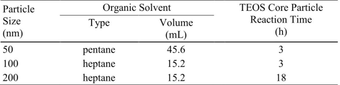
Table 2.1 Variable synthetic parameters for each
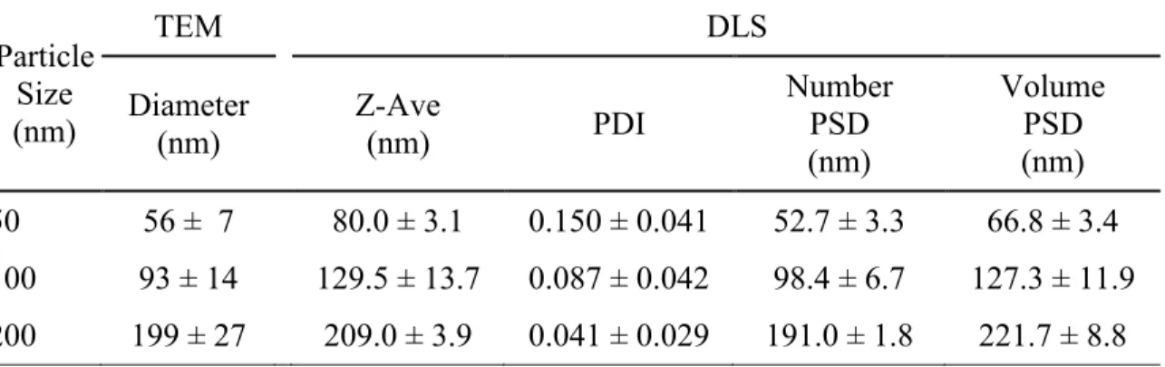
amine-functionalized nanoparticle size ...64 Table 2.2 Particle size as determined by transmission electron
microscopy (TEM) and dynamic light scattering (DLS) ...74 Table 2.3 Zeta potential values of AHAP/TEOS and
N-diazeniumdiolate AHAP/TEOS silica nanoparticles as
determined by Laser Doppler Velocimetry ...75 Table 2.4 Nitrogen content, NO release properties, and extent of
amine to N-diazeniumdiolate conversion for the
amine-functionalized silica nanoparticles ...83 Table 2.5 Minimum bactericidal concentration of NO-releasing
AHAP/TEOS of each size after 2 and 24 h incubation with
P. aeruginosa ...86 Table 2.6 Viability of P. aeruginosa following treatement with blank
(i.e., in PBS only) or control (i.e., non-NO-releasing control AHAP/TEOS particles). Initial bacteria concentration is
106 CFU/mL ...87 Table 2.7 Concentration of NO corresponding to the particle
concentrations tested in cytotoxicity assays ...95 Table 3.1 Atomic concentrations (%) and C/N ratios of unmodified
AHAP and surface-modified AHAP silica nanoparticles as
determined with X-ray photoelectron spectroscopy (XPS) ...118 Table 3.2 Amine to N-diazeniumdiolate conversion efficiency of
surface-modified AHAP particles using sodium methoxide
in either a 1:9 mixture of MeOH in DMF or pure MeOH ...121 Table 3.3 Nitric oxide release characterization of unmodified
AHAP/NO and surface-modified AHAP/NO silica nanoparticles, including maximum instantaneous NO concentration ([NO]max), time required to reach [NO]max
(tmax), total amount of NO released ([NO]T), and the time
required for the systems to release half of [NO]T (t1/2) ...123
Table 3.4 Characterization of Tecoplast electrospun polymer fibers doped with unmodified and modified NO-releasing silica particles including nitric oxide release, and particle
Table 5.1 Zeta potential measured from AHAP and QA-modified
AHAP particle solutions ...177
Table 5.2 Nitric oxide release properties of unmodified and QA-modified silica nanoparticles, including total NO release ([NO]T), maximum instantaneous concentration of NO
LIST OF FIGURES
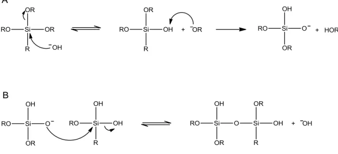
Figure 1.1 (A) Hydrolysis and (B) condensation reactions involved in
the sol–gel process under basic conditions ...3
Figure 1.2 Silane precursors for sol–gel chemistry can be (A)
monofunctional, (B) bifunctional, (C) trifunctional, or (D)
tetrafunctional ...5
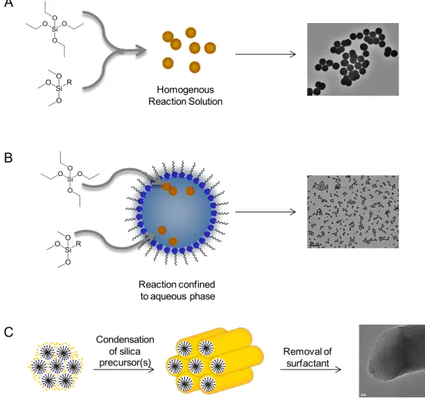
Figure 1.3 Synthetic techniques for synthesizing silica nanoparticles for drug delivery include (A) the Stöber method, (B) the reverse microemulsion technique, and (C)
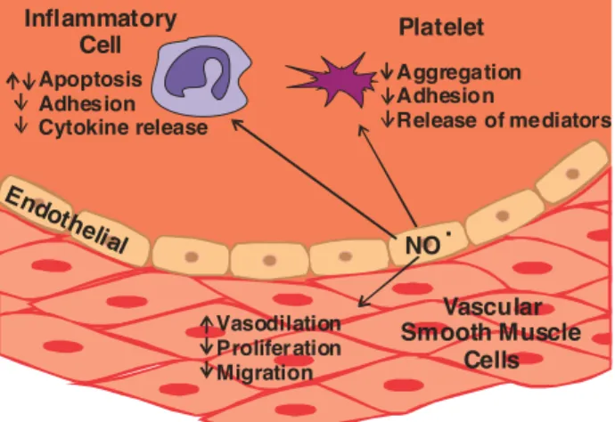
surfactant-templated synthesis ...7 Figure 1.4 Schematic of nitric oxide’s role in the vascular endothelium
and its effects on cellular activities ...23 Figure 1.5 The dual role of nitric oxide in cancer biology ...25 Figure 1.6 The numerous antibacterial mechanisms of nitric oxide and
its byproducts (A) lead to decreased bacterial viability and decreased adhesion on NO-releasing surfaces (B) compared to control surfaces (C). Images of bacteria were obtained
using atomic force microscopy ...26
Figure 1.7 Reactions for S-nitrosothiol formation and degradation ...30 Figure 1.8 The two proposed mechanisms of N-diazeniumdiolate
formation involve (A) the formation of the NO dimer or (B) the formation of nitrosamine anion. (C) The
decomposition of NONOates via proton-initiated
decomposition ...31 Figure 1.9 Aminosilanes (A-G) and tetraalkoxysilanes (H-I) used to
synthesize hybrid silica particles. (A) N-
(6-aminohexyl)aminopropyltrimethoxysilane (AHAP), (B)
N-(2-aminoethyl)-3-aminopropyltrimethoxysilane (AEAP), (C) (aminoethylaminomethyl)phenethyltrimethoxysilane (AEMP) (D) 3-methylaminopropyltrimethoxysilane (MAP), (E) N-(2-aminohexyl)-11-aminoundecyl-
trimethoxysilane (AEAUD), (F) 3-methylaminopropyl-trimethoxysilane (MAP), (G) (3-trimethoxysilane)-diethylenetriamine (DET), (H) tetramethoxysilane
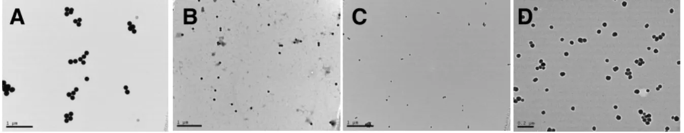
Figure 2.1 Transmission electron micrographs of silica particles resulting from the addition of (A) 0, (B) 27, (C) 53, (D) 77,
or (E) 100 mol% aminosilane (balance TEOS) ...71
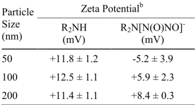
Figure 2.2 TEM micrographs of (A) 200, (B) 100, and (C, D) 50 nm AHAP/TEOS silica nanoparticles. Scale bar is 1 µm for
A–C and 0.2 µm for D ...77 Figure 2.3 Solid state CP/MAS 29Si NMR of (A) 50 nm, (B) 100 nm,
and (C) 200 nm AHAP/TEOS nanoparticles. The T-band (ca. -70 ppm) represents silicon atoms that are bound to three oxygen atoms (i.e., AHAP). The Q-band (ca. -100 ppm) indicates silicon atoms with four siloxane bonds (i.e.,
TEOS) ...79
Figure 2.4 Real time NO release profile for 50 (black), 100 (red), and 200 (blue) nm NO-releasing AHAP/TEOS particles from t=0 to t=24 h. Inset: NO release from t=0 to t=0.2 h, corresponding to the shortest time period at which particle-bacteria association was investigated with confocal
microscopy ...84 Figure 2.5 Scanning confocal images of P. aeruginosa treated with 50
nm RITC-modified NO-releasing AHAP/TEOS nanoparticles. Brightfield, fluorescence (RITC), and overlay images were acquired (A) 0 (addition of particles), (B) 2.4, (C) 6.4, (D) 19.5, and (E) 39 min after addition of
10 μg mL-1 nanoparticles. Scale bars are 10 µm ...89 Figure 2.6 Scanning confocal images of P. aeruginosa treated with 50
nm RITC-modified NO-releasing AHAP/TEOS nanoparticles. Brightfield image (A) was acquired before addition of 10 μg mL-1 nanoparticles and fluorescence overlay image (B) was acquired after 10 min incubation
with nanoparticles. Scale bars are 5 µm ...90
Figure 2.8 Cytotoxicity of control and NO-releasing particles against L929 mouse fibroblast cells as measured using the MTS assay after (A) 2 h exposure at 0 (white), 0.4 (light grey), 0.8 (grey), 1.6 (dark grey), and 3.2 (black) mg mL-1 and after (B) 24 h exposure at 0 (white), 0.1 (light grey), 0.2
(grey), 0.4 (dark grey), and 0.8 (black) mg mL-1 ...94 Figure 3.1 Structures of silanes of increasing hydrophobic character:
(A) ethyltrimethoxysilane, (B) isobutyltrimethoxysilane, (C) octadecyltrimethoxysilane, and (D)
(heptadecafluoro-1,1,2,2-tetrahydrodecyl)trimethoxysilane ...111
Figure 3.2 Scanning electron micrographs of (A) unmodified AHAP (d=148±23 nm), (B) ethyl-AHAP (d=146±22 nm), (C) butyl-AHAP (d=144±18 nm), (D) octadecyl-AHAP (d=152±17 nm), and (E) heptadecafluoro-AHAP
(d=152±22 nm). Scale bar = 1 µm ...113
Figure 3.3 Solid state direct polarization-magic angle spinning (DP-MAS) 13C NMR spectrum of (A) unmodified AHAP and (B) ethyl-, (C) butyl-, (D) octadecyl-, and (E)
heptadecafluoro-modified AHAP silica nanoparticles ...115
Figure 3.4 Solid state direct polarization-magic angle spinning (DP/MAS) 19F NMR spectrum of heptadecafluoro-AHAP
silica nanoparticles ...116
Figure 3.5 Derived count rate from dynamic light scattering analysis of unmodified and surface-modified AHAP particles (0.5
mg/mL) in phosphate buffered saline ...120 Figure 3.6 Scanning electron microscopy images of Tecoplast
electrospun polymer fibers doped with 5 wt% (A,F) AHAP/NO (d=318±90 nm), (B,G) ethyl-AHAP/NO (d=353±185 nm), (C,H) butyl-AHAP/NO (d=427±141 nm), (D,I) octadecyl-AHAP/NO (d=299±101 nm), and (E,J) heptadecafluoro-AHAP/NO (d=309±176 nm) silica
nanoparticles. Scale bars = 5 µm for A–E and 2 µm for F–J ...125 Figure 4.1 Synthesis of mesoporous silica nanoparticles (MSNs)
involves the condensation of silane precursors around micelle structures to yield porous silica nanoparticles. Following surfactant removal, organosilanes can be condensed onto the MSNs through reaction with surface
Figure 4.2 Scanning electron micrographs of mesoporous silica nanoparticles synthesized via a surfactant templated
approach ...147 Figure 4.3 Solid state CP/MAS 29Si NMR spectrum of
MOM-Pip/NO-modified MSNs. The T-band (ca. -60 ppm) indicates silicon atoms bound to three oxygens (i.e., MOM-Pip/NO-TMS), and the Q-band (ca -100 ppm) represents silicon
atoms bound to four oxygens (i.e., TEOS) ...148 Figure 4.4 UV-Vis absorbance spectrum of MOM-Pip/NO-modified
particles with an maximum absorbance at 220 nm indicating the presence of the MOM-protected
diazeniumdiolates ...150 Figure 4.5 Nitric oxide release from MOM-Pip/NO-MSNs. Total NO
storage is 2.5 µmol/mg as determined by elemental analysis ...151
Figure 4.6 Nitric oxide release in 10 vol% BHI in PBS from dental composites doped with 1 wt% AHAP/NO (squares) and
MOM-Pip/NO (triangle) particles...153
Figure 4.7 Viability of adhered S. mutans to control and
particle-doped composites following 24 h incubation in 10 vol%
BHI in PBS ...155
Figure 4.8 Atomic force micrographs of (A) control, (B), AHAP/NO-doped, and (C) MOM-Pip/NO-doped composites, (D) representative height trace from center section of each image (indicated by horizontal red line), and (E) rms
surface roughness determined from four 2 µm2 areas ...156
Figure 5.1 Scanning electron micrographs of (A) AHAP (d=180±26 nm), (B) methylQA (d=181±27 nm), (C) butylQA (d=187±23 nm), (D) octylQA (d=185±26 nm), and (E) dodecylQA (d=187±24 nm) nanoparticles. Scale bar = 500
Figure 5.4 Minimum bactericidal concentrations (MBC24h) against (A)
S. aureus and (B) P. aeruginosa for non-NO-releasing (solid) and NO-releasing (hashed) AHAP (red), methylQA (green), butylQA (blue), octylQA (magenta), dodecylQA (cyan). Treatment with a 50:50 (w/w) mixture of dodecylQA and AHAP/NO particles is shown in yellow
(hashed). ...182 Figure 5.5 Change in bacterial viability of (A) S. aureus and (B) P.
aeruginosa following exposure to sublethal doses of dodecylQA and/or AHAP/NO nanoparticles either
simultaneously or at 30 min intervals ...185 Figure 5.6 Confocal microscopy images of S. aureus exposed to (A)
dodecylQA/NO and (B) AHAP/NO particles exhibit green fluorescence due to intracellular NO (DAF) and red fluorescence due to compromised membrane (PI). Scale bar
= 1 µm ...186 Figure 5.7 Percent viability of L929 mouse fibroblasts cells following
24 h exposure to non-releasing (solid) and NO-releasing (hashed) AHAP (red), methylQA (green), butylQA (blue), octylQA (magenta), and dodecylQA (cyan) particles compared to control (untreated) cells with the numbers corresponding to the MBC24h against S. aureus
and P. aeruginosa (in mg/mL dose) ...189 Figure 6.1 Transmission electron micrograph of Fe2O3 nanocrystals
(diameter = 7 nm) synthesized via the thermal degradation
of iron pentacarbonyl in dioctyl ether and oleic acid ...204 Figure 6.2 The addition of n-octyltrimethoxysilane to the surface of
oleic acid-capped Fe2O3 nanocrystals via van der Waals interactions allows for their stability in aqueous media and
promotes the formation of a silica shell ...206 Figure 6.3. Transmission electron micrograph of magnetic
NO-releasing silica particles composed of Fe2O3 core and a
LIST OF SCHEMES
Scheme 2.1 Synthesis of amine-functionalized silica nanoparticles via a reverse microemulsion. Step 1 involves micelle formation. Step 2 is the addition of tetraethoxysilane (TEOS) to the emulsion to form monodisperse “seed” particles. Lastly, step 3 is the subsequent addition of TEOS and AHAP that
co-condense to form the AHAP/TEOS silica nanoparticles ...72
Scheme 2.2 N-Diazeniumdiolate formation on aminosilane-modified
silica nanoparticle ...81
Scheme 3.1 Organosilanes are grafted onto the surface of hybrid silica
nanoparticles through a condensation reaction with surface
silanols on the particles and methoxides on the organosilanes ...110
Scheme 4.1 Synthesis of O2-methoxymethyl
1-(piperazin-1-yl)diazen-1-ium-1,2-diolate (MOM-Pip/NO) ...142
Scheme 4.2 Synthesis of O2-methoxymethyl
1-(4-(3-
(trimethoxysilyl)propyl)piperazin-1-yl)diazen-1-ium-1,2-diolate (MOM-Pip/NO-TMS) ...143
Scheme 5.1 Quaternary ammonium (QA) epoxides were synthesized
via reaction of epichlorohydrin with a dimethylalkylamine and subsequently reacted with the primary amines on the surface of AHAP/TMOS particles to yield
QA-functionalized silica nanoparticles ...164
Scheme 5.2 N-Diazeniumdiolate NO donors were formed on secondary
amines within the particle scaffold upon exposure to high pressures of NO in the presence of a base (e.g.,
NaOSiMe3). In the presence of a proton source (e.g., H2O),
these NO donors breakdown to regenerate the parent amine
LIST OF ABBREVIATIONS AND SYMBOLS
~ approximately
% percentage(s)
%Econv conversion efficiency
°C degree(s) Celsius
g times the force of gravity
[…] concentration
17FTMS (heptadecafluoro-1,1,2,2-tetrahydodecyl)trimethoxysilane
µg microgram(s)
µL microliter(s)
µm micrometer(s)
µM micromolar
µmol micromole
λ wavelength
AEAP N-(2-aminoethyl)-3-aminopropyltrimethoxysilane
AEAUD N-(2-aminohexyl)-11-aminoundecyl-trimethoxysilane
AEMP (aminoethylaminomethyl)phenethyltrimethoxysilane
AFM atomic force microscope/microscopy
Ag+ silver ion
AgNO3 silver nitrate
AHAM N-(6-aminohexyl)aminomethyltrimethoxysilane
AHAP N-(6-aminohexyl)aminopropyltrimethoxysilane
AHAP/NO N-diazeniumdiolate-modified
ATCC American Type Culture Collection
atm atmosphere(s)
BAP 3-butylaminopropyltrimethoxysilane
BET Brunauer-Emmett-Teller
BHI brain heart infusion
BTMOS isobutyltrimethoxysilane
butyl-AHAP isobutyltrimethoxysilane-modified AHAP particles
butylQA dimethylbutyl-quaternary ammonium
C. albicans Candida albicans
ca. approximately
Ca2+ calcium ion
CaCl2 calcium chloride
CDI collagen density index
CFU colony forming units
cGMP cyclic guanosine monophosphate
CHCl3 chloroform
cm centimeter(s)
CO2 carbon dioxide
CP cross polarization
DLS dynamic light scattering
DMF N,N-dimethylformamide
DMAc N,N-dimethylacetamide
DNA deoxyribonucleic acid
dodecylQA dimethyldodecyl-quaternary ammonium
DP direct polarization
E. coli Escherichia coli
ECM extracellular matrix
e.g. for example
EDRF endothelium-derived relaxation factor
eNOS endothelial nitric oxide synthase
EPR enhance permability retention effect
ESEM environmental scanning electron microscopy/microscope
ESI/MS electrospray ionization mass spectrometry
et al. and others
etc. and so forth
ethyl-AHAP ethyltrimethoxysilane-modified AHAP particles
EtOH ethanol
Fig. figure
h hour(s)
H2O water
HCl hydrochloric acid
heptadecyl-AHAP (heptadecafluoro-1,1,2,2-tetrahydrodecyl)-trimethoxysilane-modified AHAP particles
ICP-OES inductively coupled plasma atomic emission spectroscopy
IgG immunoglobulin G
iNOS inducible nitric oxide synthase
IR infrared
kg kilogram
kHz kilohertz
KOH potassium hydroxide
kV kilovolts
LMW low molecular weight
m meter(s)
M molar
MΩ megaohm(s)
MAP 3-methylaminopropyltrimethoxysilane
MAS magic angle spinning
MBC minimum bactericidal concentration
MeOH methanol
methylQA trimethyl-quaternary ammonium
mg milligram(s)
MHz megahertz
mol mole(s)
mol% percent of total moles
MOM-Pip/NO O2-methoxymethyl 1-(piperazin-1-yl)diazen-1-ium-1,2-diolate
MOM-Pip/NO-TMS O2-methoxymethyl 1-(4-(3-(trimethoxysilyl)propyl))piperazin-1-yl)diazen-1-ium-1,2-diolate
MPTMS 3-mercaptopropyltrimethoxysilane
MRSA methicillin-resistant Staphylococcus aureus
MSN mesoporous silica nanoparticle
MTS
(3-(4,5-dimethylthiazol-2-yl)-5-(3-carboxymethoxyphenyl)-2-(4-sulfophenyl)-2H-tetrazolium
N.A. numerical aperture
N2 nitrogen gas
NaCl sodium chloride
NaOMe sodium methoxide
NaOH sodium hydroxide
NH4OH ammonium hydroxide
nm nanometer(s)
nmol nanomole(s)
NMR nuclear magnetic resonance spectroscopy
nNOS neuronal nitric oxide synthase
NO nitric oxide
NO2 nitrogen dioxide
NO2- nitrite
[NO]max maximum NO flux
NONOate N-diazeniumdiolate
NOS nitric oxide synthase
O2 oxygen gas
octadecyl-AHAP octadecyltrimethoxysilane-modified AHAP particles
octylQA dimethyloctyl-quaternary ammonium
OH- hydroxide ion
ONOO- peroxynitrite
P. aeruginosa Pseudomonas aeruginosa
PBS phosphate buffered saline, pH 7.4
PDI polydispersity index
PEG poly(ethylene glycol)
pH -log of proton concentration
PI propidium iodide
pmol picomole(s)
ppb parts per billion
ppm parts per million
PROLI/NO N-diazeniumdiolate-modified L-proline
PU polyurethane
PVC poly(vinyl) chloride
S. mutans Streptococcus mutans
SEM scanning electron microscope/microscopy
SS solid state
t1/2 half-life
t time
tmax time to max NO flux
TEOS tetraethoxysilane
TEM transmission electron microscope/microscopy
THF tetrahydrofuran
TMOS tetramethoxysilane
TSA tryptic soy agar
TSB tryptic soy broth
U.S. United States
UV ultraviolet
UV-Vis ultraviolet-visible spectroscopy
v/v volume/volume
VEGF vascular endothelial growth factor
wt% percent by weight
wt/wt weight/weight
Chapter 1:
Designing Silica Particles for the Delivery of Therapeutic Nitric Oxide
A wide range of nitric oxide (NO)-releasing materials have emerged as potential
therapeutics that exploit NO’s vast biological roles. Macromolecular scaffolds, such as silica
nanoparticles, are particularly promising due to their ability to store and deliver larger NO
payloads in a more controlled and effective manner compared to low molecular weight NO
donors. In this introductory chapter, the synthesis and design of silica particles for drug
delivery is presented, followed by a discussion on the development of NO-releasing silica
nanoparticles for therapeutic applications.
1.1 Overview of silica nanoparticles for drug delivery
Over the past forty years, nanotechnology has revolutionized scientific research,
impacting the world in ways that parallel the development of electricity, biotechnology and
digital information.1 Drawn by the fascinating properties exhibited by materials in the
nanometer regime (≤100 nm), research in nearly all area of science have directed some focus
spread use arises from well-defined and tunable structures that can be tailored towards a
desired therapeutic application. Indeed, any number of functionalities may be incorporated
into the silica network through facile sol–gel chemistry. Physical properties such as particle
size, shape, and porosity can also be easily tuned, allowing researchers to investigate their
influence on drug delivery (e.g., cellular uptake, clearance/fate, and aggregation).
Furthermore, silica’s inherent biocompatibility and water-solubility provide two distinct
advantages when designing practical drug delivery scaffolds.
1.1.1 Sol–gel chemistry
The sol–gel process to form silica-based materials involves the hydrolysis and
condensation of silane precursors to form a solid network.6 In basic conditions, hydrolysis
occurs with the displacement of a labile ligand on the silicon atom by a hydroxyl ion (Figure
1.1A). The hydrolyzed monomer then undergoes a condensation reaction with a second
hydrolyzed molecule to form a siloxane bond (Figure 1.1B). Hydrolysis and condensation
continue, forming a colloidal suspension (sol) that cross-links to form a solid network
suspended in a continuous liquid phase (gel). Depending on the reaction conditions, the
condensed products resulting from the sol–gel process can range from highly porous,
nanocrystalline materials to dense amorphous networks. In addition to synthetic control,
other advantages of sol–gel chemistry include mild reaction conditions, low-temperature
preparation, and easy purification.7
The type of material that results from sol–gel processing is dependent on the type of
precursor and the pH of the reaction. Precursors can be mono-, bi-, tri- or tetrafunctional
more of the hydrolyzable groups have been replaced with a non-hydrolyzable organic ligand
are called organosilanes. The substituents on the central silicon atom govern the rate of
hydrolysis and condensation through both steric and inductive effects. Larger or branched
ligands hinder the access of nucleophiles to the silicon atom, thereby slowing the rate of
hydrolysis. The incorporation of an electron-donating alkyl group also contributes to slower
hydrolysis as the central silicon atom has a higher electron density and is therefore less
electrophilic.
An acid or base catalyst is used to promote hydrolysis and condensation as well as
govern the structure of the condensed product.6 In acid-catalyzed reactions, the rates of
hydrolysis are fast and the rates of condensation are slow. Acidic conditions with low water
to silane ratios favor the formation of linear and branched polymers that interpenetrate with
each other to form films. Alternatively, base-catalyzed sol–gel reactions with high water to
silane ratios promote slow rates of hydrolysis and rapid condensation, leading to highly
branched clusters that do not entangle. The preparation of silica particles is thus usually base
catalyzed. Base-catalyzed hydrolysis and condensation of a single tetrafunctional silane,
such as tetraethoxysilane (TEOS), represents the simplest synthesis of silica particles. To
broaden their applicability, it is often desirable to vary the chemical and physical
characteristics of silica particles. Thus, modified sol–gel techniques have been developed
that allow for precise control over particle growth to result in materials with specific
properties. The two general classes of silica particles are nonporous silica particles,
synthesized by the Stöber method or the reverse microemulsion technique, and mesoporous
silica particles, prepared via a surfactant-templated method.
condensation of silane precursors in a solution of water, an alcohol solvent (typically ethanol)
and a base catalyst.8 A short-chain alcohol solvent is necessary to create a homogenous
reaction solution as the tetraalkoxysilane is immiscible with water. The Stöber method yields
monodisperse, spherical particles typically in the micron to submicron size range and is
scalable to increase throughput. The Stöber method can be conducted in a “one-pot” reaction
where all reactants are combined simultaneously. Alternatively, a seeded-growth method
utilizes small particles that are initially grown and used as seeds upon which larger particles
form following multi-step addition of more silane precursors. Lastly, a semi-batch approach
can be used where one reactant (e.g., silane or catalyst) is added at a constant (typically slow)
rate to a vessel containing the other reactants. As will be discussed in Section 1.3.1, each of
these synthetic strategies allows for particle size to be tuned by changing the concentration of
water or reactants, reaction temperature, and solvent type. Particles with specific organic
functionalities can be created by using a corresponding silane precursor, the details of which
are discussed in Section 1.4.1.
The growth of monodisperse particles via the Stöber method can be described using
the LaMer theory.9,10 These models relate the concentration of hydrolyzed silane
([monomer]) to reaction time and the number of particles formed. As hydrolysis reactions
occur, the [monomer] increases with time until it reaches a critical concentration
([monomer]nucleation) where initial sites of particle formation (i.e., nuclei) form. While
[monomer] ≥ [monomer]nucleation, hydrolyzed silanes can either react with each other to form
new nuclei or add to already formed nuclei. Eventually, [monomer] falls below that of the
sequential addition of hydrolyzed monomers. To achieve monodisperse particle populations
Condensation of silica
precursor(s) Removal of surfactant C
B A
Homogenous Reaction Solution
Reaction confined to aqueous phase
Figure 1.3 Synthetic techniques for synthesizing silica nanoparticles for drug delivery
surfactant-most nuclei are created at the same time and experience the same growth histories. The
Stöber method typically produces particles larger than 100 nm as the reaction conditions do
not stabilize nuclei, causing them to aggregate and form larger particles.
The reverse microemulsion technique is a method to produce silica particles less than
100 nm with improved monodispersity.11 In contrast to the Stöber method where the sol–gel
process occurs throughout the bulk of the reaction solution, the reverse microemulsion
confines particle formation to the aqueous interior of micelles stabilized in an organic solvent
(Figure 1.3B). Surfactants used to form the micelles can be cationic (e.g.,
cetyltrimethylammonium bromide), anionic (e.g., sodium bis(2-ethylhexyl) sulfosuccinate),
or nonionic (e.g., Triton X-100 or polyoxyethylene nonylphenyl ether). Typically a
co-surfactant, such as 1-hexanol, is employed to stabilize the micelles. The interior of the
micelles is composed of water and the base catalyst and act as nanoreactors for silica
nanoparticle synthesis.
In a reverse microemulsion system, particle formation is also described according to
the LaMer theory but is believed to occur at a much slower rate.12 Upon addition to the
reaction mixture, the unhydrolyzed silanes become dispersed throughout the organic phase.
As they slowly come in contact with the micelles, the silanes are hydrolyzed and enter the
micelle’s aqueous interior.13 Nucleation occurs when [monomer] reaches the critical
concentration (e.g., [monomer]nucleation) within the micelle. Similar to the Stöber method, the
decrease in [monomer] after the onset of the nucleation phase occurs mainly through the
diffusion-controlled growth of particles and nuclei. However, the diffusion-controlled
growth occurs at a much slower rate in a microemulsion due to the micellar structure’s effect
a greater number of particles and smaller particle sizes. Furthermore, the nuclei formed in a
reverse microemulsion are believed to be stabilized by the surfactant molecules, thus the
extent of nuclei aggregation is lower than in the Stöber method. Particle growth is confined
to the micelle, thus particle size can be finely tuned by controlling micelle size. For example,
the water to surfactant ratio, surfactant to solvent ratio, the type of organic solvent, and
reaction time all affect the resulting particle size. Functionalized particles can be obtained by
the addition of organosilanes to the emulsion. The major drawbacks to synthesizing silica
particles via a reverse microemulsion are low yields and extensive purification steps.
Particularly for biomedical applications, complete removal of the potentially cytotoxic
surfactant is necessary and requires copious washing with a variety of alcohols. The ability
to achieve small particles with narrow size distributions is the main advantage over the
Stöber method.
The two previously discussed techniques for sol–gel chemistry result in nonporous
silica particles. More recently, mesoporous silica particles (MSN) have been designed as a
strategy to achieve greater functionalization potential and drug-loading capacity.14 The
surface area of MSNs is typically >900 m2/g, while that of Stöber and reverse
microemulsion-prepared silica particles is typically 5–200 m2/g. The pores of MSNs typically
have a large volume (>0.9 cm3/g) with tunable sizes of narrow distributions (2–10 nm).
Once the siloxane polymerization is complete, the surfactant molecules are removed either
by solvent extraction with acidic ethanol or by calcination to reveal empty channels
throughout the silica network.
1.1.2 Drug delivery
Silica-based nanomaterials are a popular scaffold for drug delivery due to their low
cytotoxicity, chemical stability and high capacity for functionalization.4 Compared to “soft”
nanoparticles (e.g., dendrimers, polymers, and vesicles), silica exhibits increased stability at
high temperatures and varied pHs and is more resistant to mechanical stresses and
hydrolysis-induced degradation
Ideally, a successful drug delivery scaffold would be biocompatible and capable of
high drug loading.14 Delivery to a specific site (e.g., cell type, tissue, or organ) is a desirable
trait for most therapeutic applications and can be achieved using targeting moieties, as will
be discussed later. The major challenge in developing drug-releasing nanoparticles is the
need for a scaffold that exhibits zero drug release prior to arrival at the site of interest and
then provides complete delivery of drug payload. Furthermore, the rate of drug release from
the nanoscaffold often governs its biological effect,16 thus triggerable release of therapeutics
is also desirable. In general, achieving these goals with soft nanomaterials is difficult due to
uncontrollable leaching and limited functionalization ability.14
The two strategies that are used to load drugs onto/into silica particles include
covalent incorporation and non-covalent encapsulation. Covalent incorporation provides a
more secure drug storage method that decreases the chance of premature release; however, a
notable release triggers for covalently loaded drugs include redox chemistry, enzymatic
cleavage, pH, photolysis and thermolysis.3 Redox chemistry is a common strategy for
achieving intracellular drug delivery due to the reducing environment present inside cells.
Drugs can be tethered to organically modified silica scaffolds via reduction-sensitive
linkages such as disulfide17 or organometallic18 bonds. Hydrolysable or enzymatically
cleavable bonds such as esters, carbonates, carbamates, hydrazones and amides are also
common methods of covalent incorporation into silica.19 Alternatively, pH-sensitive
functionalities can be incorporated into the silica network that result in drug release once the
scaffold enters an area of a certain pH. For example, the interior of endosomes is
characterized by a lower pH than that of extracellular fluid (pH 5–6.5 vs 7.4, respectively).
Thus, incorporating groups that are labile at lower pH values allows for selective drug release
within cells. Light-responsive materials have been designed where the bond tethering the
drug to the scaffold is photo-labile such that drug release can be triggered with light
irradiation. Gold nanoparticles can be also be used for drug release as they will absorb light
and convert the energy to heat to trigger thermolytic cleavage. While UV, visible, and
near-IR active materials have been developed, near-near-IR irradiation (650–900 nm) is the most
promising photo-trigger as it is only minimally absorbed by skin and tissue. When using
covalent incorporation for drug delivery, one must ensure that the chemical modifications
diffusion by placing the porous particles in highly concentrated drug solutions; however, the
upper limit of loading is relatively low for this method. Alternatively, electrostatic
interactions between the negatively charged silica scaffold and positively charged drug
molecules can be used to achieve higher loading. Increased loading of neutral or negatively
charged drugs proves more difficult. Liu et al. reported a strategy to coerce negatively
charged drugs into MSNs through the formation of “protocells”. 20 In this method, a
positively charged liposome is fused around the negatively charged silica particle in a
solution containing the drug. The electrostatic interactions between the negatively charged
drug and the positively charged liposome cause the drug to be forced into the MSNs as the
liposome encapsulates the silica.
When drugs are not covalently bound to the scaffold, premature release represents a
major obstacle.14 Stability of the drug within the scaffold can be promoted through
non-covalent interactions such as electrostatic or hydrogen-bonding. For example, increased drug
loading efficiency and extended release profiles of ibuprofen-loaded MSNs were achieved
with aminosilane-modified MSNs.21 The hydrogen-bonding interactions between the
protonated primary amines (at pH 7) tethered to the scaffold and the carboxylate groups of
ibuprofen aided in drug loading stabilization.
Another method for inhibiting the release of encapsulated drugs from mesoporous
particles is through the use of “gatekeepers”, whereby zero premature release of drug is
achieved by “plugging” the entrances of pores with protecting groups after drug loading.14, 22
The aforementioned strategies for releasing covalently bound drugs have been translated to
the gatekeeping concept. For example, Lai et al. blocked the entrances to the channels of
with disulfide linkages.23 Reduction of the disulfide bond released the gatekeeping particle
from the pore, allowing the encapsulated drug to diffuse out. Other metallic nanoparticles as
well as dendrimers and bulky organic molecules have also been employed as gatekeepers
with release stimuli ranging from light to oscillating magnetic fields.3, 22
In summary, silica-based nanoparticles offer a stable and chemically flexible scaffold
that can be loaded with high concentrations of drug. The numerous opportunities for drug
loading and release mechanisms are unique to these materials as such functionalities are not
easily incorporated into soft materials.
1.1.3 Influence of nanoparticle properties on nanoparticle-cell interactions
Efficient drug delivery is governed by the extent of direct interaction between the
nanoparticle carrier and the targeted cells. Although the release of drug into the environment
surrounding the target may still result in some therapeutic effect, diffusion of the drug away
from the cell would necessitate a higher dosage to elicit the same therapeutic response if the
drug were delivered directly into the cell. Additionally, some drugs may be unable to
permeate the cell membrane unassisted, thus nanoparticle carriers aid in intracellular
delivery. Nanoparticle characteristics (i.e., size, shape, and surface chemistry) govern
particle-cell interactions both in terms of association and uptake. As such, much effort has
lead to nanoparticle association. Indeed, positively charged particles generally exhibit
greater association and subsequent internalization into mammalian cells than neutral or
negatively charged particles.24 Similarly, the extent of association between positively
charged particles and bacteria cells is greater than that of negatively charged particles.25 In
addition to carrying a negative charge, the surface of both prokaryotes and eukaryotes also
express hydrophobic character due to membrane lipids. As such, cells have exhibited an
affinity for particles expressing lipophilic ligands, such as fatty acids and fatty amines.26 Due
to the lipid bilayer comprising the cellular envelope, hydrophobic effects will also regulate
whether a nanoparticle simply adsorbs to the membrane or is capable of penetrating into the
interior of the cell.27 Lastly, nanoparticle-cell association can be greatly enhanced by
tethering molecules onto the particle that bind to cell surface receptors. For example, the
inclusion of carbohydrates onto nanoparticle surfaces was found to enhance association with
Escherichia coli by binding to the FimH adhesion protein in the bacterium’s pili.28 To date,
several targeting moieties have been designed to increase the association of nanoparticles
with both prokaryotic and eukaryotic cells, the details of which are discussed in Section
1.1.4.
For eukaryotic cells, once an association event has occurred cellular uptake may
ensue. The process by which matter is taken into mammalian cells is called endocytosis.
Phagocytosis and pinocytosis are the two most prominent endocytic mechanisms for
eukaryotic cells. Large particles are taken up by phagocytosis (“cell eating”), while small
particles, fluids, and solutes are taken up by pinocytosis (“cell drinking”). Phagocytosis
mostly occurs with macrophages and polymorphonuclear neutrophils, as these immune cells
internalization strategy for all types of mammalian cells and is the generally accepted method
by which particles are internalized by eukaryotic cells.5, 14, 29 The four main mechanisms for
pinocytosis include macrophinocytosis, clathrin-mediated endocytosis, caveolae-mediated
endocytosis, and clathrin- and caveolae-independent endocytosis.29 Macrophinocytosis is an
actin driven process where protrusions form, collapse, and then fuse onto the cell membrane
to form large endocytic vesicles with diameters of 0.5–10 µm. In clathrin-mediated
endocytosis, a coated pit is created by an assembly of clathrin, forming a vesicle ~120 nm in
size. In caveolae-mediated endocytosis, flask-shaped invaginations are formed by the protein
caveolin, and internalized vesicles (~60 nm) are directed to the endoplasmic reticulum and
the nucleus. Clathrin- and caveolin-independent endocytosis occur through cholesterol-rich
microdomains on the plasma membrane called lipid rafts (40–50 nm). In general, uptake of
silica particles into eukaryotic cells occurs through clathrin-coated endocytosis pathways
although the inclusion of certain ligands may direct other mechanisms of endocytosis.
Although endocytosis is a necessary function for eukaryotes, an endocytosis-like
mechanism has not been identified for prokayrotes.30, 31 Yet, the presence of nanoparticles in
the interior of bacteria cells has been observed.32-35 It is believed that the route of ingress of
nanoparticles into uncompromised bacterial cells may be through pores present in the outer
membrane.25 Since these pores are used to secrete large proteins, only particles of very small
membranes. Thus, it is hypothesized that the particles entered the cellular interior only after
the membrane had been compromised.25
The mechanism and extent of cellular uptake is greatly affected by the physical and
chemical characteristics of the nanoparticle scaffold. It is generally believed that efficient
uptake by non-phagocytic cells requires that the particle must be on the submicron scale,14
although some have reported the internalization of particles as large as 5 µm.29 Overall,
smaller particles are able to accumulate in the cellular interior to a greater extent than larger
particles. For example, the uptake of 50 nm MSNs by HeLa cells was 4, 20, and 11 times
greater than that of 110, 170, and 280 nm particles, respectively.37 Similarly, the uptake of
nonporous silica particles also exhibited a size-dependency with 23 nm particles being
uptaken to a greater extent than larger 85 nm particles.38 The kinetics of internalization was
also observed to be size dependent, with larger sized particles experiencing a slower rate of
uptake. After 60 min incubation, the 85 nm nonporous particles mostly remained physically
adsorbed to the cell surface, while the majority of the smaller 23 nm particles were
distributed throughout the cytosol. 38 Similarly, a greater concentration of smaller metallic
particles has been observed in the interior of bacteria cells compared to that of larger
particles.39 Due to the lack of endocytic mechanisms in prokaryotic cells, this size
dependence is likely the result of Brownian motion across a damaged membrane. A
dependence of bactericidal efficacy on particle size is evident and is the focus of Chapter 2.
Particle shape has also been observed to impact drug delivery efficiency by affecting
particle-cell adhesion strength, internalization rate, cytotoxicity, circulation time and
biodistribution.29, 40, 41 For example, higher aspect ratio MSNs were found to be internalized
Furthermore, high aspect ratio particles resulted in a greater impact on cell proliferation,
apoptosis, cytoskeleton formation, adhesion and migration. In vivo experiments revealed
that particle shape also governs biodistribution as MSNs with an aspect ratio of 1.5
accumulated in the liver, while those with an aspect ratio of 5 distributed in the spleen.41
Furthermore, the lower aspect ratio MSNs experienced a more rapid clearance rate. Lu et al.
observed a similar trend with antimicrobial MSNs, where those of higher aspect ratio
exhibited a greater therapeutic impact.42 It was hypothesized that higher aspect ratios allows
for greater surface contact between MSNs and the microbes, which in turn allowed for more
efficient drug delivery directly to the bacterial membrane.
In addition to physical characteristics, different surface chemistries present on the
particle scaffold may influence the mechanism of cellular uptake. For example, amine and
guanidinium-functionalized MSNs are reportedly taken up by clatherin- and
caveolae-independent mechanisms, compared to the usual clathrin-coated endocytosis of silica
particles.43 The uptake mechanism greatly impacts the effectiveness of the drug as it dictates
the localization of the internalized nanoparticle. Drug delivery to the cytosol of the cell is
most ideal, but typically internalized particles remain trapped in endosomes. Methods for
triggering endosomal release have been achieved by modifying the surface of the particle.43
For example, Verma et al. found that particles modified with alternating anionic and
proteins from adsorbing to the particle.24 Minimal protein adsorption results in decreased
phagocytic cellular uptake and increased circulation time. PEG-modified particles are
typically used for antitumor therapies where the passive targeting of tumor cells is achieved
through the enhanced permeation and retention (EPR) effect.
Uptake of nanoparticles is most commonly monitored by flow cytometry,
transmission electron microscopy, and confocal fluorescence microscopy.45, 46
Internalization versus external association can be differentiated via a number of analytical
techniques. In flow cytometry, the particles must be fluorescent in order to visualize their
location on/in the cell. Particle internalization can be identified by the presence of
fluorescence after copiously washing the cells or by employing extracellular fluorescence
quenchers. The use of transmission electron microscopy (TEM) is advantageous as there is
no need to modify the particles to allow for visualization. The localization of particles can be
elucidated with TEM by using a microtome to obtain 100 nm thin slices of the specimen that
can be imaged individually. Stains are normally used for TEM imaging to increase the
contrast between different cellular compartments. However, TEM imaging requires fixation
of the specimen, which can lead to artificially increased uptake. Alternatively, the use of
Z-stack imaging with confocal microscopy allows for photographic “slices” to be taken along
the z-axis of live cells in solution. Thus, real-time uptake and localization information can
be obtained. The presence of particles in the interior or exterior of the cells is determined by
identifying the location along the z-axis where fluorescence is the greatest. For now, the
resolution of the Z-stack method limits this technique to investigating internalization by
1.1.4 Targeting strategies
The efficiency of drug delivery can be enhanced by increasing the probability and
rate of an interaction between the nanoparticle and the intended tissue or cell. Targeted
delivery of nanoparticles can be achieved passively through processes such as the enhanced
permeability and retention (EPR) effect or actively through the use of targeting or directing
moieties.
The EPR effect has been used for targeting nanoparticles to tumor sites by taking
advantage of the fact that solid tumors and inflamed tissue have a more leaky vasculature
than healthy tissue.2 As nanoparticles circulate throughout the vasculature, they are more
likely to extravasate into cancerous tissue than normal tissue. This phenomenon represents a
passive strategy for the selective delivery of nanoparticles to cancerous tissue over normal
tissue. Nanoparticles that utilize the EPR for selectivity must be between 100–300 nm and
experience long circulation life-times.4, 47 Micron-sized particles are quickly cleared by
active phagocytosis of the reticuloendothelial system, while particles with diameters below
100 nm are insufficient due to rapid renal clearance. Increased circulation times can be
achieved by modifying particle surfaces with PEG, which prevents non-specific binding of
proteins and macrophages. While the EPR effect allows for passive targeting of
nanoparticles to tumor sites, active targeting strategies allow for increased internalization and
that nanoparticles loaded with a chemotherapeutic agent and smaller magnetite (Fe3O4)
nanoparticles could be magnetically directed to a tumor site and resulted in a 15-fold higher
drug concentration at the tumor site compared to freely circulating drug.51 Additionally,
once the nanoparticle carrier has reached the targeted site, the application of an oscillating
magnetic field can be used for heat generation to trigger drug release.52, 53
Other active targeting strategies involve incorporating ligands on the particle surface
that may increase the affinity of the intended cell for the particle by seeking a particular
aspect that is more present on the targeted cell than other cells. The most direct method of
active targeting is to employ molecules that bind to receptors on the targeted cell surface.47
For example, proteins, such as antibodies and glycoproteins, have been tethered to particle
surfaces for active targeting. Transferrin and transferrin receptor antibodies have proven
effective antitumor targeting moieties as the majority of cancer cells overexpress transferrin
receptors.54 Conjugating nanoparticles with the secondary human immunoglobulin (IgG)
that targets protein A on the bacteria cell wall resulted in a significant increase in binding of
the nanoparticles to bacteria compared to unmodified controls.55, 56 Moreover, the addition of
the glycoprotein D-mannose onto nanoparticles was also found to increase binding to the
bacteria cell wall compared to unmodified controls.28, 57 Avidity, the strength of multiple
bonding interactions, can be enhanced through the use of larger multivalent targeting
moieties; however, sterics limits the number of groups that can be tethered to the particle
surface. Smaller biomolecules have been employed to increase the probability of binding to
the target. One such type of small biomolecules are aptamers, which are a recently
developed targeting strategy composed of single-stranded oligonucleotides such as DNA or
acid targets including peptides, proteins, and even whole cells with high affinity and
specificity. Other small molecules such as carbohydrates, vitamins, or peptides can also be
tethered to the particle surface to actively target tumor cells.5, 47 For example, the addition of
folic acid ligands allows for high-affinity binding to the folate receptor that is overexpressed
on numerous types of cancer cells compared to normal cells.59 The fusion of nanoparticles to
the cell membrane (and subsequent uptake into the cytoplasm) can be further enhanced by
incorporating cell-penetrating peptides such as RGD, allatostatin 1, PLL, and arginine-rich
peptides.60 The active targeting of prokaryotic cells has also been investigated by tethering
the clinically used antibiotic glycopeptide vancomycin to nanoparticles for targeting
Gram-positive bacterium. Vancomycin binds to the terminal peptide (D-alanyl-D-alanine) on the
cell wall of Gram-positive bacteria through five hydrogen-bonds, increasing the avidity of
the bacterium for the vancomycin-modified nanoparticles.61, 62
In addition to increasing the efficiency of drug-delivering silica particles, the
incorporation of targeting strategies also lowers the potential toxicity associated with
exposure of non-diseased tissue, decreasing side-effects. For example, the addition of folic
acid ligands to silica particles was shown to increase preferential uptake by cancer cells up to
five times that of normal cells.63
processes. First identified for its cardiovascular role,64-66 NO is now known to play key roles
in human physiology and pathophysiology, including cancer biology, the innate immune
response, as well as the wound healing cascade.67-72 Due to the fastidious nature of NO
chemistry and biology, a thorough knowledge of NO’s physiological effects is vital to
designing successful therapies.
1.2.1 The biological and therapeutic roles of nitric oxide
In the vascular endothelium, NO is generated to maintain proper blood flow and
pressure.73 When NO is produced from vascular endothelial cells, it influences the cellular
activities of smooth muscle cells, platelets, and immune cells (Figure 1.4). After generation,
NO diffuses into vascular smooth muscle cells and reacts with the iron of soluble guanylate
cyclase. This activation of guanylate cyclase results in the production of cyclic guanosine
monophosphate (cGMP), leading to relaxation of the smooth muscle cells and an overall
dilation of blood vessels. Deficiencies in NO occur when the endothelium is injured or not
functioning properly as is the case for several cardiovascular conditions, including
atherosclerosis, heart failure, hypertension, arterial thrombotic disorders, coronary heart
disease, and stroke.72 The administration of exogenous NO or the upregulation of
endogenous NO production has vasoactive effects for treating ischemic heart disease, heart
failure, and hypertension.74
In cancer biology, NO is often described as a “double-edged sword” serving as either
a tumor progressor or suppressor based on concentration and duration of exposure (Figure
1.5).75 High NO concentrations produce reactive nitrogen species, which along with reactive
NO. Platelet Inflammatory
Cell
Apoptosis Adhesion Cytokine release
Aggregation Adhesion
Release of mediators
Vascular Smooth Muscle
Cells
Vasodilation Proliferation Migration En
doth elial
Figure 1.4 Schematic of nitric oxide’s role in the vascular endothelium and its effects on
nitrosylation of enzymes, impaired cellular function, enhanced inflammatory reactions,
inhibited mitochondrial respiration and tumor cell apoptosis.76 Alternatively, low
concentrations of NO present anti-apoptotic effects and promote angiogenesis thereby
increasing nutrient delivery and facilitating tumor growth. All three isoforms of nitric oxide
synthase (NOS) have been found in human solid tumors and are typically at higher levels
than in normal tissue, indicating that NO may be a mediator of tumor progression.77
Alternatively, the production of NO is part of the innate antitumor immune response
mechanism of macrophages. This complex relationship between NO concentration and
tumor development/regression has ignited much research into both pro- and anti-NO cancer
therapies. Anti-NO therapies are essentially NOS inhibitors, which have shown to decrease
endogenous NO levels and subsequently decrease tumor growth.75 Use of NOS inhibitors
requires long term, systemic administration that can cause hypertension and tumor regrowth
if treatment is halted prior to complete eradication.78 Pro-NO cancer therapies aim to
increase NO concentrations at the tumor site to cause apoptosis and/or necrosis of cancer
cells.77
Nitric oxide is also a potent antimicrobial agent, released from inducible nitric oxide
synthase (iNOS) in macrophages to eliminate pathogens.79 As depicted in Figure 1.6, NO
exhibits antimicrobial effects both alone and upon reaction with oxygen or reactive oxygen
intermediates (e.g., superoxide and hydrogen peroxide) to form other antimicrobial species
including peroxynitrite, RSNOs, nitrogen dioxide, dinitrogen trioxide, and dinitrogen
tetroxide.79 These reactive species can then interact with microbial proteins, DNA and
metabolic enzymes, ultimately disrupting vital cellular structures and functions and leading
Gram-Tumor Regression Tumor Progression
High [NO]
Mitochondrial and DNA damage Oxidative and nitrosative
Low [NO]
Mutation Migration Proliferation
NO-Treated Bacteria Healthy Bacteria
NO O2
O2
NO2
O2
-.
O2
-. OONO
- HOONO
HOONO
N O2 3
.
NO.
NO .
Membrane Proteins
OONO- Tyrosine
Nitration NO.
NO.
H+
NO + OH2
. . Me mbrane P roteins NO. NO. NO. NO. NO.
Cys Nitrosation of AminesThiol Nitros ation
Tyr Met Trp DN A Cleavage In tr a c e llu la r Ox id a ti v e S tr e s s Ex tr a c e ll u la r Ni tr o s a ti v e S tr e ss Ba ct e ri a l Me m b ra n e + + Lipid Peroxidation A B C