POLYMERIC PRINTHYDROGEL NANOPARTICLES AS A DELIVERY PLATFORM FOR
SUBUNIT VACCINE ANTIGENS AND ADJUVANTS
Sarah Nicole Mueller
A dissertation submitted to the faculty of the University of North Carolina at Chapel Hill in partial fulfillment of the requirements for the degree of Doctor of Philosophy
in the Department of Chemistry.
Chapel Hill 2014
Approved by:
Joseph M. DeSimone
Alexander V. Kabanov
Sergei S. Sheiko
Jenny P.Y. Ting
ABSTRACT
Sarah Nicole Mueller: POLYMERIC PRINTHYDROGEL NANOPARTICLES AS A DELIVERY
PLATFORM FOR SUBUNIT VACCINE ANTIGENS AND ADJUVANTS
(Under the direction of Joseph M. DeSimone)
Vaccines consisting of purified soluble antigens rather than killed or attenuated whole
pathogens have shown great promise in increasing vaccine safety. However, these subunit
vaccines (proteins, DNA, polysaccharides, lipids) are susceptible to degradation and are usually
less immunogenic than whole pathogen vaccines. Subunit vaccines have shown increased
efficacy when delivered in particulate form compared to soluble form. Previous research,
however, has been limited by particle fabrication methods that are often incapable of yielding
homogeneous particles and are incompatible with industrial scale-up. The use of Particle
Replication in Non-wetting Templates (PRINT®) technology avoids these issues, allowing for precise control over particle size, shape, composition, and surface characteristics. In addition,
PRINT is a highly scalable, GMP compliant process. Herein, PRINT is employed to fabricate
polymeric hydrogel nanoparticles for the delivery of novel pro-adjuvants and protein antigens in vitro and in vivo. The model protein antigen, ovalbumin (OVA), was directly conjugated to the surface of nanoparticles through a poly(ethylene glycol) (PEG) linker. Surface presentation of
OVA led to antigen processing and presentation by antigen presenting cells and elicited robust
immune responses. The linker chemistries utilized for this model antigen are applicable to a
range of clinically relevant vaccine antigens, with studies toward a dengue virus vaccine and an
influenza vaccine in preliminary phases. Resiquimod, a toll-like receptor 7/8 agonist and vaccine
loaded nanoparticles were capable of steadily releasing the original, active adjuvant when
exposed to endosomal pH (pH 5), while protecting the adjuvant from premature release at
physiological pH (pH 7.4). This allowed for intracellular delivery of resiquimod and limited
systemic exposure. Therefore, PRINT nanoparticles can be formulated into potent particulate
vaccines for controlled and efficiently co-delivery of adjuvants and antigens. Overall, these
ACKNOWLEDGEMENTS
I would sincerely like to thank my friends and family who made all of this possible. I
would especially like to thank Dr. Joseph DeSimone who has served as my mentor for the last
four and a half years. We’re probably the first Hokie and Wahoo who have learned to
successfully work together. To all of the wonderful core facilities staff and scientists at UNC, I
would have been lost without your assistance and guidance. To the DeSimone lab members and
alums, you all have made the late nights and long weekends so much more pleasant. I would
have pulled out all of my hair after year one without your support in the lab and in activities
outside of the lab as well. Special thank you to the soon to be Drs. Dominica H.C. Wong and
Tessa Carducci – it’s not often you find women in science who can show you how to run an
NMR in the morning and watch The Bachelor with you at night. I appreciate all of the science
and non-science conversations we’ve had, especially ones about the science of girly things like
gel manicures. To another soon to be Dr. Kevin Reuter – my partner in crime for all of this.
You’ve pushed me to be a better scientist, and I hope I have encouraged and inspired you as
much as you have done for me. To my parents – thank you for only mentioning once or twice a
year how I should have gotten a job instead of going to graduate school and for not kicking me
off of the family cellphone plan. Hopefully this will all be worth it in the end and I’ll be making
the big bucks, so I can support you when you’re old with an unlimited supply of Zaxby’s. To my
sisters and brother – how did I get two degrees in the time it took the three of you to get one???
Just kidding though, I love you all and hope none of you ever have to go to graduate school.
TABLE OF CONTENTS
LIST OF FIGURES…..………...…………... x
LIST OF TABLES…………..………...………...…….….... xii
LIST OF SCHEMES…………..………...………...……. xiii
LIST OF ABBREVIATIONS AND SYMBOLS………...………...………. xiv
CHAPTER 1 VACCINATION AND THE POTENTIAL OF PARTICLE MEDIATED IMMUNITY ... 1
1.1 Modern Vaccination Strategies and Subunit Vaccines ... 1
1.2 Activating Toll-Like Receptors as Vaccine Adjuvants ... 3
1.3 Current Strategies for Particle-Based Delivery Vehicles for Subunit Vaccines ... 5
1.4 Fabrication of Nanoparticles Via PRINT Technology – Controlling Size, Shape, Composition, and Surface Properties ... 8
1.5 References ... 12
CHAPTER 2 MANIPULATING PHYSICOCHEMICAL PROPERTIES OF POLYMERIC HYDROGEL PARTICLES TO ENHANCE LYMPHATIC TRAFFICKING AND IMMUNOGENICITY OF A MODEL SUBUNIT VACCINE ... 15
2.1 Introduction ... 15
2.2 Results and Discussion ... 19
2.2.1 Lymphatic Trafficking of Bare Particles ... 19
2.2.2 Conjugation of Model Antigen and Surface Characteristics ... 23
2.2.3 Influence of Particle Drainage on Immune Response ... 35
2.3 Conclusions ... 39
2.4.1 Materials ... 40
2.4.2 Animals ... 41
2.4.3 Fabrication of Hydrogel NPs via the PRINT Process ... 41
2.4.4 Nanoparticle Characterization ... 43
2.4.5 Lymphatic Drainage Studies ... 43
2.4.6 Ex Vivo Imaging ... 43
2.4.7 Flow Cytometry ... 44
2.4.8 In Vivo CD4+ T Cell Proliferation. ... 44
2.4.9 Complement Activation. ... 44
2.4.10 Confocal Microscopy. ... 45
2.4.11 Immunizations and Antibody ELISA. ... 45
2.5 References ... 47
CHAPTER 3 APRO-ADJUVANT APPROACH TO ACHIEVE CONTROLLED DELIVERY OF VACCINE COMPONENTS VIA PRINTNANOPARTICLES ... 51
3.1 Introduction ... 51
3.2 Results and Discussion ... 54
3.2.1 Synthesis and Characterization of R848 Pro-Adjuvant Monomer ... 54
3.2.2 Release of R848 from R848-NPs ... 55
3.2.3 In Vitro Analysis of R848 ... 60
3.2.4 R848-NPs as a Vaccine Adjuvant In Vivo ... 64
3.3 Conclusions ... 69
3.4 Materials and Methods ... 70
3.4.1 Materials ... 70
3.4.3 Synthesis of Pro-Adjuvant ... 70
3.4.4 Fabrication of Hydrogel NPs via PRINT Process ... 72
3.4.5 Characterization of R848-NPs ... 73
3.4.6 In Vitro Studies. ... 73
3.4.7 Serum Cytokine Study ... 74
3.4.8 Immunizations and Antibody ELISA. ... 75
3.5 References ... 76
CHAPTER 4 PRINTBASED VACCINES FOR CLINICALLY RELEVANT DISEASE MODELS ... 79
4.1 Introduction ... 79
4.1.1 Dengue Virus as a Global Threat and Current Vaccine Strategies ... 79
4.1.2 Influenza Virus: Overcoming Barriers Towards Total Immunization ... 82
4.2 Results and Discussion ... 86
4.2.1 Conjugation of Dengue Virus Envelope Protein to PRINT NPs ... 86
4.2.2 Vaccination with Soluble Versus Particulate Dengue Recombinant E Protein Antigen ... 88
4.2.3 PRINT-Based Vaccine Against Hemagglutinin Protein for Influenza Virus ... 91
4.3 Conclusion ... 91
4.4 Materials and Methods ... 92
4.4.1 Materials ... 92
4.4.2 Dengue E Protein Characterization ... 92
4.4.3 Animals ... 93
4.4.4 Fabrication of Hydrogel NPs via the PRINT Process and Protein Conjugation ... 93
4.4.5 Nanoparticle Characterization ... 95
4.5 References ... 97
CHAPTER 5 FUTURE DIRECTIONS AND SUMMARY ... 102
5.1 Future Directions ... 102
5.1.1 Combining Vaccine Antigens and Adjuvants into a Single Particle Formulation .. 102
5.1.2 Exploring the Synergy Between Multiple Adjuvants in Augmenting Vaccine Efficacy ... 105
5.2 Summary ... 109
5.2.1 Manipulating Physicochemical Properties of Polymeric Hydrogel PRINT Nanoparticles to Enhance Lymphatic Trafficking and Immunogenicity of a Model Subunit Vaccine ... 109
5.2.2 A Pro-Adjuvant Approach to Achieve Controlled Delivery of Vaccine Components via PRINT Nanoparticles ... 110
5.2.3 PRINT Based Vaccines for Clinically Relevant Disease Models ... 110
5.3 References ... 112
LIST OF FIGURES
Figure 1.1 Particle Replication in Non-wetting Templates.. ... 10
Figure 2.1 Lymphatic drainage and cell uptake of blank hydrogel particles ... 21
Figure 2.2 Localization of NPs in the PLNs was confirmed by confocal microscopy ... 21
Figure 2.3 Flow cytometry of cells from lymph nodes resected 48 hours after injection.. ... 23
Figure 2.4 Total drainage OVA-loaded hydrogel of NPs in lymph nodes. ... 26
Figure 2.5 80×180 nm PEG(500)OVA NPs drained rapidly to the lymph nodes accumulated over 48 h. ... 27
Figure 2.6 Persistent delivery of antigen to B cells in soluble form or by hydrogel NPs. ... 28
Figure 2.7 80×180 nm NPs are efficiently taken up by key antigen presenting cells ... 30
Figure 2.8 Uptake of 80×180 nm PEG(500)OVA particles by dendritic cells ... 30
Figure 2.9 Hydrogel NPs activate complement system ... 32
Figure 2.10 Analysis of LN DC subsets by flow cytometry. ... 34
Figure 2.11 In vivo CD4+ OT-II T cell proliferation ... 36
Figure 2.12 Hydrogel vaccine elicits higher antibody titers than soluble antigen with or without Alum adjuvant. ... 37
Figure 2.13 Size rather than PEG linker length dramatically influences IgG response.. ... 39
Figure 3.1 Release profile for R848 from Me2ProR848-NPs at varying pH ... 58
Figure 3.2 Release of R848 from Me2ProR848-NPs at pH 5 vs. pH 7.4. ... 59
Figure 3.3 Release profile for R848 from Me2- and Et2ProR848-NPs at pH 5. ... 60
Figure 3.4 Activity of Me2- and Et2ProR848 NPs compared to free R848. ... 61
Figure 3.5 Activation of RawBlue cells after dosing of R848 and silanol-R848 together. ... 63
Figure 3.6 Cytotoxicity of Me2Pro-R848 particles and monomer ... 64
Figure 3.8 R848-NPs significantly improve production of OVA-specific antibodies
compared to soluble administration. ... 66
Figure 3.9 R848-NP formulation shows dose sparing effects for adjuvant dosage compared to soluble administration. ... 67
Figure 3.10 R848-NP formulation shows dose-sparing effects in antigen dosing compared to soluble R848 administration.. ... 67
Figure 3.11 R848-NP dosage shows OVA-specific antibodies persisting in the blood out to at least 120 days.. ... 68
Figure 4.1 DV2-specific IgG antibody response after prime and boost dose. ... 89
Figure 4.2 Antibody response to soluble DV2. ... 90
Figure A.1 MS spectrum of Me2ProR848 dissolved in methanol………...115
Figure A.2 1H NMR Spectrum of Me2ProR848 dissolved in CD2Cl2 ...……….116
Figure A.3 Two Dimensional NMR of Me2ProR848 dissolved in CD2Cl2……….117
Figure A.4 MS spectrum of Et2ProR848 dissolved in methanol……….118
Figure A.5 1H NMR spectrum of Et2ProR848 dissolved in deuterated methanol………...119
Figure A.6 LC-MS analysis of Me2ProR848-NP degradation products………..120
LIST OF TABLES
Table 1.1 Toll-Like Receptors and their mode of action ... 5
Table 2.1 Characterization of bare NPs ... 20
Table 2.2 Characterization of OVA-conjugated NPs ... 25
Table 2.3 Composition of hydrogel particles ... 40
Table 3.1 Composition of Pro-Adjuvant NPs and Blank NPs ... 56
Table 3.2 Characterization of ProAdjuvant NPs ... 56
Table 3.3 Half-life of Me2ProR848-NPs in situ ... 59
Table 3.4 Half maximal effective concentration (EC50) for R848 and ProR848 formulations .... 62
Table 4.1 Low OVA:NP ratio reactions ... 87
Table 4.2 Composition of PRINT NPs ... 94
LIST OF SCHEMES
Scheme 3.1 Synthesis of R2ProR848 ... 55
LIST OF ABBREVIATIONS AND SYMBOLS
1k, 5k molecular weight 1000 g/mol, 5000 g/mol
14k 1,400
ADE antibody-dependent enhancement
AEM 2-aminoethyl methacrylate
Alum aluminum salts
APC antigen presenting cell
BCA assay bicinchoninic acid assay
BMDC bone marrow derived dendritic cell
°C degrees Celsius
CD2Cl2 deuterated dichloromethane
CD3OD deuterated methanol
c-di-GMP cyclic dimeric guanosine monophosphate
CLR C-type lectin receptor
CpG CpG Oligodeoxynucleotide
CSM cure-site monomer
CYD yellow fever 17D
DEET diethyltoluamide
DENV dengue virus
DC Dendritic cell
DLN draining lymph node
DMF Dimethylformamide
DV2 dengue virus, serotype 2
E dengue virus envelope protein
EC50 Half maximal effective concentration
ECM extracellular matrix
EDC (1-ethyl-3-(3-dimethylaminopropyl)carbodiimide
ELISA enzyme-linked immunosorbent assay
Et2ProR848 diethyl bis-silyl ether acrylate R848
FBS fetal bovine serum
FDA USA Food and Drug Administration
flu influenza
GBS group B Streptococcus antigen GLA Glucopyranosyl Lipid Adjuvant
GMP good manufacturing practice
h hour
HA influenza hemagglutinin A protein
HEA Hydroxyethyl acrylate
HIV human immunodeficiency virus
HP(250)A hydroxy tetraethylene glycol acrylate, molecular weight = 250 g/mol
HPLC high performance liquid chromatography
HPV human papillomavirus
HRMS high resolution mass spectrometry
ICMV interbilayer-crosslinked multilamellar vesicles
IL interleukin
ISCOM immunostimulating complex
LA live attenuated
LC-MS liquid chromatography-mass spectrometry
LN Lymph node M1, M2 influenza matrix proteins
LPS lipopolysaccharide
LTQFT Linear ion trap – Fourier transform mass spectrometry
MeOH methanol
Me2ProR848 dimethyl bis silyl ether acrylate R848
MHz megahertz
mL milliliter, 10-3 liters
mM millimolar
min minute
MP microparticle
MPLA monophosphoryl lipid A
MPS mononuclear phagocyte system
MS mass spectrometry
MS microsphere
MW molecular weight
NA influenza neuraminidase protein
NHS N-Hydroxysuccinimide
NLR nod-like receptor
NMR nuclear magnetic resonance
NP nanoparticle
OCT optimal cutting temperature
OD optical density
OVA ovalbumin
PAMP pattern associated molecular pattern
PC dioleoylphosphatidylcholine
PEG Poly(ethylene glycol)
PEGDA poly(ethylene glycol) diacrylate, molecular weight = 700 g/mol
PFPE perfluoropolyether
PLGA poly(lactic-co-glycolic acid)
PLN Popliteal lymph node
Poly(I:C) polyinosinic:polycytidylic acid
PRINT particle replication in non-wetting templates
prM dengue virus premembrane protein
PRR pattern recognition receptor
psi pounds per square inch
PVOH poly(vinyl alcohol)
R any alkyl group
recE recombinant dengue virus envelope protein
RLR Rig-1-like receptor
RPM rotations per minute
SiO2 silicon dioxide
TLR toll like receptor
TMB 3,3′,5,5′-tetramethylbenzidine
TNF tumor necrosis factor
TPO 2,4,6 trimethylbenzoyl diphenylphosphine oxide
UV ultra-violet
VLP virus-like particle
µL microliter, 10-6 liters µm micron; 10-6 meters
CHAPTER 1 VACCINATION AND THE PROMISE OF PARTICLE MEDIATED IMMUNITY
1.1 Modern Vaccination Strategies and Subunit Vaccines
Vaccination has revolutionized preventative medicine, providing the opportunity to avert
deadly diseases rather than scrambling to treat them once they have infected a patient or
community. The tremendous success of vaccination, however, has humble origins. Although a
process known as variolation was practiced as least as early as the eleventh century in China and
India,1 true vaccination was not developed until Edward Jenner’s work in the late 1790s.2 Jenner discovered that inoculating a person with material from the cowpox infection of a milk maid
would result in immunity against smallpox. Advances on this initial discovery paved the way for
further development of the vaccine, eventually leading to the total eradication of smallpox in
1980.2
Since the time of Jenner, vaccines have been developed against nearly thirty different
diseases.3 Most vaccines fall into one of two categories of whole-pathogen vaccines: inactivated or live attenuated. Live attenuated vaccines are synthesized by growing a virus in cell culture or
another host organism (eggs are widely used) and passaging it through multiple generations until
the virus has adapted to growing in non-human cells. The attenuated virus is rendered less
capable of infecting humans, but is still able to replicate, though to a much lesser extent.1 Because they are still able to replicate in the body, attenuated vaccines are very immunogenic,
contraindicated for immunocompromised patients because they carry the risk for the attenuated
pathogen to revert back to its original, infectious form.5,6
Inactivated virus vaccines are fabricated by taking a virus and totally inactivating it by
exposure to heat, ultra-violet radiation, or chemicals such as formaldehyde. The killed virus is
unable to replicate in vivo but still contains the virus capsid structure, which is recognized by immune cells.1 Inactivated vaccines are non-replicating, thereby avoiding the risk of infection, but are also less immunogenic, requiring regular booster doses.4 They are, however, safer for dosing in immunocompromised patients.
The next generation of vaccines is based on dosing pathogen subunits rather than whole
pathogens. The subunits used represent the antigenic portions of the pathogen – surface proteins,
DNA, polysaccharides, lipids – recognized by the immune system, instructing it to mount an
immune response. Subunits can be either purified from the whole pathogen or synthesized in a
lab. Purified subunits rely on growing large amounts of the pathogen, as with whole pathogen
vaccines, leading to a reliance on the supply chain of cell or animal culture as well as being
limited by the time it takes to grow the pathogen. They are potentially safer than whole pathogen
vaccines, as the majority of the pathogen has been discarded. On the other hand, synthetic
subunits can be made relatively quickly and easily without having contact with infectious
material. Synthetic subunits can be expressed in high quantities in cell culture or fabricated de novo by different instruments.
As vaccines becomes less and less similar to the original pathogen, their immunogenicity
are quickly cleared and degraded by the body. Different formulation and delivery techniques are
being investigated to boost the immunogenicity of protein subunits as will be discussed below.
1.2 Activating Toll-Like Receptors as Vaccine Adjuvants
In order to improve the immunogenicity of vaccines, whole pathogens and antigen
subunits are often formulated with one or more adjuvant. The purpose of an adjuvant is to
modulate the response against a vaccine antigen while reducing the amount of antigen required
in induce immunity.7 The most widely used category of adjuvants is aluminum salts – (Al(OH)3),
aluminum phosphate (AlPO4) or alum (AlK(SO4)2·12H2O).8,9 Aluminum salts, generally referred
to as Alum, have been in use in vaccines since the 1920s. The mechanism of action for Alum is
not well known, but studies have shown that adsorption of antigens to Alum helps to create a
depot effect at the site of vaccine administration, allowing for prolonged release of antigen.10 Additionally, Alum may facilitate the production of other pro-inflammatory signals, leading to
immune cell recruitment and enhanced uptake and presentation of antigen.10
Current adjuvant development strategies have focused on the rational design of molecules
to mimic biologically conserved pathogen associated molecular patterns (PAMPs). PAMPs are
molecular “danger signals” that are common to viruses and bacteria but are not found in higher
organisms, indicating to the immune system that they belong to a non-self invader. PAMPs are
recognized by a broad class of immune cell receptors called pattern recognition receptors (PRRs),
trans-membrane proteins found on the cell surface, in endosomal compartments, and in the
cytosol. PRRs are further broken down into toll-like receptors (TLRs), C-type lectin receptors
(CLRs), nod-like receptors (NLRs), and Rig-1-like receptors (RLRs) depending on the type of
pattern they recognize and the structure of the receptor. While all of these may be viable targets
There are ten TLRs that have been identified in humans and thirteen in mice, each
recognizing a different PAMP.11 Table 1.1 outlines some of the TLRs that have been studied for vaccine applications, the PAMP recognized, and the adjuvant(s) used as ligands for those TLRs.
When incorporating TLR ligands into vaccine formulations, these molecules activate the immune
system by mimicking the danger signals presented by pathogens, augmenting the immune
response against weakly immunogenic antigens. This strategy has been used in only a few
vaccines available in the USA: human papillomavirus vaccine (Cervarix®, GlaxoSmithKline) and hepatitis B virus vaccine (Fendrix®, GlaxoSmithKline), which include a combined adjuvant, AS04, made up of MPLA (monophosphoryl lipid A) and Alum.12 Many other TLR adjuvants and adjuvant systems have been examined at various stages of pre-clinical and clinical trials.7,13,14 Wide spread adoption of these vaccine adjuvants has been delayed due to the potential side
effects and toxicity associated with systemic exposure to these potent compounds.15–18
Formulating adjuvants into particulate delivery vehicles, thereby directing uptake and release
towards immune cells, may mitigate the effects of systemic exposure to both adjuvants and the
Table 1.1 Toll-Like Receptors and their mode of action13,19,20
Toll-Like Receptor PAMP recognized Adjuvant/Ligand
TLR1/TLR2 Gram-positive and gram-negative bacteria components: di- and triacetylated lipoproteins, peptidoglycans,
lipopolysaccharides
PAM3CAG
TLR2/TLR6
FSL-1 (synthetic diacetylated lipoprotein)
TLR3 Viral double stranded RNA, tRNA, siRNA Poly (I:C)
TLR4
Structural component of gram-negative bacteria: lipopolysaccharides
LPS, MPLA
TLR5 Gram-positive and gram-negative bacterial flagellum Flagellin
TLR7
Single stranded RNA, Imidazoquinolines, guanosine analogs
R848, imiquimod, loxoribine, 3M-019, 3M-052
TLR8 Single stranded RNA, Imidazoquinolines R848
TLR9 Bacterial DNA CpG ODN
1.3 Current Strategies for Particle-Based Delivery Vehicles for Subunit Vaccines
As detailed above, dosing soluble protein antigens and adjuvants both present challenges.
By allowing these pathogen subunits to distribute throughout the body, as opposed to targeting
them to the immune system, the bioavailability for uptake by immune cells declines and potential
for off-target inflammatory effects grows. In order to formulate these subunits in a manner
resembling their natural presentation by pathogens, thereby facilitating a more robust immune
major categories of vaccine delivery vehicles include virus-like particles (VLPs), lipid-based
particles, and polymer-based particles.13,14,21
Virus-like particles (VLPs) closely resemble natural pathogens. VLPs are self-assembled
nanoparticles made up of the capsid proteins of non-pathogenic viruses or assemblies of the
antigenic protein itself.22–24 The HPV vaccines Cervarix and Gardasil® (Merck) are based on proteins from several strains of HPV that self-assemble into VLPs.25 Relatively few antigenic proteins self-assemble into VLPs, so non-immunogenic VLPs can be used to deliver vaccine
subunits carried in their interior or displayed on their surface.26 This second category of VLPs is more widely applicable, but carries the potential for “viral interference” where patients develop
antibodies against the vaccine vector rather than or in addition to the target antigen.27
Lipid-based particles include liposomes, immunostimulating complexes (ISCOMs), and
interbilayer-crosslinked multilamellar vesicles (ICMVs). These particle types are made up of
lipid bilayers, which provide the opportunity to trap hydrophilic cargo in the particle core or
between the layers of the particle and incorporate hydrophobic cargo within the lipid bilayers.
ISCOMs are cage-like particles that spontaneously form when cholesterol, phospholipids, and
the saponin adjuvant Quil A are combined in the correct ratio and act as antigen carriers as well
as adjuvants.14,24 ICMVs have the additional benefit of having crosslinks between lipid bilayers, increasing the long term stability of the particles.28 These versatile lipid-based particles have been used to deliver multiple different antigens for diseases ranging from influenza to malaria to
cancer.13,29
There is a wide variety of polymers that have been studied for use as vaccine delivery
(e.g. chitosan, heparin), biodegradable polymers (e.g. poly(lactic-co-glycolic acid), poly(ε-caprolactone)), and non-degradable, biocompatible polymers (e.g. poly(ethylene glycol)),
poly(methyl methacrylate)).21,30,31 Each category of polymers has different properties that can be exploited depending on the needs of a delivery system. For example, hydrophobic polymers like
PLGA (poly(lactic-co-glycolic acid)) can be used to efficiently encapsulate hydrophobic antigens and adjuvants while hydrophilic polymers like PEG (poly(ethylene glycol) can be used to
encapsulate hydrophilic vaccine components. Additionally, polymeric systems can be modified
with various functional handles for post-fabrication chemistries. Biopolymers and biodegradable
polymers can be degraded by changes in pH, reducing environments found within the endosome
and cytosol, or enzymes in the body; non-degradable particles are often cleared more slowly by
the liver or kidneys.
Several major techniques are used for fabricating polymeric nano- and microparticles.
One of the most utilized techniques is emulsion polymerization.14,21,32–35 Emulsion polymerization uses hydrophobic monomers and polymerizes them within amphipathic
surfactant micelles.34,36 Hydrophobic cargos can be physically entrapped or covalently conjugated into the polymer core while the corona of surfactant molecules provide additional
stability. Hydrophilic antigens and adjuvants may also be conjugated to the surface of the
particles via the hydrophilic portions of the surfactant molecules.34 Other particle fabrication techniques include precipitation,37 electrospray,38 and dendrimer formation.39 Unique among the available particle fabrication technologies, PRINT – Particle Replication in Non-wetting
Templates – allows for fabrication of nano- and microparticles with discrete size, shape,
1.4 Fabrication of Nanoparticles via PRINT Technology –
Controlling Size, Shape, Composition, and Surface Properties
PRINT (Particle Replication in Non-wetting Templates) is a unique particle molding
technique combining lithographic methods from the semiconductor industry with the
non-wetting properties of fluorinated polymers. Previous research on nano- and microparticle
delivery vehicles for vaccine applications has been limited by particle fabrication methods that
are often incapable of yielding homogeneous particles and are incompatible with industrial
scale-up. The use of PRINT technology avoids these issues, allowing for precise control over particle
size, shape, composition, and surface characteristics. 41 In addition, PRINT is a highly scalable, GMP compliant process.
PRINT was first developed in the DeSimone Group in the mid-2000s and has led to the
subsequent formation of Liquidia Technologies, a start-up company focused on commercializing
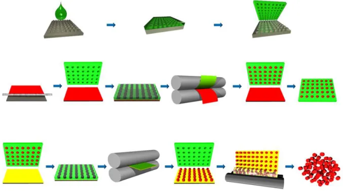
PRINT technology. As pictured below in Figure 1.1, fabrication of particles via PRINT begins
with a silicon wafer patterned with the feature size and shape of interest using traditional
photolithography techniques. Low-surface energy perfluoropolyether (PFPE) is then applied to
the silicon master template and chemically cross-linked to create a flexible elastomeric film with
nano- or micron-sized cavities, known as a PRINT mold. The low surface energy of the PFPE
allows for it to wet the entire surface of the mater template, resulting in faithful reproduction of
the features. Additionally, the chemical resistance of PFPE prevents deformation of the PRINT
mold when exposed to the organic solutions used in making monomer and polymer films, aiding
in the fidelity of the produced particles to the original master template.42 The PRINT mold is then filled using a thin film of the monomer or polymer solution of the desired composition for
the final particles. The versatility of PRINT technology allows for particles to be fabricated from
other FDA approved materials, proteins, and chemotherapeutics.40,41,43–45 Capillary action is harnessed to fill nano-sized cavities while mechanical force aids in filling larger features. The
non-wetting nature of PFPE prevents excess monomer or polymer from creating an
interconnecting flash layer or scum layer, resulting in individual particles. When using a
monomer solution, the monomer filled mold is photocured by brief irradiation by ultra-violet
light, resulting in cross-linked particles. Particles can be removed from the PRINT mold by
mating the filled mold with a sacrificial harvesting layer made of water soluble polymer capable
of forming hydrogen bonds with the newly fabricated PRINT particles. Common harvesting
layers are composed of poly(vinyl alcohol) (PVOH) and Plasdone™. Running the mold and harvesting layer through a heated laminator allows for the transfer of particles to the harvesting
layer, leaving an array of particles on the sacrificial harvesting layer and an empty mold. The
harvesting layer is then dissolved away using water or another appropriate solvent, yielding a
Figure 1.1 Particle Replication in Non-wetting Templates. A silicon wafer (gray) is patterned with nano- or micron-sized features through photolithography methods. PFPE (green) is dispersed across the silicon wafer and chemically cross-linked, yielding an elastomeric PRINT mold. A monomer or polymer thin film (red) is spread across a sheet of poly(ethylene terephthalate) (PET) and mated to the PRINT mold. Upon passing the mold and film through a laminator nip, the mold is filled with the particle material of choice. The filled mold is cured if necessary before being mated to a sacrificial harvesting layer (yellow) and passed again through a heated laminator. The mold is then removed from the harvesting sheet, revealing an array of individual particles. The sacrificial harvesting layer is then dissolved in a bead of water, revealing a nearly monodisperse solution of PRINT particles.
For vaccine applications, PRINT allows for imitation of natural pathogens in particle size,
shape, and surface display of vaccine subunits. Previous work has shown that PLGA-based
PRINT particles can be used to effectively adsorb influenza antigen, leading to an increased
immune response compared to dosing the trivalent inactivated virus vaccine.46 Investigation into this promising strategy is ongoing; however, changing the particle matrix from PLGA to a PEG
based hydrogel system presents new opportunities for vaccine subunit delivery. The functional
diversity of monomers that can be incorporated into the hydrogel particles affords the
opportunity to conjugate vaccine subunit cargos throughout the particle matrix as well as
conjugate cargos to the surface of the particles. The inert hydrogel matrix results in particles that
mirroring pathogen presentation of antigens and adjuvants, PRINT provides an opportunity to
explore the relationship between particle size, shape, and surface characteristics, antigen and
adjuvant trafficking and presentation, and the subsequent immune response. How these
characteristics work together to stimulate the immune system, toward the goal of formulating
1.5 References
(1) Plotkin, S. A. Nat. Med.2005, 11, S5–11.
(2) Riedel, S. Proc. (Bayl. Univ. Med. Cent).2005, 18, 21–25.
(3) Vaccines and Preventable Diseases http://www.cdc.gov/vaccines/vpd-vac/ (accessed Sep 10, 2014).
(4) Vaccines
http://www.niaid.nih.gov/topics/vaccines/understanding/pages/typesvaccines.aspx (accessed Sep 12, 2014).
(5) Zbinden, D.; Manuel, O. Immunotherapy2014, 6, 131–139.
(6) Ison, M. G.; Michaels, M. G. Am. J. Transplant2009, 9 Suppl 4, S166–72.
(7) Levast, B.; Awate, S.; Babiuk, L.; Mutwiri, G.; Gerdts, V.; van Drunen Littel-van den Hurk, S. Vaccines2014, 2, 297–322.
(8) Baylor, N. W.; Egan, W.; Richman, P. Vaccine2002, 20 Suppl 3, S18–23. (9) Gupta, R.; Rost, B.; Relyveld, E.; Siber, G. Vaccine Des.1995, 6, 229–248. (10) Marrack, P.; McKee, A. S.; Munks, M. W. Nat. Rev. Immunol.2009, 9, 287–293. (11) Kawai, T.; Akira, S. Cell Death Differ.2006, 13, 816–825.
(12) Didierlaurent, A. M.; Morel, S.; Lockman, L.; Giannini, S. L.; Bisteau, M.; Carlsen, H.; Kielland, A.; Vosters, O.; Vanderheyde, N.; Schiavetti, F.; Larocque, D.; Van Mechelen, M.; Garçon, N. J. Immunol.2009, 183, 6186–6197.
(13) Demento, S. L.; Siefert, A. L.; Bandyopadhyay, A.; Sharp, F. A; Fahmy, T. M. Trends Biotechnol.2011, 29, 294–306.
(14) de Temmerman, M.-L.; Rejman, J.; Demeester, J.; Irvine, D. J.; Gander, B.; De Smedt, S. C. Drug Discov. Today2011, 16, 569–582.
(15) Ahmed, S. S.; Plotkin, S. A; Black, S.; Coffman, R. L. Sci. Transl. Med.2011, 3, 93rv2. (16) Fife, K. H.; Meng, T.-C.; Ferris, D. G.; Liu, P. Antimicrob. Agents Chemother.2008, 52,
477–482.
(18) Dockrell, D. H.; Kinghorn, G. R. J. Antimicrob. Chemother.2001, 48, 751–755. (19) Belz, G.; Smith, C.; Bharadwaj, M.; Rice, A.; Jackson, D. Cytotherapy2004, 6, 88–98. (20) Storni, T.; Kündig, T. M.; Senti, G.; Johansen, P. Adv. Drug Deliv. Rev.2005, 57, 333–
355.
(21) Ferreira, S. A; Gama, F. M.; Vilanova, M. Nanomedicine2013, 9, 159–173. (22) Noad, R.; Roy, P. Trends Microbiol.2003, 11, 438–444.
(23) Grgacic, E. V. L.; Anderson, D. A. Methods2006, 40, 60–65.
(24) Zhao, L.; Seth, A.; Wibowo, N.; Zhao, C.-X.; Mitter, N.; Yu, C.; Middelberg, A. P. J.
Vaccine2014, 32, 327–337.
(25) Kang, S.-M.; Song, J.-M.; Quan, F.-S.; Compans, R. W. Virus Res.2009, 143, 140–146. (26) Tissot, A. C.; Renhofa, R.; Schmitz, N.; Cielens, I.; Meijerink, E.; Ose, V.; Jennings, G.
T.; Saudan, P.; Pumpens, P.; Bachmann, M. F. PLoS One2010, 5, e9809.
(27) Khanam, S.; Rajendra, P.; Khanna, N.; Swaminathan, S. BMC Biotechnol.2007, 7, 10. (28) Moon, J. J.; Suh, H.; Bershteyn, A.; Stephan, M. T.; Liu, H.; Huang, B.; Sohail, M.; Luo,
S.; Um, S. H.; Khant, H.; Goodwin, J. T.; Ramos, J.; Chiu, W.; Irvine, D. J. Nat. Mater.
2011, 10, 243–251.
(29) Moon, J. J.; Suh, H.; Li, A. V; Ockenhouse, C. F.; Yadava, A.; Irvine, D. J. Proc. Natl. Acad. Sci. U. S. A.2012, 109, 1080–1085.
(30) Socha, M.; Bartecki, P.; Passirani, C.; Sapin, A; Damgé, C.; Lecompte, T.; Barré, J.; El Ghazouani, F.; Maincent, P. J. Drug Target.2009, 17, 575–585.
(31) Passirani, C.; Barratt, G.; Devissaguet, J.-P.; Labarre, D. Life Sci.1998, 62, 775–785. (32) Hirosue, S.; Kourtis, I. C.; van der Vlies, A. J.; Hubbell, J. A; Swartz, M. A. Vaccine
2010, 28, 7897–7906.
(33) Ilyinskii, P. O.; Roy, C. J.; O’Neil, C. P.; Browning, E. A; Pittet, L. A; Altreuter, D. H.; Alexis, F.; Tonti, E.; Shi, J.; Basto, P. A; Iannacone, M.; Radovic-Moreno, A. F.; Langer, R. S.; Farokhzad, O. C.; von Andrian, U. H.; Johnston, L. P. M.; Kishimoto, T. K. Vaccine
2014, 32, 2882–2895.
(35) Akagi, T.; Baba, M.; Akashi, M. In Polymers in Nanomedicine; Kunugi, S.; Yamaoka, T., Eds.; Springer Berlin Heidelberg, 2012; pp. 31–64.
(36) Rehor, A.; Tirelli, N.; Hubbell, J. A. Macromolecules 2002, 35, 8688–8693.
(37) John, A. L. S.; Chan, C. Y.; Staats, H. F.; Leong, K. W.; Abraham, S. N. Nat. Mater.
2012, 11, 1–8.
(38) Duong, A. D.; Sharma, S.; Peine, K. J.; Gupta, G.; Satoskar, A. R.; Bachelder, E. M.; Wyslouzil, B. E.; Ainslie, K. M. Mol. Pharm.2013, 10, 1045–1055.
(39) Kaminskas, L. M.; Porter, C. J. H. Adv. Drug Deliv. Rev.2011, 63, 890–900.
(40) Perry, J. L.; Herlihy, K. P.; Napier, M. E.; Desimone, J. M. Acc. Chem. Res.2011, 44, 990–998.
(41) Rolland, J. P.; Maynor, B. W.; Euliss, L. E.; Exner, A. E.; Denison, G. M.; DeSimone, J. M. J. Am. Chem. Soc.2005, 127, 10096–10100.
(42) Rolland, J. P.; Hagberg, E. C.; Denison, G. M.; Carter, K. R.; De Simone, J. M. Angew. Chemie2004, 116, 5920–5923.
(43) Kelly, J. Y.; DeSimone, J. M. J. Am. Chem. Soc.2008, 130, 5438–5439. (44) Perry, J.; Reuter, K.; Kai, M.; Herlihy, K. Nano Lett.2012, 1–16.
(45) Enlow, E. M.; Luft, J. C.; Napier, M. E.; DeSimone, J. M. Nano Lett.2011, 11, 808–813. (46) Galloway, A. L.; Murphy, A.; DeSimone, J. M.; Di, J.; Herrmann, J. P.; Hunter, M. E.;
Kindig, J. P.; Malinoski, F. J.; Rumley, M. A; Stoltz, D. M.; Templeman, T. S.; Hubby, B.
Nanomedicine2013, 9, 523–531.
CHAPTER 2 MANIPULATING PHYSICOCHEMICAL PROPERTIES OF POLYMERIC HYDROGEL
PARTICLES TO ENHANCE LYMPHATIC TRAFFICKING AND IMMUNOGENICITY OF A MODEL
SUBUNIT VACCINE
2.1 Introduction
Draining lymph nodes (LNs) are the primary site of action for initiating adaptive
immunity, where cells that initiate adaptive immune responses (T and B cells) meet antigen or
antigen-loaded antigen presenting cells (dendritic cells, macrophages).1,2 To activate B cells and generate a robust humoral response, two signals are required: direct crosslinking of B cell
receptors by antigens, and co-stimulatory signals from CD4+ T cells (e.g. cytokines and
CD40/CD40L binding).3,4 Antigen presenting cells, especially dendritic cells (DCs) are critical in priming T cells to provide helper signals to B cells.4–9 Because of the myriad activities of the immune system that take place in the lymph nodes, recent literature has focused on delivering
vaccines directly to the draining LNs.4,6,7,9,10 By targeting the draining LNs vaccine uptake by antigen presenting cells (APCs), APC maturation, and cross presentation to T and B cells may all
occur in close proximity, thus increasing the potency of the resulting response.
Utilizing purified and synthetic pathogen subunits (peptides, polysaccharides, lipids,
DNA, etc.) for vaccination has become an increasingly attractive option due to significantly
improved safety profiles compared to whole pathogen-based vaccines. However, limited clinical
efficiently captured by immune cells, are susceptible to non-specific degradation and metabolism
in vivo and are subject to rapid clearance.10–13 Particle mediated delivery has shown great potential for subunit vaccine development and has gained increasing attention.6,10–12,14–23 Size, shape, and surface properties of particle vectors can be manipulated to target key APCs and
promote cell uptake of antigens via phagocytosis, or facilitate self-drainage and direct delivery of
vaccine to lymph node-resident immune cells.10–13,24 Surface display of antigens on particle carriers may allow multivalent interaction with B cells that mimics presentation by natural
pathogens, enabling more efficient cross-linking of cognate B cell receptors, thereby increasing
potency of these agents10,17,25 and achieving dose sparing effects.14,25
Many parameters of particulate vaccine carriers (charge, size, and surface properties)
may all contribute to the quality of the resulting immune response. Previous work on lymphatic
trafficking of nanoparticles (NPs) has focused on delivery of cancer therapeutics or diagnostic
molecules to aid in LN imaging;21,26–31 only recently have the effects of lymphatic trafficking on the efficacy of novel, particulate vaccines been examined.16,19,20,23,32,33 Cationic NPs have been found to have higher uptake in vitro and facilitate a more favorable endosomal pH environment, mitigating premature degradation of acid-sensitive antigens.34 Conversely, in vivo results favor anionic NPs for higher rates of NP trafficking and subsequent uptake by APCs. This critical
difference between in vitro and in vivo results may be due to the composition of the extracellular matrix (ECM) in the interstitial space of the lymphatic system: collagen fibers and highly anionic
glycosaminoglycans.35 The high density of negative charge may lead to aggregation of cationic NPs at the injection site while the electrostatic repulsion allows anionic NPs to move through the
Size plays perhaps the largest role in the trafficking of NPs from the site of injection
through the lymphatic system. Lymphatic trafficking of many sizes and compositions of
spherical NPs have been examined including PLGA NPs (size 50 – 200 nm),38 liposomes (single and multi-lamellar),37 dendrimers (size 5 – 15 nm),29 star polymers (size 10 – 25 nm),30
polystyrene beads (size 20 – 2000 nm),19 and other polymeric NPs (size 25 – 3700 nm)32,39 (all sizes reported as diameter). The ideal NP size to harness interstitial flow and passively target the
LNs appears to be between 20 nm, under which NPs are likely to enter directly into systemic
circulation,29,38 and 100 nm, above which size NPs become stuck at the site of injection and rely on cell-mediated trafficking to the LNs.10 Within this size range, the optimal NP size appears to be widely system dependent.19,21,36,39
PEGylation (surface conjugation or coating with chains of poly(ethylene glycol)) is
commonly used to conjugate various cargos to nanoparticles, polymers, and biologics. For the
purpose of developing a vaccine carrier, the level of PEGylation used to attach the vaccine cargo
to the nanoparticle carrier should take in to consideration the effects PEG length and PEGylation
density may have on lymphatic drainage and uptake by immune cells. PEGylation has been
studied extensively in the search for long-circulating NPs, capable of avoiding detection and
clearance by the mononuclear phagocyte system (MPS). Studies have found that while a dense
layer of long PEG chains is necessary to maximize circulation time, the desired effects of low
protein binding and decreased phagocytic uptake are seen at even low PEG densities and chain
lengths.40,41 PEGylation has also been studied for applications in mucosal penetration for
intranasal delivery of therapeutics:42 PEG coating is believed to interact with the mucus layer in a way that prevents NP aggregation, facilitating NP transport. This finding may be applied to the
findings agree that PEGylation increases lymphatic drainage from the injection site throughout
various densities and molecular weights of PEG,20–23,27,31,33,43 but findings are conflicting
regarding how this affects uptake of PEGylated NPs in the LNs. A study with 300 nm liposomes
indicated that PEGylation can not only increase drainage and uptake, but facilitate deeper NP
penetration into all regions of the LNs.20 Others have found that PEGylation actually decreases retention of NPs in the LNs and uptake by APCs.21,33 Further examination of the effects of PEGylation on vaccine delivery would be greatly beneficial in the design of future vaccine
delivery vehicles.
In addition to PEGylation, other surface characteristics can have dramatic effects on how
the body interacts with NP vaccine carriers. The complement system is the body’s first line of
defense against invading pathogens, linking innate and adaptive immunity and playing an
important role in peripheral lymph nodes to enhance B and T cell responses.52 Hubbell and co-workers reported that nanoparticles can be engineered to activate the complement system and
improve immune responses to vaccines.53,54 By utilizing a hydroxyl-terminated monomer versus a methoxy-terminated monomer as the primary component of NPs, particles can be designed to
activate the complement system and facilitate a stronger immune response against antigen.
Herein we present a versatile vaccine delivery platform based on hydrogel particles made
of hydroxy-poly(ethylene glycol) (PEG) via PRINT technology (Particle Replication in
Non-Wetting Templates),44–46 a unique mold-based particle fabrication process. The highly tunable nature of PRINT allows for a great degree of control over NP size, aspect ratio, charge, and
surface functionality, facilitating a systematic study of these effects on NP trafficking through
vaccine carrier has the capacity to deliver subunit vaccine components to the draining LNs in a
sustained manner and elicit robust antigen-specific humoral immune response.
2.2 Results and Discussion
Nanoparticle delivery of protein subunit vaccines to the lymph nodes allows antigens to
interact directly with the immune system. Additionally, surface display of protein antigens
similar to antigen presentation by natural pathogens may boost therapeutic efficacy of these
subunits to the levels associated with whole pathogen vaccines without the related safety
concerns.4,8,47 This study aimed to evaluate PRINT nanoparticles of various size, aspect ratio, and surface characteristics for their ability to traffic through the lymphatic system and explore
their use for antigen delivery in vaccine applications.
2.2.1 Lymphatic Trafficking of Bare Particles
A delivery vector that traffics quickly and efficiently to the draining lymph nodes would
be beneficial for delivering antigens and/or adjuvants to B cells and other antigen presenting
cells (APCs) resident in the lymph nodes. A panel of rod/cylindrical PRINT NPs of different size,
aspect ratio, and surface charge (Table 2.1) were injected subcutaneously in mice and screened
for their ability to drain to the popliteal lymph nodes (PLNs). Rod shaped NPs may have added
benefits over traditional spherical NPs in terms of cellular uptake as well as an increased surface
area for cargo loading.45 An 80×180 nm rod has over 3 times the volume of a spherical NP with the same diameter and 2.75 times the surface area. Consistent with literature, we found that
sub-100 nm diameter,16,19,36,48–50 anionic28–30,37,50 NPs were able to traffic quickly to the PLN, with an increase in NP concentration over 48 hours (Figure 2.1). In contrast, all other NPs, regardless of
size and charge, generally remained at the site of injection with less than 0.2% of the injected
of anionic 80×180 nm NPs populated the B cell-follicular area, which may provide opportunity
for antigen presentation to B cells and development of a humoral immune response (Figure 2.2).
Larger and cationic NPs showed little accumulation in the LNs.
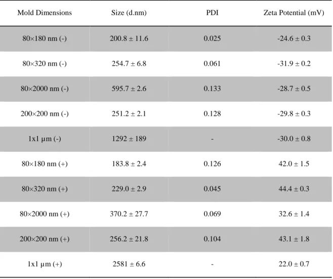
Table 2.1 Characterization of bare NPs
Mold Dimensions Size (d.nm) PDI Zeta Potential (mV)
80×180 nm (-) 200.8 ± 11.6 0.025 -24.6 ± 0.3
80×320 nm (-) 254.7 ± 6.8 0.061 -31.9 ± 0.2
80×2000 nm (-) 595.7 ± 2.6 0.133 -28.7 ± 0.5
200×200 nm (-) 251.2 ± 2.1 0.128 -29.8 ± 0.3
1x1 µm (-) 1292 ± 189 - -30.0 ± 0.8
80×180 nm (+) 183.8 ± 2.4 0.126 42.0 ± 1.5
80×320 nm (+) 229.0 ± 2.9 0.045 44.4 ± 0.3
80×2000 nm (+) 370.2 ± 27.7 0.069 32.6 ± 1.4
200×200 nm (+) 256.2 ± 21.8 0.104 43.1 ± 1.8
Figure 2.1 Lymphatic drainage and cell uptake of bare hydrogel particles. Mice were injected with 50 μg NPs in hind footpads. Draining PLNs were resected at indicated time points and examined for particle fluorescence by IVIS imaging. Error bars stand for SEM, N ≥ 4. **, p < 0.01 by one-way ANOVA compared to all other groups at 48 hours time point.
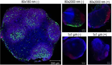
Figure 2.2 Localization of NPs in the PLNs was confirmed by confocal microscopy at 48 hours (80×180 nm NPs) or 72 hours (80×2000 nm and 1×1 μm NPs) post subcutaneous injections of different size and charge NPs. Blue: nuclei (DAPI); red: B cell (B220+); green: particles.
Flow cytometry was performed to determine which cell populations within the PLNs took
up NPs. For all examined APC types (dendritic cells (DCs), macrophages, B cells, and
cell population that took up NPs (Figure 2.3). For all NP types, anionic NPs were present in a
higher percentage of cells than their cationic counterpart. Similar to total lymphatic drainage,
size dependence was observed with 80×180s being most efficiently internalized by all cell types
examined. Strikingly, although less than 2% of total injected 80×180 nm NPs trafficked to the
PLNs at 48 hours, an average of 20% of DCs in the LNs took up anionic 80×180 NPs,
confirming that particle of this size, charge, and aspect ratio is able to target DCs.
Anionic 80×180 nm NPs, the best self-draining particle type, was chosen for further
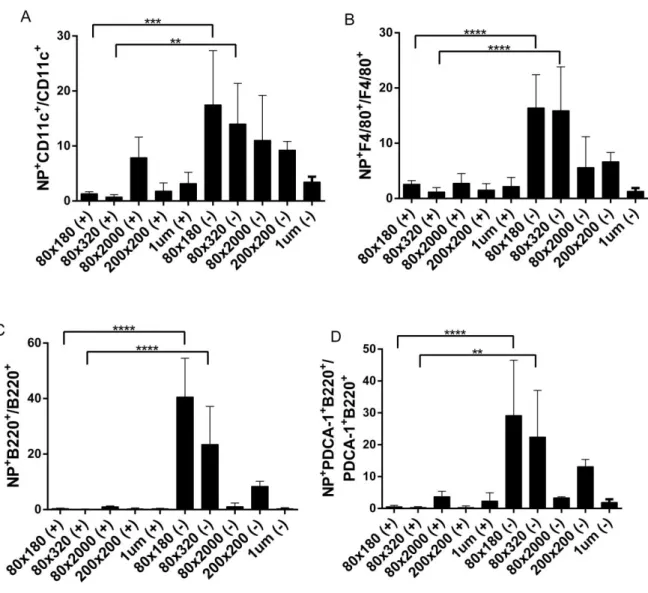
Figure 2.3 Flow cytometry of cells from lymph nodes resected 48 hours after injection. Data shown as NPpositive cells/total cell population×100 to demonstrate percent of each cell type that had taken up NPs. A) Dendritic cells, DC11c+, B) macrophages, F4/80+, C) B cells, B220+, D) plasmacytoid DCs, PDCA-1+B220+. *, p < 0.05; **, p < 0.01; ***, p < 0.001; ****, p < 0.0001 by unpaired t test. N ≥ 3.
2.2.2 Conjugation of Model Antigen and Surface Characteristics
Surface display of antigens greatly increases the chances of direct antigen presentation to
B cells, facilitating a more robust antibody response. To test immunogenicity of antigen
delivered by the hydrogel NP carrier, a model protein antigen ovalbumin (OVA) was covalently
glycol) (PEG)-based linkers, a common bioconjugation technique used to control the distance
between ligands and NPs. PEGylation is frequently used to increase circulation half-life of small
molecule drugs, biologics, and nanoparticles by decreasing the binding of serum proteins and
opsonins, thus decreasing recognition by the mononuclear phagocyte system (MPS).51 For vaccine carriers, PEGylation may enhance drainage of NPs from the site of injection to the
lymph nodes by blocking interactions with the extracellular matrix (ECM); however, a high level
PEGylation, especially with high molecular weight PEG, could be undesirable as it may prevent
NP uptake by phagocytic APCs.6 In order to examine the effect of PEG linker length on lymphatic drainage and cell uptake, OVA was conjugated to the surface of NPs via large,
medium or small linkers: 5000 Da molecular weight PEG (PEG(5k)), 500 Da molecular weight
PEG (PEG(500)), or a direct amide bond from protein to NP (PEG(0)) respectively. After
conjugation of antigen, all NPs remained very well dispersed with polydispersity index (PDI)
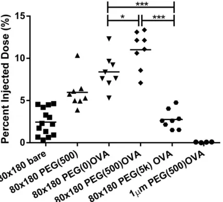
below 0.15 (Table 2.2). Forty-eight hours post-injection, significantly more PEG(500)OVA NPs
reached PLNs as compared to the PEG(5k)OVA and PEG(0)OVA NPs (Figure 2.4). Surface
modification with the long PEG(5k) linker was apparently not favorable for lymphatic drainage.
Further comparison with bare NPs and no-OVA PEG(500) NPs indicated that the increase in
trafficking for the PEG(500)OVA NPs came from the synergy between PEG(500) and OVA,
rather than either component alone. PEGylation with a dense layer of short PEG(500) may
stabilize the NPs under physiological conditions and decrease interactions with the ECM, while
longer PEG(5k) may have a greater chance of becoming entangled with the biopolymers in the
ECM.6,35 Additionally, compared to 80×180 nm PEG(500)OVA NPs, the 1 μm PEG(500)OVA NPs showed poor lymphatic trafficking on par with the bare 1 µm NPs (Figure 2.1); conjugation
that the size of particles is an essential determinant for lymphatic drainage patterns of particle
vectors, which can be further modulated by different lengths of PEG linkers.
Table 2.2 Characterization of OVA-conjugated NPs
Particle Type Size (d.nm) PDI Zeta Potential (mV)
OVA Loading (µg/mg NP)
80×180 nm bare 200.8 ± 11.6 0.025 -24.6 ± 0.3 –
80×180 nm PEG(0)OVA
246.8 ± 1.2 0.139 -33.6 ± 1.2 30-100
80×180 nm PEG(500)OVA
192.0 ± 2.1 0.044 -39.3 ± 1.6 30 – 90
80×180 nm PEG(5k)OVA
191.3 ± 0.6 0.076 -27.5 ± 0.3 10 – 100
1×1 μm PEG(500)OVA
1459 ± 189.4 – -7.0 ± 0.5 10 – 100
1×1 μm PEG(5k)OVA
Figure 2.4 Total drainage OVA-loaded hydrogel of NPs in lymph nodes. 50 μg fluorescently labeled 80×180 nm hydrogels were subcutaneously injected into footpads of balb/c mice, and draining popliteal LNs were collected at 48 h, and imaged with IVIS Lumina. *, p < 0.05; ***, p < 0.001 by one-way ANOVA. N = 8-14.
The best draining particles, 80×180 nm PEG(500)OVA NPs, also showed rapid
trafficking and were present in the PLN in as short as five minutes after injection, with the
concentration of NPs in the PLN continuously increasing over forty-eight hours (Figure 2.5). At
forty-eight hours, NP trafficking reached 10 % of total injected dose, 5× higher than bare anionic
Figure 2.5 80×180 nm PEG(500)OVA NPs drained rapidly to the lymph nodes accumulated over 48 h. 50 μg fluorescently labeled 80×180 nm hydrogels were subcutaneously injected into footpads of balb/c mice. Draining popliteal LNs were collected at the indicated time points and imaged with IVIS Lumina. Error bars stand for SEM. N = 4-8.
In order to efficiently elicit an immune response, an effective NP delivery vector should
ideally be able to ensure the antigen arrives at the site of action without being degraded or
released prematurely. To compare the drainage of NP bound OVA to that of free OVA, we
tagged the NPs and OVA with two different fluorophores. Free OVA (red) drained rapidly and
was observed in the PLN two hours after injection, but was no longer detectable at twenty-four
hours (Figure 2.6). This is consistent with literature indicating that soluble proteins are subject to
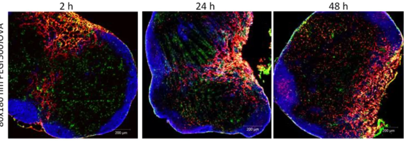
quick lymphatic clearance.13 For 80×180 nm PEG(500)OVA NPs, particles and OVA (shown in yellow as overlapping of green NPs and red OVA) also drained quickly, as seen previously with
the trafficking experiments, and were co-localized in the subcapsular regions of the PLN two
hours after injection. NP-OVA (yellow) stayed in the PLN much longer than soluble OVA (red)
(yellow) versus NPs alone (green) decreased over time, presumably due to OVA cleavage and
degradation by proteases. More importantly, cleaved OVA (red) selectively accumulated in the B
cell follicles and the presence of OVA in this region persisted for up to 15 days. A similar
phenomenon was also observed for 80×180 nm PEG(0)OVA (Figure 2.6). This observation
indicates that in general 80×180 nm hydrogel NPs are able to efficiently deliver antigen to the
LNs and support sustained presentation of antigen to B cells. The longer residence time of
NP-conjugated OVA in the PLN may help increase the interaction between antigen and B cells and
LN-resident APCs compared to free OVA, resulting in an enhanced antibody response.
In addition to the delivery of antigens to B cells and crosslinking of cognate B cell
receptors, eliciting a potent humoral response and B cell memory also requires help from CD4+ T cells,24 therefore good vaccine carriers need to be able to deliver antigens to APCs and prime T cells efficiently. Analysis of draining LNs by flow cytometry showed that 48 hours post
subcutaneous dosing, 80×180 nm hydrogels with OVA linked through all three linker lengths
reached 10-20% of the DCs, and 10-35% of the macrophages in the PLNs while 1 μm
PEG(500)OVA NPs were found in less than 2% of DCs or macrophages (Figure 2.7), indicating
that the 80×180 nm NPs may efficiently deliver antigens to key APCs. Although the total
drainage to LNs of these three NPs with various linker lengths (Figure 2.4) did not directly
correlate with the uptake of NPs by cells in the PLNs, both results suggest that a long PEG linker
is less favorable for antigen delivery to immune cells. The co-localization of the 80×180 nm
PEG(500)OVA NPs with DCs was also observed by confocal microscopy analysis of sectioned
draining LNs (Figure 2.8), indicating that these NPs are able to access all regions of the PLNs
where B cell and T cell activation can occur, facilitating activation of both humoral and cellular
Figure 2.7 80×180 nm NPs are efficiently taken up by key antigen presenting cells (DCs and macrophages) in LNs 48 h post subcutaneous injection, as analyzed by flow cytometry. *, p < 0.05; **, p < 0.01; ***, p < 0.001; , p < 0.001 compared to all other groups, analyzed by unpaired t-test. N ≥ 4.
Figure 2.8 Uptake of 80×180 nm PEG(500)OVA particles by dendritic cells were confirmed by confocal
microscopy at indicated time points post subcutaneous injections of particles. Blue: B cell (B220+); green: dendritic cells (CD11c+); red: particles.
The complement system acts not only as the first line of defense for the body, but also
links innate and adaptive immunity and plays an important role in peripheral lymph nodes to
pathways: classical, lectin, and alternative; however, all three pathways share a common step –
activating the central component C3. Hubbell and co-workers reported that nanoparticles can be
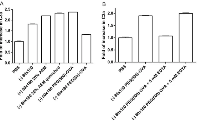
engineered to activate the complement system and improve immune responses to vaccines.53,54 Here we show that PRINT hydrogel NPs activate the complement system, as indicated by
increase in the conversion of C3 to C3a (Figure 2.9a). Both bare and OVA-conjugated NPs
promoted the conversion of C3 to C3a, suggesting that activation may result from the NP
composition rather than post-fabrication modifications to the NPs. However, surface
modification with long chain PEG may reduce the capacity of the NPs to activate the
complement system, possibly due to a higher degree of shielding of the NP surface groups that
would otherwise interact with components in the complement system. Furthermore, EDTA
(ethylenediaminetetraacetic acid) but not EGTA (ethylene glycol tetraacetic acid) blocked the
conversion of C3 to C3a (Figure 2.9b), indicative of complement activation via the alternative
pathway rather than the classical pathway. These results demonstrate that in addition to the
efficient LN targeted delivery of antigen, PRINT hydrogel NP vaccine vectors may potentially
Figure 2.9 Hydrogel NPs activate complement system. Serum from C57BL/6 mice was incubated with a) 0.5 mg/ml NPs and b) 1.2 mg/ml NPs for 50 min at 37ºC. Conversion from C3 to C3a was assayed by ELISA. The data represent one of two similar individual experiments; bars are average of two replicate wells in each experiment.
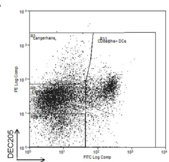
Lymph nodes are home to a large population of DCs, especially CD8α+
DCs which have
been shown to be the most efficient DCs in antigen cross-presentation.6,9,55 In addition, there are other major DC subsets including migratory Langerhans cells and dermal DCs, normally resident
in distal areas of the body, as well as LN resident double negative DCs as defined by surface
markers CD8 and DEC20522 (Figure 2.10a). Subsequent analysis of draining LNs showed that initially 80×180 nm PEG(500)OVA NPs distributed in all four different subsets of DCs
somewhat evenly with an increase in the percentage of LN resident CD8α+ DCs over a 30-minute period (Figure 2.10b). This suggests self-draining NPs may be efficiently taken up by LN
resident CD8α+
NP+ migratory dermal DCs increased, likely due to continuous uptake of NPs by dermal DCs at the injection site followed by cell mediated transport to PLNs. This is further verified by the
presence of NPs in the PLNs at as early as 5 minutes post injections (Figure 2.5): cell-mediated
delivery of NPs has been shown to occur over several hours to days.3,49 These results
demonstrate that the 80×180 nm hydrogel NPs can be taken up by various DC subsets, with a
high percentage of CD8α+
DCs and dermal DCs internalizing NPs, potentially preparing them
Figure 2.10 A) Analysis of LN DC subsets by flow cytometry. Single cell suspension of LNs was stained with anti-mouse CD11c, CD8α and DEC205. Cells gated on CD11c+ were shown. The CD11c+ DC populations are defined as: CD8α+ DC (CD11c+CD8α+), DN DC (CD11c+DEC205-CD8α-), Langerhans cell (CD11c+DC205hiCD8α-), and dermal DC (CD11c+DEC205intCD8α-). B) Uptake of NPs by various DC subsets in draining LNs, with an increase in the percentage of migratory DCs over time. 50 μg fluorescently labeled 80×180 nm hydrogels were
subcutaneously injected into footpads of C57BL/6 mice. Draining popliteal LNs were collected at the indicated time points analyzed via flow cytometry.
B
2.2.3 Influence of Particle Drainage on Immune Response
The increased lymphatic drainage and the ability to access to key APCs present an
opportunity for the 80×180 nm PEG(500)OVA particles to enhance immunogenicity of subunit
vaccines.
To further explore the T cell priming ability of the 80×180 nm PEG(500)OVA NPs, an in vivo T cell proliferation assay was performed using CD4+ OT-II cells that recognize OVA-derived epitope OVA323-339. As displayed in Figure 2.11, immunizations with 80×180 nm
PEG(500)OVA NPs loaded with just 1 µg of OVA effectively stimulated the proliferation of
CFSE (carboxyfluorescein diacrylate succinimidyl ester)-labeled CD4+ OT-II T cells, causing a dilution of the fluorescent dye, while no proliferation was seen in mice that were untreated or
dosed with 1 µg soluble OVA. Together with the flow cytometry data (Figure 1.7. and 1.10b),
we can deduce that the 80×180 nm PEG(500)OVA NPs are effectively taken up by APCs where
Figure 2.11 In vivo CD4+ OT-II T cell proliferation. Hydrogel-mediated delivery of antigen is more efficient in stimulating CD4+ T Cell proliferation than soluble antigen. N = 3-4.
To test immunogenicity of antigen delivered by this NP vector, mice were vaccinated
against OVA delivered either in soluble form or conjugated to NPs as described previously. The
display of antigen on the NP surface may increase the chance of direct present of antigen to B
cells, although this strategy may be less protective to the antigen than encapsulation techniques.
The immune response to free versus particulate OVA was evaluated following a prime-boost
regimen. Seven days after the boost dose, mice immunized with 80×180 nm PEG(500)OVA NPs
showed a tenfold increase in OVA-specific IgG production compared to free OVA and free OVA
+ bare NPs (p < 0.05, Figure 2.12a), whereas the NPs that were co-injected with free OVA did
not augment the immune response. This data suggests that covalent conjugation to the NP vector
Untreated
Soluble OVA
is necessary for enhanced immunity. NP-OVA was compared to free OVA plus the adjuvant
alum, the standard of care for adjuvanted vaccines.57 Free OVA + alum elicited higher antibody titers than NP-OVA; however, NP-OVA + alum gave a significant increase in antibody response
compared to free OVA + alum (Figure 2.12b), indicating this NP-based vector for antigen
delivery may be able to further improve the antibody response against protein antigen in
adjuvanted vaccines. Previous work has shown that the PRINT hydrogel NPs induce no
inflammatory response on their own;58 therefore the major advantage of the NP vector most likely comes from its efficient delivery of antigen to immune system rather than direct
immunomodulating ability.
Figure 2.12 Hydrogel vaccine elicits higher antibody titers than soluble antigen with or without Alum adjuvant. Mice were immunized on day 0 and again on day 21 with 5 μg OVA, soluble or conjugated to PRINT hydrogel NPs. OVA-specific IgG in plasma were examined by ELISA. *, p < 0.05; **, p < 0.01 by unpaired t-test. Error bars stand for SEM. N = 5.
To examine the correlation between trafficking and immune response, we investigated
anti-OVA antibody production after OVA delivery via 80×180 nm NPs with various PEG linker
NP trafficking (Figure 2.4), PEG linker length appeared to have no statistical effect on
antigen-specific IgG production (Figure 2.13a). All linker lengths showed a tenfold increase in
OVA-specific IgG production compared to free OVA, but the IgG levels were equivalent among the
NP groups. However, the size of the NPs used to deliver OVA appeared to have a more dramatic
effect on the total IgG. The antibody response against the 80×180 nm PEG(500)OVA NPs was
over 1000 times higher than the response to the 1 μm PEG(500)OVA NPs (p < 0.05, Figure
2.13b). Remarkably, IgG response to 1 μm PEG(500)OVA NPs was even lower than soluble
OVA, strongly suggesting that drainage of vaccine carrier and antigen interaction with
LN-resident B cells are crucial to eliciting a humoral response. It is likely that there is a threshold
amount of antigen needed in the lymph nodes for initiating a humoral immune response. This
level may be sufficiently reached by the 80×180 nm NPs, including the relatively low
self-draining 80×180 nm PEG(5k)OVA NPs, while the 1 μm NPs do not appear to deliver enough
Figure 2.13 Size rather than PEG linker length dramatically influences IgG response. A, Length of PEG linker for OVA conjugation does not affect IgG response. b, Large 1 µm NPs elicit lower IgG production than soluble administration or smaller 80×180 nm NPs. Mice were immunized as in Figure 2.12 and plasma IgG was evaluated by ELISA. *, p < 0.05 by unpaired t-test. Data represent two or three individual experiments of N = 4. Error bars stand for SEM.
2.3 Conclusions
In conclusion, we have designed and optimized a versatile vaccine delivery platform
based on PRINT NPs. We demonstrate that the size, aspect ratio, charge, and surface
characteristics of NPs are all important in improving the lymphatic trafficking of NPs and their
subsequent uptake by key APCs. Anionic sub-100 nm hydrogel NPs loaded with a model antigen
showed high levels of self-drainage and were able to efficiently deliver antigen to B cells and
major APCs, inducing antigen-specific humoral and cellular responses superior to free antigen
alone. The simplicity of the chemistries used in antigen conjugation confers versatility to this