i
MODELING THE CONSEQUENCES OF EPIDERMAL GROWTH FACTOR RECEPTOR INHIBITION ON CARDIAC DEVELOPMENT, FUNCTION, AND
HOMEOSTASIS
Cordelia Johnson Barrick
A dissertation submitted to the faculty of the University of North Carolina at Chapel Hill in partial fulfillment of the requirements for the degree of Doctor of Philosophy in the
Curriculum of Toxicology
Chapel Hill 2007
Approved by: David Threadgill
Curtis Harper William Coleman
ii
ABSTRACT
Cordelia Johnson Barrick: MODELING THE CONSEQUENCES OF EPIDERMAL GROWTH FACTOR RECEPTOR INHIBITION ON CARDIAC DEVELOPMENT,
FUNCTION, AND HOMEOSTASIS
(Under the direction of David Threadgill)
The epidermal growth factor receptor (EGFR/ERBB1) is the prototypical and first discovered
member of the ERBB family of receptor tyrosine kinases. As transmembrane receptors, their
primary function is to translate extracellular signals into cellular response. Signaling is
initiated through binding by members of the EGF ligand family, which induces receptor
homodimerization or heterodimerization with other ERBB receptors (ERBB2, ERBB3 or
ERBB4). Activation of downstream cytoplasmic signaling pathways occurs, leading to
alterations in biological responses such as cellular proliferation, survival, motility, and
adhesion. As EGFR is expressed in most developing and adult tissues, misregulation or
dysfunction of EGFR activity severely impacts embryonic viability, tissue maintenance and
multiple disease processes. Since EGFR was first proposed as a cancer drug target over
twenty years ago, substantial research has defined a central role for aberrant ERBB signaling
in cancer and led to the design of targeted therapies that effectively inhibit receptor activity.
However, significant cardiotoxicity was observed in clinical trials targeting the closely
related ERBB2 receptor, necessitating further studies on the role of ERBB signaling in
cardiac development and function. Genetic ablation of any of the ERBB receptors, select
ligands, or ligand-processing enzymes results in severe congenital cardiac defects, often
iii
ligand neuregulin-1 (NRG1) supports cardiomyocyte survival, while GPCR mediated
transactivation of EGFR likely plays a significant role in cardiac hypertrophy and
hypertension. These discoveries have fostered interest in novel therapies targeting the EGFR
signaling pathway for the treatment of several common cardiovascular diseases. However,
the effects of chronic EGFR inhibition on cardiovascular homeostasis have not been
evaluated. Through the use of mouse models, we demonstrated that genetic or
pharmaceutical repression of EGFR signaling significantly altered cardiac function and
homeostasis. Since the cardiac phenotype of mice harboring a hypomorphic mutation in Egfr
was strongly dependent on genetic background, we broadly localized quantitative trait loci
(QTL) which modulate the cardiac phenotype. These studies should be useful in predicting
degenerative cardiac changes associated with EGFR inhibition which may be overlooked in
short term clinical trials, and may advance understanding of the role of EGFR in cardiac
iv
v
ACKNOWLEDGEMENTS
Graduate school requires a special combination of good luck, tenacity and personal
motivation.
I was fortunate to join a laboratory headed by David Threadgill, who gives his students an
extraordinary amount of freedom. As a result, there is always an interesting and eclectic mix
of ongoing research and graduate students in the Threadgill laboratory. Choosing one’s
research project is a bit like picking a piece of chocolate out of a Whitman’s sampler box,
except that there is less certainty about what is really in the center of the chocolate. Is that
brown piece that appears so appetizing really a truffle? Or, when you pick it up, does it smell
bad? When making this choice, I was also lucky, as I inherited a well-tended project from
Reade Roberts, an exceptional scientist and friend. I was also privileged to meet and work
closely with Susan Smyth while she was at UNC-Chapel Hill. Susan has been a source of
encouragement, a fantastic mentor, and has contributed greatly to my overall development as
a scientist. As far as tenacity is concerned, my innate stubbornness has proved to be an asset
in completion of this research project. The unexpected loss of my father to cardiovascular
disease has motivated me in my research efforts, far beyond completion of the doctoral
degree.
I am especially grateful to my husband, Brian Barrick, my family, and friends for their love
vi
TABLE OF CONTENTS
LIST OF TABLES………..……….……..…...ix
LIST OF FIGURES………....xi
LIST OF ABBREVIATIONS………..xiv
Chapter Page I. THE ROLES OF EGFR/ERBB1 SIGNALING IN CARDIAC DEVELOPMENT AND DISEASE………...…..1
Introduction………....2
Outline of heart development………...5
ERBB signaling in cardiac development……….…..8
EGFR signaling in cardiac development………..……….9
ERBB signaling in cardiac disease………...…………..….11
EGFR signaling in cardiac disease………...……...13
Conclusions………...15
vii
II. CARDIAC RESPONSE TO PRESSURE OVERLOAD IN 129S1/SVIMJ AND
C57BL/6J MICE: TEMPORAL AND BACKGROUND DEPENDENT DEVELOPMENT OF CONCENTRIC LEFT VENTRICULAR
HYPERTROPHY………...…29
Abstract………...………29
Introduction………...………..30
Materials and Methods………...……….33
Results………...………... 37
Discussion………...………....43
References………...………....60
III. EPIDERMAL GROWTH FACTOR RECEPTOR IS REQUIRED TO PREVENT LEFT VENTRICULAR HYPERTROPHY, CARDIAC FAILURE AND CALCIFIC VALVULAR AORTIC STENOSIS IN C57BL/6J BUT NOT 129S1/SVIMJ MICE………...65
Abstract………...………....65
Introduction………...………...66
Materials and methods……...………...…..67
Results………...………..72
Discussion………...………79
References………...………97
IV. CHRONIC REPRESSION OF EGFR ACTIVITY LEADS TO CARDIAC DYSFUNCTION IN C57BL/6J MICE………...102
Abstract………...………..102
ntroduction………...………...……..102
viii
Results………...………....108
Discussion………...………..113
References……….125
V. GENETIC MODIFIER LOCI AFFECTING LEFT VENTRICULAR HYPERTROPHY IN THE EGFR WA2 MOUSE MODEL OF AORTIC STENOSIS...131
Abstract……….131
Introduction………...132
Materials and methods………...134
Results………...136
Discussion………...143
References……….169
VI. CONCLUSIONS AND FUTURE DIRECTIONS………173
ix
LIST OF TABLES
Table
1-1 Phenotypes resulting from genetic ablation of ERBB receptors,
ERBB ligands or ligand processing enzymes ……….………18
2-1 Echocardiographic analysis in baseline, sham, and banded mice………50
2-2 Percent change in echocardiographic parameters compared
to baseline five weeks post surgery………...………..51
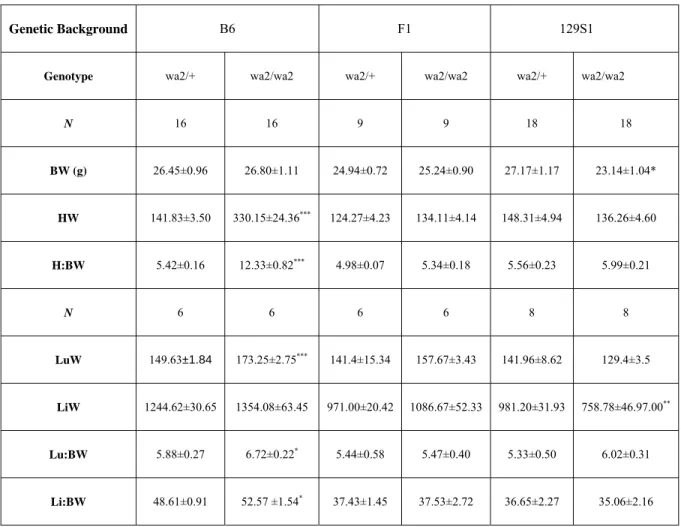
2-3 Comparison of organ weights between SHAM and TAC treated mice ………..52
2-4 Blood pressure measurements in conscious ten week-old male mice ………53
2-5 Percent change over baseline in echocardiographic parameters
five weeks post-surgery in conscious mice………..…54
3-1 Organ weights from three-to five month old Egfr wa2littermates………86 3-2 Measurements of cardiac function from
three-to five month old Egfr wa2littermates……….90 3-3 Blood pressure measurements from
three-to five month old Egfr wa2littermates……….91 4-1 Dietary exposure to AG1478 significantly reduces
polyp count in ApcMin/+ mice………..………118
4- 2 Organ and body weights of EGFR inhibitor exposed mice
compared to controls…….……….121
4-3 Echocardiographic parameters measured at baseline and after approximately 90 days on respective diets………...122
5-1 Informative markers used for SNP genotyping……….146
x
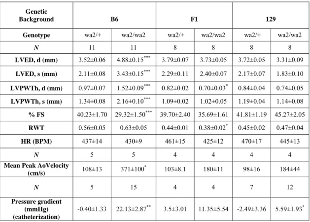
5-3 Echocardiographic parameters in three-to five month old
Egfr wa2littermates……….157 5-4 Representative echocardiographic parameters and organ weights from F2 -Egfr wa2/wa2
mice………164
xi
LIST OF FIGURES
Figure
1-1 ERBB ligand binding specificity is depicted
for the four ERBB receptors ……….………...17
1-2 Developmental stages of heart development………..……...20
1-3 Developmental stages of cardiac valve development………...…….21
2-1 B6 and 129S1 male mice have innate differences in cardiac morphology……….…...49
2-2 Comparison of mean cardiomyocyte area
across treatment group and genetic background………...55
2-3 Comparison of severity and location of cardiac fibrosis
by treatment and genetic background………....………...56
2-4 Inflammatory infiltrate is associated with
fibrosis in B6 TAC hearts………..………....57
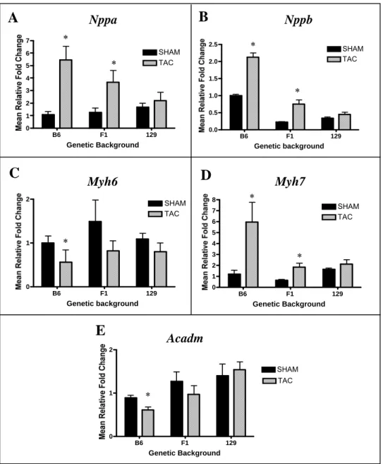
2-5 Comparison of relative expression of cardiac hypertrophy
markers in LV by treatment and genetic background………….…………...………...58
2-6 129S1 genetic modifiers delay transition to
decompensated heart failure ……….………….……….………..59
3-1 Survival curve, gross and histological comparison of hearts in B6 Egfr wa2littermates and comparison of normalized
heart weights in Egfr wa2 littermates………...………..………….……….85 3-2 Western blot and densitometry analysis of total EGFR and
phospho-ERK1/2 from B6 and 129S1 Egfr wa2
littermates………...……….…….….…87
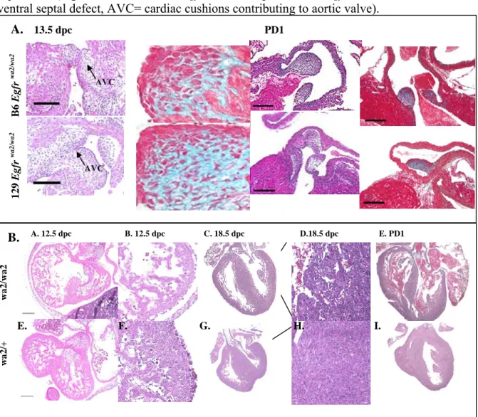
3-3 Comparison of congenital cardiac defects
in B6 and 129 Egfr wa2 embryos……….……...88
3-4 Comparison of cardiomyocyte size and cardiac fibrosis
in B6 Egfr wa2 littermates……….…………...89 3-5 Comparison of aortic cusp thickness and correlation of aortic
xii
3-6 Representative Doppler tracings from B6 Egfr wa2 littermates
and correlation between pressure gradients and mean cusp thickness
in Egfr wa2littermates………...……….93 3-7 Histological comparison of markers for cellular proliferation
in aortic cusps from B6 and 129S1 Egfr wa2 littermates………..……….94 3-8 Histological comparison of markers for altered extracellular matrix composition,
calcification and inflammation in aortic cusps
from B6 and 129S1 Egfr wa2 littermates……….…………..95
3-9 Timeline comparing observed cardiac phenotypes
in B6 and 129S1 Egfr wa2mice……….………..………..96
4-1 Immunoblot analysis of liver lysates from wild-type B6 mice
exposed to the EGFR small molecule inhibitor AG-1478 in AIN 93G diet
or AIN 93G diet for three months………...………...119
4-2 Effects of dietary exposure to EGFR inhibitors
on weight gain in B6 female mice……….………....120
4-3 Pathological changes in the hearts of B6 mice
chronically exposed to EGFR inhibitors………123
4-4 Valvular changes in the hearts of EGFR inhibitor-exposed B6 mice
compared to mice fed normal chow………...124
4-5 Mean thickness of aortic valves and relative fold changes
in gene expression in the LV of B6 male mice chronically exposed
to AG-1478 compared to controls………...125
5-1 Comparison of mean heart weight, mean body weight,
and normalized heart weight by genetic background in Egfr wa2/wa2 mice………...158 5-2 Distribution of heart weight and body weight in F2 progeny
and comparison of these parameters to B6, F1 (B6x129S1)
and 129S1 Egfr wa2/wa2 mice……….…………...159
5-3 Representative histological sections from the hearts of
F2 Egfr wa2/wa2 three-month old littermates………..………...…161
5-4 Single marker association tests for a whole genome scan
xiii
5-5 Single marker association tests for a whole genome scan
performed on F2 male progeny ……….163
5-6 Single marker association tests for heart weight in F2 male mice
using body weight (BW) as a covariate……….………164
5-7 Interval mapping and plot showing the relationship between body weight and genotype at markers
on chromosome 9 and chromosome 12 in F2 male progeny………..……165
5-8 Single marker association test results for a whole genome scan
performed on F2 female mice……….………166
5-9 Confidence interval for QTLs on chromosome 9 and 16
xiv
LIST OF ABBREVIATIONS
AKT protein kinase B
ANOVA analysis of variance
AREG amphiregullin
AS aortic stenosis
BTC betacellulin
dpc days post coitus
DTR/HB-EGF diphtheria toxin receptor
ECC endocardial cushions
ECM extracellular matrix
EGF epidermal growth factor
EGFR epidermal growth factor receptor
EH essential hypertension
EMT endocardial mesenchymal transdifferentiation
EPGN epigen
EREG epiregulin
FS fractional shortening
GPCR G protein coupled receptor
H&E hemotoxylin and eosin
HR heart rate
HW heart weight
xv
LV left ventricle
LVED,d left ventricular end diastolic diameter
LVED,s left ventricular end systolic diameter
LVH left ventricular hypertrophy
LVMI left ventricle mass index
LVPWTh,d left ventricular posterior wall thickness, diastole
LVPWTh,s left ventricular posterior wall thickness, systole
MAP mean arterial pressure
MAPK mitogen-activated protein kinase
NRG neuregulin
RCE restraint and cold exposure
TAC transverse aortic constriction
TGFA transforming growth factor alpha
TKI tyrosine kinase inhibitor
TTE transthoracic echocardiography
1
CHAPTER 1
THE ROLES OF EGFR/ERBB1 SIGNALING IN CARDIAC DEVELOPMENT AND DISEASE
Abstract
The epidermal growth factor receptor (EGFR/ERBB1) is the prototypical and first
discovered member of the ERBB family of receptor tyrosine kinases. As transmembrane
receptors, their primary function is to translate extracellular signals into cellular response.
Signaling is initiated through by members of the EGF ligand family, which induces receptor
homodimerization or heterodimerization with other ERBB receptors (ERBB2, ERBB3 or
ERBB4). Activation of downstream cytoplasmic signaling pathways occurs, leading to
alterations in biological responses such as cellular proliferation, survival, motility, and
adhesion. As EGFR is expressed in most developing and adult tissues, misregulation or
dysfunction of EGFR activity severely impacts embryonic viability, tissue maintenance and
multiple disease processes. Since EGFR was first proposed as a cancer drug target over
twenty years ago, substantial research has defined a central role for aberrant ERBB signaling
in cancer and led to the design of targeted therapies that effectively inhibit receptor activity.
However, a major side effect on cardiac function was observed, necessitating further studies
on the role of ERBB signaling in cardiac development and function. Genetic ablation of any
of the ERBB receptors, select ligands, or ligand-processing enzymes results in severe
congenital cardiac defects, often causing embryonic lethality. Direct stimulation of
2
survival, while GPCR mediated transactivation of EGFR likely plays a significant role in
cardiac hypertrophy and hypertension. These discoveries have fostered interest in novel
therapies targeting the EGFR signaling pathway for the treatment of common cardiovascular
diseases. However, the effects of chronic EGFR inhibition on cardiovascular homeostasis
have not been evaluated. Here, we review current research in ERBB signaling in cardiac
development and disease, with particular emphasis on EGFR signaling. Through the use of
mouse models, we demonstrate that genetic or pharmaceutical reduction of EGFR activity
results in significant alterations in cardiac development and homeostasis; moreover the
severity of these alterations is modified by genetic background. Genetic mapping studies also
identify loci conferring susceptibility to EGFR-related degenerative cardiac pathology.
Together, these studies should be useful in predicting degenerative cardiac changes
associated with EGFR inhibition which may be overlooked in short term clinical trials.
Introduction
The EGFR (HER1) is the prototypical and first discovered member of the
ERBB/HER family of membrane receptors, which also includes ERBB2/HER2,
ERBB3/HER3 and ERBB4/HER4 [2]. These receptor tyrosine kinases have a conserved
molecular structure with an extracellular, cysteine-rich ligand-binding domain, a single
alpha-helix transmembrane domain and an intracellular domain with tyrosine kinase (TK)
activity in the carboxy-terminal tail (except for ERBB3/ HER3) [3]. Ligand binding by
members of the epidermal growth factor (EGF) family induces homodimerization or
heterodimerization with other ERBB receptors, resulting in tyrosine kinase activity [4].
Subsequently, autophosphorylation or transphosphorylation of tyrosine residues in the
3
net result of intracellular signaling cascades is altered biological processes, including
proliferation, differentiation, motility and survival [3].
The ERBB signaling pathway is complex and strongly associated with other central
intracellular and extracellular signaling pathways [6, 7]. In addition to EGF [8], there are ten
additional known ligands which bind to ERBB homo and heterodimers with differing degrees
of preference, including transforming growth factor-α (TGF-α) [9], amphiregulin (AREG)
[10, 11], diphtheria toxin receptor/ heparin-binding, EGF-like growth factor (HB-EGF/DTR)
[12], betacellulin (BTC) [13, 14], epiregulin (EREG) [15], epigen (EPG) [16], and
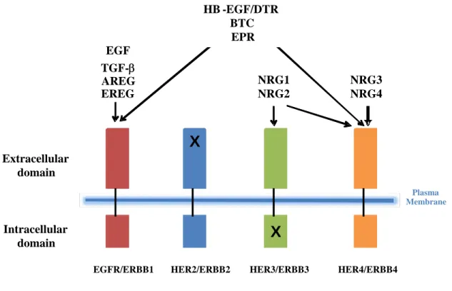
neuregulins (NRG1-4) [17-19]. As illustrated in Figure 1-1, EGF, TGF-α, and AR show
specificity to EGFR, and HB-EGF/DTR, EPR and BTC bind to both EGFR and ERBB4.
NRG1 and NRG2 bind to both ERBB3 and ERBB4, while NRG3 and NRG4 are specific to
the ERBB4 receptor. Adding to the complexity, ERBB2 has no known ligand but enhances
and stabilizes dimerization with other ERBBs, while ERBB3 is catalytically inactive [20-23].
Receptor homodimer or heterodimer combinations induce phosphorylation of unique tyrosine
sites which in turn serve as docking sites for specific SH-2 adaptor proteins that initiate
intracellular signaling cascades. The Ras- and Shc-activated mitogen-activated protein kinase
(MAPK) pathway is a target of all ERBB receptors, while the PI(3)K-activated AKT and
p70S6K/p85S6K pathways are downstream of only some ERBB dimers [7]. Activation of
signaling networks translates in the nucleus into distinct transcriptional programs involving
the proto-oncogenes FOS, JUN and MYC, and several transcription factors, including SP1 and
EGR1. An additional control of biological outcomes is signal duration. The kinetics of signal
termination is dependent upon ligand-mediated receptor endocytosis and receptor
4
Cross activation by other receptor families also contributes to ERBB signaling
intricacy. For example, members of the G protein coupled receptor superfamily (GPCR)
transactivate EGFR via metalloproteinase activation and subsequent cleavage and
extracellular release of EGF-like ligands [24]. This is thought to be how GPCR agonists,
such as angiotensin II (ANG-II), endothelin-1 (ET-1) and thrombin elicit growth effects via
activation of the MAPK pathway, circumventing the lack of intrinsic tyrosine kinase activity
of their cognate receptors [25, 26]. Recently, it was proposed that in the heart, ERBB2 forms
a heterocomplex with GPCRs in a ligand-dependent fashion, leading to a direct activation of
MAPK signaling [27].
ERBB signaling in cardiac development, function and homeostasis. Gene targeting
studies in mice demonstrate that all four ERBB receptors, select ligands and
ligand-processing enzymes are required for normal cardiac development (summarized in Table 1-1).
Mice lacking Erbb2, Erbb4, and Nrg1 all die around 10.5 days post-coitus (dpc) primarily
due to defects in cardiomyocyte development [28-30]. While Erbb3 -/- mice have normal
cardiomyocyte development, defective cardiac valve formation and function results in
embryonic lethality by 13.5 dpc [31]. Mice nullzygous for Egfr show strain-dependent
lethality varying from early pre-implantation to three weeks after birth [32-34]. On the CD-1
background, which supports survival to term, Egfr null mice have cardiac valve enlargement
in addition to severe neurological and gastrointestinal defects [35, 36]. Of the ten known
ligands, only genetic ablation of Nrg1 or Dtr/Hb-egf affects cardiovascular development. The
precursor, membrane-bound form of DTR is cleaved by members of the “α disintegrin and
metalloprotease” [37] family of convertases, resulting in the shedding of soluble
5
heterodimers. Dtr/Hb-egf -/- mice exhibit severe defects in heart chamber and valve formation
[35, 38], while mice lacking members of the ADAM family (tumor necrosis factor-alpha
converting enzyme (TACE) or ADAM19) show similar valve phenotypes [1, 35]. Homozygous deletion of other ligands individually, or even combined
Egf/Areg/Tgf-α deletion, does not result in embryonic lethality or cardiovascular defects, suggesting
significant signaling pathway redundancy in development [39, 40]. Cumulatively, these
knock-out models suggest distinct, chronological roles of ERBB receptors and ligands during
cardiovascular development, with NRG-1-ERBB2/4 signaling being of central importance in
cardiomyocyte development and maturation, while DTR/HB-EGF and/or NRG-1 activation
of EGFR, ERBB3 (and possibly ERBB2) being central to normal cardiac valve development.
In order to define these roles in context, a brief outline of cardiac development follows.
Outline of heart development:
A. Cardiac crest formation. At approximately 7 days post-coitus (dpc) in the mouse,
cardiac progenitor cells derived from the mesoderm and neural crest migrate towards the
anterior and anterio-lateral portion of the embryo to form the “cardiac crescent” or
primary heart field. Specification of the myocardial and endocardial cell lineages occurs
during this stage (Figure 2A) [41].
B. Formation of the primitive heart tube. By 8.0 dpc in the mouse, the cardiac crescent
has fused at the ventral midline to form a heart tube, consisting of an incomplete outer
layer of myocardium and an inner lining of endocardial cells separated by an extensive
extracellular matrix (ECM) referred to as the cardiac jelly (Figure 1-2B). At this stage,
the forming heart is centrally located within the embryo and is bilaterally symmetrical.
6
the middle region will give rise to a common ventricle, and the conus truncus, with
contributions from a secondary heart field, will give rise to the outflow tract of the heart
[41].
C. Cardiac looping. A crucial remodeling process of the heart tube known as cardiac
looping occurs during 8.5-10.5 dpc in the mouse (Figure 1-2C). This positions the atrial
region of the tube posterior to the common ventricle, setting the stage for the formation
and maturation of the four chambers as well as establishment of separate pulmonary and
systemic circulation (Figure 1-2C).
D. Cardiac maturation. Maturation of the heart occurs in multiple stages: (1) formation
of trabeculated myocardium, (2) formation of endocardial cushions (ECC), which
contribute to septation of the heart and cardiac valves and (3) compaction of the spongy
myocardium coinciding with establishment of the coronary circulation. Ventricular
trabeculation, whereby myocardial cells are activated by endocardium-derived signals to
proliferate and invade the interior of the cardiac chambers, begins soon after right
forward looping of the heart tube (Figure 1-2D). This process serves primarily as a
means to increase myocardial oxygenation in the absence of coronary circulation and is
necessary for circulation in the early stages of cardiac morphogenesis. Coinciding with
ventricular septation, trabeculae start to compact at their base adjacent to the outer
compact myocardium, adding to its thickness (beginning at Figure 1-2E). In the mouse,
this process is fairly rapid, occurring between 13 and 14 dpc with the establishment of
coronary circulation.[42]Non-compaction of the myocardium, even in small localized
regions of the ventricular wall, results in serious functional consequences and is
7
E. Cardiac valve development. Formation of the endocardial cushion (ECC) tissues is
extremely important for cardiac morphogenesis, as they provide the “glue” for virtually
all of the septal structures in the heart [42]. Dysmorphogenesis of the ECC or failure of
proper fusion are generally thought to play a major role in the etiology of congenital
heart defects [43]. ECC formation is characterized by endothelial-mesenchymal
transdifferentiation (EMT), where a subset of endocardial cells in the cushion-forming
regions delaminates and invades the cardiac jelly shortly after cardiac looping (Figure
1-3A). These cells proliferate and complete their differentiation into mesenchymal cells,
forming cushions that subsequently give rise to the septa of the four-chambered heart as
well as the cardiac valves. ECC formation is complete by 12.5 dpc in the mouse, and is
followed by a valvular remodeling stage in which cell proliferation ceases and apoptosis
increases, together remodeling the cushions into slender valve leaflets by 15.5 dpc. In
normal mouse and human cardiac valve development, cell proliferation is significant
during cushion development, decreases significantly during late embryonic development
and early postnatal life (coinciding with valve remodeling), and is undetectable in adult
valves [44].
Proportional valve growth after birth is largely attributable to increased ECM
production. Stratification of the ECM into three overlapping layers also begins in valve
remodeling stages but is not complete until postnatal life. In aortic valves, the arterial
aspect of the cusp (fibrosa) is composed predominantly of collagen fibers; the central
aspect (spongiosa) consists largely of loosely arranged proteoglycans; while the
8
degeneration of these layers is a pathological characteristic of congenital and acquired
aortic stenosis (AS) which has profound consequences on valve function.
ERBB signaling and cardiac development. ERBB signaling is particularly important in
mid-to late gestational cardiac processes, particularly in cardiomyocyte trabeculation and
cardiac valve development, both of which depend upon reciprocal signaling between
endocardial cells, ECM and cardiomyocytes to regulate cellular proliferation, differentiation
and invasion. As mentioned previously, mice lacking ERBB2, ERBB4, and NRG1 die
around 10.5 dpc, primarily due to reduced blood flow resulting from a lack of ventricular
trabeculae [28-30]. In wild-type 9.5/10.5 dpc embryos, ERBB2 and ERBB4 expression is
specific to developing cardiomyocytes,whereas NRG is localized to the endocardium.
Although ERBB2 is the preferred dimerization partner for ERBB3 andERBB4 upon NRG
stimulation, ventricular trabeculation is relatively normal in Erbb3-/- mice [31]. Since neither
ERBB2 nor ERBB4 can compensate for the loss of the other receptor in the heart, cardiac
NRG-1 signaling likely requires ERBB2/4heterodimers. Moreover, genetic rescue of
Erbb2-/- and Erbb4-/- embryos by myocardial expression of Erbb2 or Erbb4 cDNA under the control of cardiac specific promoters circumvents cardiac defects and embryonic lethality
[45, 46]. These data are consistent with a paracrine model whereby NRG1 is released by
endocardial cells, diffuses through the cardiac jelly and activates ERBB2/4 receptors
localized on cardiomyocytes, thus triggering proliferation and invasion to form ventricular
trabeculae.
During cardiac development, ERBB3 expression appears to be restricted to
endocardial cushion mesenchyme [30]. Embryos lacking ERBB3 have thinned ECC
9
lethality at 13.5 dpc due to inability to sustain cardiac function [31]. While ERBB3 on its
own lacks biological activity, NRG-1 activation of ERBB2/3 heterodimers generates a potent
signal [47]. Although not as severe as the Erbb3 -/- phenotype, Nrg1-/- or Erbb2 -/-embryos
also have underdeveloped ECC at 10.5 dpc, suggesting that NRG1-ERBB2/3 signaling plays
a role in early stages of cellular proliferation and differentiation in ECC formation. Recent
work has uncovered additional modulators of ERBB3 activity in this developmental context.
The extracellular matrix component hyaluronic acid appears to regulate ERBB2/3 signaling
during cushion formation, while the transcription factors GATA binding protein 4 (GATA-4)
and SRY (sex determining region Y)-box 9 (SOX9) are upstream mediators of ERBB3
expression in differentiated mesenchymal cells [48-50].
EGFR signaling and cardiac development. Mice lacking EGFR, its ligand DTR/HB-EGF
or the convertases TACE or ADAM19, have enlarged, hyperplastic cardiac valves [1, 35, 36,
38]. While these mutant embryos exhibit enlargement of all four cardiac valves, embryos
homozygous for the hypomorphic waved-2 mutation have hyperplastic semilunar (SL, i.e.
aortic and pulmonic) valves, suggesting there is greater requirement for EGFR signaling in
SL valve development [35, 36]. Exposure to EGFR inhibitors or knockdown of Egfr
expression in developing zebrafish results in a narrowed outflow tract, ventricular and atrial
enlargement, and decreased circulation, suggesting that the cardiac phenotype resulting from
disrupted EGFR signaling is conserved across species [51].
Cardiac cushions form normally in Dtr/Hb-egf -/-embryos, but are only modestly
condensed during remodeling stages (12.5-14.5 dpc) and thus remain significantly enlarged
compared to wild-type controls by 15.5 dpc [35]. Aberrant mesenchymal cell proliferation,
10
embryos have similarly enlarged, hyperplastic valves at 15.5 dpc, suggesting that impaired
remodeling also underlies valve enlargement in these mutants [35]. During the valve
remodeling stage in wild-type embryos, and TACE expression is detected throughout the
heart but enriched in cardiac cushions while DTR/HB-EGF is expressed exclusively by the
endocardial cells lining the developing valves, but not in differentiated mesenchymal cells
[35, 38]. Additionally, mice with Dtr knocked-out in endothelial and smooth muscle cell
lineages, or homozygous for an uncleavable form of DTR/HB-EGF, have a similar valve
phenotype [52, 53]. Together, this strongly suggests a paracrine model where DTR is
processed and released from the endocardial cells by TACE or ADAM19, diffuses through
the extracellular matrix, and activates EGFR on invading, differentiating cells to negatively
regulate proliferation.
Several findings suggest that signaling through EGFR may counterbalance bone
morphogenic protein (BMP) signaling to suppress mesenchymal cell proliferation. Genetic
studies using knockout mouse models established that endocardial outflow track cushion
growth is largely controlled by the amount of BMP signaling, with diminished and excessive
signaling correlating to hypo and hyperplastic cushions, respectively [54, 55]. Since the
enlarged valves of Dtr/Hb-egf -/- embryos have dramatic increases in activated BMP
signaling effectors SMAD1/5/8 and since EGFR downregulates BMP signaling by
inactivating SMAD1 in vitro, EGFR activation by TACE-derived soluble DTR may normally
limit BMP signaling during the transition from cushion formation/growth to valve
remodeling [46]. Potential binding partners for EGFR in this process include ERBB3 or
ERBB4 since both are expressed in remodeling valves. Since Erbb4 -/- embryos rescued by
11
embryos have defective cushion formation, ERBB3 seems a likely heterodimer candidate in
DTR/EGFR signaling during valve remodeling.
More recently, phospholipase C epsilon (PLCε), a phosphoinositide-specific
phospholipase C, was proposed as a downstream effector of EGFR signaling in valve
remodeling [56]. Mice homozygous for a targeted, inactivating mutation in Plcε (PlcεΔX/ΔX)
have hyperplastic semilunar valves, ventricular dilation, aortic stenosis and aortic
regurgitation. Increased SMAD1/5/8 activity is detected in the remodeling valves of Plcε
ΔX/ΔX 15.5 dpc embryos. The authors conclude that PLCε may play an important role in
EGFR-mediated negative regulation of the SMAD1/5/8 activation at a late stage of valve
remodeling [56].
ERBB signaling in cardiac disease. A role for ERBB signaling in adult cardiac
homeostasis and heart disease is also emerging. Three of the four receptors (EGFR, ERBB2
and ERBB4) remain expressed in the adult human and rodent heart; among these ERBB4
appears to be the most abundant [57-60]. The expression and activity of ERBB2 and ERBB4
receptors is depressed in clinical and experimentally induced heart failure [61-63] and
signaling via NRG-1/ERBB2/4 activation is critical for neonatal and adult cardiomyocyte
survival and growth [57, 64-67]. The importance of this signaling pathway in cardiac
homeostasis was not fully appreciated until the unexpected cardiotoxicity reported in breast
cancer clinical trials using trastuzmab (Herceptin, Genetech San Francisco, CA), a
humanized monoclonal antibody designed to repress ERBB2 activity by blocking the ligand
binding site [66, 68-70]. While approximately 8% of patients had cardiac changes with
12
anthracycline, a commonly used chemotherapeutic drug, developed dilated cardiomyopathy.
Mouse models with ventricular specific deletion of Erbb2 or Erbb4 have normal cardiac
development, but progressive postnatal degeneration that mimics the cardiotoxicity observed
in clinical trials [71-73]. Erbb2 conditional knock out (CKO) mice have up to 70% reduction
of cardiomyocyte ERBB2 expression, with no alterations in ERBB4 expression. These mice
develop ventricular dilation, reactivation of embryonic gene expression, increased
normalized heart weight, and depressed cardiac contractility. Erbb4 CKO mice have almost
80% reduction in ventricular ERBB4 protein levels, no alterations in ERBB2 protein levels,
and cardiac phenotypes that match Erbb2 CKO mice as well as significant mortality by one
year of age [73]. Although the authors do not report cardiomyocyte apoptosis in Errb2 or
Erbb4 CKO hearts, isolated cardiomyocytes from Erbb2 CKO mice display increased sensitivity to the anthracycline doxorubicin, consistent with the enhanced cardiotoxicity
observed with concurrent trastuzmab therapy [72]. In addition, overexpression of the
anti-apoptotic gene Bcl2/1 partially rescues ventricular dilation and contractility in Erbb2 CKO
mice, suggesting decreased cell survival contributes to cardiac dysfunction.
Consistent with these results, mice lacking one copy of Nrg1 (Nrg1+/-) have decreased
survival due to heart failure when exposed to doxorubicin [74]. Cardiomyocyte
anti-apoptotic ERBB signaling involves activation of the serine/threonin kinase AKT, which
attenuates apoptosis by phosphorylating, and thus inactivating, a variety of pro-apoptotic
proteins. Doxorubicin-treated Nrg1 +/- mice have significantly decreased levels of
phosphorylated ERBB2, AKT, and ERK-1/2 compared to similarly exposed wild type
controls [74]. Conversely, short term intravenous administration of a recombinant NRG1
13
injury models (infarct-, viral-, anthracyline, and pacing models) in rodents and dogs [64].
Consequently, there is interest in exploiting this pathway as a novel therapy for heart failure
or as means to prevent chemotherapy-associated cardiotoxicity [75].
EGFR signaling in cardiac disease. Signaling through the EGFR has been shown to
modulate pathological processes underlying common cardiovascular diseases, including
cardiomyocyte apoptosis and hypertrophy, fibrosis and hypertension [26, 37, 76-79]. In vitro
studies of isolated cardiomyocytes demonstrate that exposure to EGF increases
cardiomyocyte contractile frequency [80], while EGF or DTR/HB-EGF induce hypertrophy
and cardiac fibroblast proliferation [81] [82-84]. On a molecular level, these changes are
accompanied by increased protein synthesis, MAPK activation and increased transcription of
early response genes, suggesting that ligand-dependant EGFR activation can directly
influence cardiac remodeling.
Several GPCR agonists, such as angiotensin II (ANGII), endothelin-1 (ET-1) and
thrombin, provoke cardiomyocyte hypertrophy, vascular smooth muscle proliferation,
vasoconstriction and fibrosis [26, 78, 85-91]. Since GPCRs lack intrinsic tyrosine kinase
activity, recent work proposes that these receptors “hijack” EGFR activity via activating
metalloproteases which cleave membrane-bound ligands, and in this manner access
downstream EGFR-dependent mitogenic pathways [24, 26, 92]. Cardiomyocyte hypertrophy
induced by over stimulation of the renin-angiotensin (RAS) depends upon GPCR/EGFR
transactivation [25]. Physiological actions of ANGII are mediated via the angiotensin type 1
(AT1R) and angiotensin type 2 (AT2R) GPCRs, which are expressed in cardiomyocytes. In
isolated neonatal cardiomyocytes, overexpression of AT1R stimulates robust hypertrophy
14
re-expression of atrial natriuretic peptide (ANP) [93]. These responses are blocked in vitro
by AG-1478, an EGFR inhibitor, or KB-R7785, which inhibits the metalloprotease ADAM12
[93, 94].Since treatment with KB-R7785 blocks processing of DTR/HB-EGF and attenuates
pressure-overload induced hypertrophy in vivo, disruption of GPCR/EGFR transactivation at
this junction has been proposed as a novel therapy for left ventricular hypertrophy [94].
EGFR activation is also cardioprotective against the effects of sustained β-adrenergic
stimulation and acute stress [79, 95]. Intermale fighting (IF) and restraint-and-cold (4°C)
exposure (RCE) are two mouse models of intense acute stress. Following IF, but not RCE,
plasma EGF concentrations increase up to 1,000-fold. Cardiac necrotic lesions and elevated
plasma activity of creatine kinase (CK, a biomarker for cardiac injury) are seen with RCE,
but not IF [79]. Administration of AG-1478 (25mg/kg ip) prior to IF leads to increased
plasma activities of CK and other biomarkers, while administration of endogenous EGF
significantly reduces biomarker activity in RCE mice compared to vehicle treated controls.
Co-administration of AG-1478 and EGF eliminates the cardioprotective effect in RCE mice,
providing additional evidence that EGFR activation may protect the heart against
stress-induced injury. Activation of EGFR generates a potent survival signal in cardiomyocytes and
other cell types [37]; conversely, EGFR inhibition enhances apoptosis in normal and
cancerous epithelial cells [96] and in regenerating tissue [97, 98]. Since prolonged β
-adrenergic stimulation is known to trigger cardiomyocyte apoptosis, these studies suggest
15
Conclusions
The EGFR/ERBB1 signaling pathway is complex, intricately intertwined with many
extracellular and intracellular signaling pathways, and required for normal development and
homeostasis of most tissues. Genetically engineered mouse models have advanced our
understanding of ERBB receptor function in cardiac development and predicted
cardiotoxicity arising from ERBB2/HER2 targeted therapy. The current use of EGFR
inhibitors for cancer treatment and proposal of novel therapies repressing EGFR activity are
proposed for cardiovascular diseases necessitates a better understanding of roles of this
signaling pathway in cardiac development, function and disease.
Mouse models with genetically reduced EGFR activity resulting from the waved-2
hypomorphic allele (Egfr wa2) have assisted in defining the role of EGFR in carcinogenesis;
moreover, phenotypes of Egfr wa2/wa2 mice mimic common side effects observed with
EGFR-targeted therapy [99, 100]. Despite extensive use of this mouse model, congenital valve
defects and AS were discovered rather recently. Previous studies revealed the severity and
penetrance of phenotypes associated with EGFR activity are highly dependent upon genetic
background. To determine if genetic modifiers play a similar role in EGFR-related cardiac
phenotypes, we backcrossed the mutation onto the commonly used C57BL/6J (B6) and
129S1/SvImJ (129S1) inbred strains, creating Egfr wa2 congenic lines and an F
1 Egfr wa2
population. Because we found impressive genetic-background-dependent differences in
cardiac phenotypes associated with congenitally enlarged aortic valves, we designed
experiments investigating cardiac response to similar afterload in B6, 129S1 and F1 wild-type
mice. In order to partition EGFR-related congenital defects from effects on cardiac
16
exposed to EGFR small molecule inhibitors. Finally, to broadly localize genetic modifiers
which confer susceptibility to EGFR-related cardiac hypertrophy and failure, we conducted
preliminary genetic mapping experiments. Together, these studies should be useful in
predicting degenerative cardiac changes associated with EGFR inhibition which may be
17
Figure 1-1. ERBB ligand binding specificity is depicted for the four ERBB receptors.
Epidermal growth factor (EGF), transforming growth factor alpha (TGF-a), amphiregulin (AREG), epiregulin (EREG), betacellulin (BTC), epigen (EPG), and neuregulins 1-4 (NRG1-4).
Figure 1-1. ERBB ligand binding specificity is depicted for the four ERBB receptors.
Representative ligands are epidermal growth factor (EGF), transforming growth factor alpha (TGFα), amphiregulin (AR), epiregulin (EPR), betacellulin (BTC), epigen (EPG), and neuregulins 1-4 (NRG1-4). ERBB2 has no known ligand, while ERBB3 has an inactive catalytic domain.
EGFR/ERBB1 HER2/ERBB2 HER3/ERBB3 HER4/ERBB4
Plasma Membrane
X
X
Extracellular domain
Intracellular domain
EGF
TGF-β
AREG
EREG
NRG1 NRG2
NRG3 NRG4 HB -EGF/DTR
18
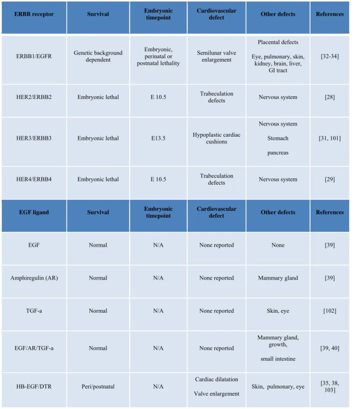
Table 1-1. Phenotypes resulting from genetic ablation of ERBB receptors, ERBB ligands or
ligand processing enzymes.
ERBB receptor Survival Embryonic timepoint Cardiovascular defect Other defects References
ERBB1/EGFR Genetic background dependent Embryonic, perinatal or postnatal lethality
Semilunar valve enlargement
Placental defects Eye, pulmonary, skin,
kidney, brain, liver, GI tract
[32-34]
HER2/ERBB2 Embryonic lethal E 10.5 Trabeculation defects Nervous system [28]
HER3/ERBB3 Embryonic lethal E13.5 Hypoplastic cardiac cushions
Nervous system Stomach pancreas
[31, 101]
HER4/ERBB4 Embryonic lethal E 10.5 Trabeculation defects Nervous system [29]
EGF ligand Survival Embryonic timepoint Cardiovascular defect Other defects References
EGF Normal N/A None reported None [39]
Amphiregulin (AR) Normal N/A None reported Mammary gland [39]
TGF-a Normal N/A None reported Skin, eye [102]
EGF/AR/TGF-a Normal N/A None reported
Mammary gland, growth, small intestine
[39, 40]
HB-EGF/DTR Peri/postnatal N/A
Cardiac dilatation Valve enlargement
19
EGF ligand
Survival Embryonic timepoint Cardiovascular defect Other defects References
Betacellulin (BTC) Normal N/A None reported None reported [35]
Epiregulin (ER) Normal N/A None reported Intestinal damage [104]
Neuregulin (NRG)-1 Embryonic lethal E 10
Trabeculation defects Cardiac conduction
Nervous system [30]
NRG-2 Normal N/A None reported Growth, reproduction [105]
Ligand processing Survival Embryonic timepoint
Cardiovascular
defect Other defects References
ADAM17(TACE ) Processing of : NRG1 and 2, HB-EGF, TGFα and AR
Perinatal/postnatal [39]
N/A
SL and AV valve enlargement
VSD
Pulmonary [35, 106]
ADAM19 Processing of: HB-EGF, NRG1, AR
Perinatal/postnatal N/A
SL and AV valve enlargement
VSD Overriding aorta Vasculature defects
None reported [1]
ADAM17/19 Embryonic lethality > E14.5
Trabeculation SL and AV valve
20
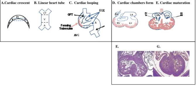
Figure 1-2. Developmental stages of heart development. A. Specification of precardiac
mesoderm and formation of cardiac crescent. B. Fusing of the cardiac mesoderm to form linear heart tube. C. Right forward lopping of the heart tube and determination of segments giving rise to cardiac chambers and cardiac valves. D. Cardiac looping leads to formation of cardiac chambers and outflow tract. An example of an E12.5 embryonic heart is shown below (E). F. Later stages of cardiac maturation include remodeling of cardiac cushions to form mature valves as well as thickening of the ventricular chamber walls. An example of an E14.5 embryonic heart is shown below (G). A, Atria; V, ventricle; CT, conus truncus; SV, sinus venous; RA, right atrium; LA, left atrium. Figures adapted from [107, 108].
E. G.
21
Figure 1-3. Developmental stages of cardiac valve development. A. Formation of the
endocardial cushions begins when a subpopulation of endocardial cells is activated by signals released by the myocardium to delaminate and invade the underlying cardiac jelly. B. These cells differentiate, migrate towards the myocardium and proliferate. C. During remodeling of the endocardial cushions, decreased proliferation and increased apoptosis results in mature slender valve leaflets. Stratification of the extracellular matrix also occurs. Molecules within the ECM and released from endocardial cells are thought to mediate this process. Figures adapted from [107, 108].
A. Cardiac cushion forms B. Cells migrate, differentiate, proliferate C. Valves remodel D. Mature valve leaflet
invading endocardial cell
Cardiac jelly
22
References
1. Zhou, H.M., et al., Essential role for ADAM19 in cardiovascular morphogenesis. Mol Cell Biol, 2004. 24(1): p. 96-104.
2. Gullick, W.J., Type I growth factor receptors: current status and future work. Biochem Soc Symp,
1998. 63: p. 193-8.
3. Wells, A., EGF receptor. Int J Biochem Cell Biol, 1999. 31(6): p. 637-43.
4. Weiss, F.U., H. Daub, and A. Ullrich, Novel mechanisms of RTK signal generation. Curr Opin Genet
Dev, 1997. 7(1): p. 80-6.
5. Schlessinger, J., Cell signaling by receptor tyrosine kinases. Cell, 2000. 103(2): p. 211-25.
6. Oda, K., et al., A comprehensive pathway map of epidermal growth factor receptor signaling. Mol Syst
Biol, 2005. 1: p. 2005 0010.
7. Yarden, Y., The EGFR family and its ligands in human cancer. signalling mechanisms and therapeutic opportunities. Eur J Cancer, 2001. 37 Suppl 4: p. S3-8.
8. Cohen, S., Isolation of a mouse submaxillary gland protein accelerating incisor eruption and eyelid opening in the new-born animal. J Biol Chem, 1962. 237: p. 1555-62.
9. Marquardt, H., et al., Rat transforming growth factor type 1: structure and relation to epidermal growth factor. Science, 1984. 223(4640): p. 1079-82.
10. Ciardiello, F., et al., Expression of cripto, a novel gene of the epidermal growth factor gene family, leads to in vitro transformation of a normal mouse mammary epithelial cell line. Cancer Res, 1991. 51(3): p. 1051-4.
11. Ciardiello, F., et al., Differential expression of epidermal growth factor-related proteins in human colorectal tumors. Proc Natl Acad Sci U S A, 1991. 88(17): p. 7792-6.
12. Higashiyama, S., et al., A heparin-binding growth factor secreted by macrophage-like cells that is related to EGF. Science, 1991. 251(4996): p. 936-9.
13. Barnard, J., Betacellulin: newest addition to the epidermal growth factor family. J Pediatr
Gastroenterol Nutr, 1993. 17(3): p. 343-4.
14. Shing, Y., et al., Betacellulin: a mitogen from pancreatic beta cell tumors. Science, 1993. 259(5101):
p. 1604-7.
15. Toyoda, H., et al., Epiregulin. A novel epidermal growth factor with mitogenic activity for rat primary hepatocytes. J Biol Chem, 1995. 270(13): p. 7495-500.
16. Kochupurakkal, B.S., et al., Epigen, the last ligand of ErbB receptors, reveals intricate relationships between affinity and mitogenicity. J Biol Chem, 2005. 280(9): p. 8503-12.
17. Carraway, K.L., 3rd, et al., Neuregulin-2, a new ligand of ErbB3/ErbB4-receptor tyrosine kinases.
Nature, 1997. 387(6632): p. 512-6.
23
19. Peles, E., et al., Isolation of the neu/HER-2 stimulatory ligand: a 44 kd glycoprotein that induces differentiation of mammary tumor cells. Cell, 1992. 69(1): p. 205-16.
20. Horan, T., et al., Binding of Neu differentiation factor with the extracellular domain of Her2 and Her3.
J Biol Chem, 1995. 270(41): p. 24604-8.
21. Kim, H.H., et al., Signal transduction by epidermal growth factor and heregulin via the kinase-deficient ErbB3 protein. Biochem J, 1998. 334 ( Pt 1): p. 189-95.
22. Sternberg, M.J. and W.J. Gullick, Neu receptor dimerization. Nature, 1989. 339(6226): p. 587.
23. Klapper, L.N., et al., The ErbB-2/HER2 oncoprotein of human carcinomas may function solely as a shared coreceptor for multiple stroma-derived growth factors. Proc Natl Acad Sci U S A, 1999. 96(9):
p. 4995-5000.
24. Daub, H., et al., Role of transactivation of the EGF receptor in signalling by G-protein-coupled receptors. Nature, 1996. 379(6565): p. 557-60.
25. Saito, Y. and B.C. Berk, Transactivation: a novel signaling pathway from angiotensin II to tyrosine kinase receptors. J Mol Cell Cardiol, 2001. 33(1): p. 3-7.
26. Shah, B.H. and K.J. Catt, Matrix metalloproteinase-dependent EGF receptor activation in hypertension and left ventricular hypertrophy. Trends Endocrinol Metab, 2004. 15(6): p. 241-3.
27. Negro, A., et al., erbB2 is required for G protein-coupled receptor signaling in the heart. Proc Natl
Acad Sci U S A, 2006. 103(43): p. 15889-93.
28. Lee, K.F., et al., Requirement for neuregulin receptor erbB2 in neural and cardiac development.
Nature, 1995. 378(6555): p. 394-8.
29. Gassmann, M., et al., Aberrant neural and cardiac development in mice lacking the ErbB4 neuregulin receptor. Nature, 1995. 378(6555): p. 390-4.
30. Meyer, D. and C. Birchmeier, Multiple essential functions of neuregulin in development. Nature, 1995. 378(6555): p. 386-90.
31. Erickson, S.L., et al., ErbB3 is required for normal cerebellar and cardiac development: a comparison with ErbB2-and heregulin-deficient mice. Development, 1997. 124(24): p. 4999-5011.
32. Miettinen, P.J., et al., Epithelial immaturity and multiorgan failure in mice lacking epidermal growth factor receptor. Nature, 1995. 376(6538): p. 337-41.
33. Threadgill, D.W., et al., Targeted disruption of mouse EGF receptor: effect of genetic background on mutant phenotype. Science, 1995. 269(5221): p. 230-4.
34. Sibilia, M. and E.F. Wagner, Strain-dependent epithelial defects in mice lacking the EGF receptor.
Science, 1995. 269(5221): p. 234-8.
35. Jackson, L.F., et al., Defective valvulogenesis in HB-EGF and TACE-null mice is associated with aberrant BMP signaling. Embo J, 2003. 22(11): p. 2704-16.
36. Chen, B., et al., Mice mutant for Egfr and Shp2 have defective cardiac semilunar valvulogenesis. Nat
24
37. Howes, A.L., et al., Galphaq expression activates EGFR and induces Akt mediated cardiomyocyte survival: dissociation from Galphaq mediated hypertrophy. J Mol Cell Cardiol, 2006. 40(5): p.
597-604.
38. Iwamoto, R., et al., Heparin-binding EGF-like growth factor and ErbB signaling is essential for heart function. Proc Natl Acad Sci U S A, 2003. 100(6): p. 3221-6.
39. Luetteke, N.C., et al., Targeted inactivation of the EGF and amphiregulin genes reveals distinct roles for EGF receptor ligands in mouse mammary gland development. Development, 1999. 126(12): p.
2739-50.
40. Troyer, K.L., et al., Growth retardation, duodenal lesions, and aberrant ileum architecture in triple null mice lacking EGF, amphiregulin, and TGF-alpha. Gastroenterology, 2001. 121(1): p. 68-78.
41. Moorman, A., et al., Development of the heart: (1) formation of the cardiac chambers and arterial trunks. Heart, 2003. 89(7): p. 806-14.
42. Wessels, A. and D. Sedmera, Developmental anatomy of the heart: a tale of mice and man. Physiol
Genomics, 2003. 15(3): p. 165-76.
43. Waller, B.F., et al., Congenital hypoplasia of portions of both right and left ventricular myocardial walls. Clinical and necropsy observations in two patients with parchment heart syndrome. Am J
Cardiol, 1980. 46(5): p. 885-91.
44. Hinton, R.B., Jr., et al., Extracellular matrix remodeling and organization in developing and diseased aortic valves. Circ Res, 2006. 98(11): p. 1431-8.
45. Woldeyesus, M.T., et al., Peripheral nervous system defects in erbB2 mutants following genetic rescue of heart development. Genes Dev, 1999. 13(19): p. 2538-48.
46. Tidcombe, H., et al., Neural and mammary gland defects in ErbB4 knockout mice genetically rescued from embryonic lethality. Proc Natl Acad Sci U S A, 2003. 100(14): p. 8281-6.
47. Sliwkowski, M.X., et al., Coexpression of erbB2 and erbB3 proteins reconstitutes a high affinity receptor for heregulin. J Biol Chem, 1994. 269(20): p. 14661-5.
48. Camenisch, T.D., et al., Heart-valve mesenchyme formation is dependent on hyaluronan-augmented activation of ErbB2-ErbB3 receptors. Nat Med, 2002. 8(8): p. 850-5.
49. Rivera-Feliciano, J., et al., Development of heart valves requires Gata4 expression in endothelial-derived cells. Development, 2006. 133(18): p. 3607-18.
50. Akiyama, H., et al., Essential role of Sox9 in the pathway that controls formation of cardiac valves and septa. Proc Natl Acad Sci U S A, 2004. 101(17): p. 6502-7.
51. Goishi, K., et al., Inhibition of zebrafish epidermal growth factor receptor activity results in cardiovascular defects. Mech Dev, 2003. 120(7): p. 811-22.
52. Nanba, D., et al., Loss of HB-EGF in smooth muscle or endothelial cell lineages causes heart malformation. Biochem Biophys Res Commun, 2006. 350(2): p. 315-21.
53. Yamazaki, S., et al., Mice with defects in HB-EGF ectodomain shedding show severe developmental abnormalities. J Cell Biol, 2003. 163(3): p. 469-75.
25
55. Delot, E.C., Control of endocardial cushion and cardiac valve maturation by BMP signaling pathways. Mol Genet Metab, 2003. 80(1-2): p. 27-35.
56. Tadano, M., et al., Congenital semilunar valvulogenesis defect in mice deficient in phospholipase C epsilon. Mol Cell Biol, 2005. 25(6): p. 2191-9.
57. Zhao, Y.Y., et al., Neuregulins promote survival and growth of cardiac myocytes. Persistence of ErbB2 and ErbB4 expression in neonatal and adult ventricular myocytes. J Biol Chem, 1998. 273(17):
p. 10261-9.
58. Zhao, Y.Y., et al., Neuregulin signaling in the heart. Dynamic targeting of erbB4 to caveolar microdomains in cardiac myocytes. Circ Res, 1999. 84(12): p. 1380-7.
59. Srinivasan, R., et al., Expression of the c-erbB-4/HER4 protein and mRNA in normal human fetal and adult tissues and in a survey of nine solid tumour types. J Pathol, 1998. 185(3): p. 236-45.
60. Fuchs, I.B., et al., Analysis of HER2 and HER4 in human myocardium to clarify the cardiotoxicity of trastuzumab (Herceptin). Breast Cancer Res Treat, 2003. 82(1): p. 23-8.
61. Uray, I.P., et al., Left ventricular unloading alters receptor tyrosine kinase expression in the failing human heart. J Heart Lung Transplant, 2002. 21(7): p. 771-82.
62. Rohrbach, S., et al., Neuregulin receptors erbB2 and erbB4 in failing human myocardium -- depressed expression and attenuated activation. Basic Res Cardiol, 2005. 100(3): p. 240-9.
63. Rohrbach, S., et al., Neuregulin in cardiac hypertrophy in rats with aortic stenosis. Differential expression of erbB2 and erbB4 receptors. Circulation, 1999. 100(4): p. 407-12.
64. Liu, X., et al., Neuregulin-1/erbB-activation improves cardiac function and survival in models of ischemic, dilated, and viral cardiomyopathy. J Am Coll Cardiol, 2006. 48(7): p. 1438-47.
65. Fukazawa, R., et al., Neuregulin-1 protects ventricular myocytes from anthracycline-induced apoptosis via erbB4-dependent activation of PI3-kinase/Akt. J Mol Cell Cardiol, 2003. 35(12): p. 1473-9.
66. Schneider, J.W., A.Y. Chang, and T.P. Rocco, Cardiotoxicity in signal transduction therapeutics: erbB2 antibodies and the heart. Semin Oncol, 2001. 28(5 Suppl 16): p. 18-26.
67. Pugatsch, T., et al., Anti-erbB2 treatment induces cardiotoxicity by interfering with cell survival pathways. Breast Cancer Res, 2006. 8(4): p. R35.
68. Schneider, J.W., A.Y. Chang, and A. Garratt, Trastuzumab cardiotoxicity: Speculations regarding pathophysiology and targets for further study. Semin Oncol, 2002. 29(3 Suppl 11): p. 22-8.
69. Ewer, M.S., et al., Cardiotoxicity in patients receiving transtuzumab (Herceptin): primary toxicity, synergistic or sequential stress, or surveillance artifact? Semin Oncol, 1999. 26(4 Suppl 12): p.
96-101.
70. Schaller, G., et al., Therapy of metastatic breast cancer with humanized antibodies against the HER2 receptor protein. J Cancer Res Clin Oncol, 1999. 125(8-9): p. 520-4.
71. Ozcelik, C., et al., Conditional mutation of the ErbB2 (HER2) receptor in cardiomyocytes leads to dilated cardiomyopathy. Proc Natl Acad Sci U S A, 2002. 99(13): p. 8880-5.
26
73. Garcia-Rivello, H., et al., Dilated cardiomyopathy in Erb-b4-deficient ventricular muscle. Am J
Physiol Heart Circ Physiol, 2005. 289(3): p. H1153-60.
74. Liu, F.F., et al., Heterozygous knockout of neuregulin-1 gene in mice exacerbates doxorubicin-induced heart failure. Am J Physiol Heart Circ Physiol, 2005. 289(2): p. H660-6.
75. Freedman, N.J. and G.S. Ginsburg, Novel--and "neu"--therapeutic possibilities for heart failure. J Am
Coll Cardiol, 2006. 48(7): p. 1448-50.
76. Zhai, P., et al., An angiotensin II type 1 receptor mutant lacking epidermal growth factor receptor transactivation does not induce angiotensin II-mediated cardiac hypertrophy. Circ Res, 2006. 99(5): p.
528-36.
77. Chan, H.W., et al., Effect of dominant-negative epidermal growth factor receptors on cardiomyocyte hypertrophy. J Recept Signal Transduct Res, 2006. 26(5-6): p. 659-77.
78. Chan, H.W., et al., Tackling the EGFR in pathological tissue remodelling. Pulm Pharmacol Ther,
2006. 19(1): p. 74-8.
79. Pareja, M., et al., Activated epidermal growth factor receptor (ErbB1) protects the heart against stress-induced injury in mice. Am J Physiol Regul Integr Comp Physiol, 2003. 285(2): p. R455-62.
80. Rabkin, S.W., The effect of alteration of extracellular Na+ or Ca2+ and inhibition of Ca2+ entry, Na(+)-H+ exchange, and Na(+)-Ca2+ exchange by diltiazem, amiloride, and dichlorobenzamil on the response of cardiac cell aggregates to epidermal growth factor. Exp Cell Res, 1990. 188(2): p. 262-6.
81. Perrella, M.A., et al., Regulation of heparin-binding epidermal growth factor-like growth factor mRNA levels by hypertrophic stimuli in neonatal and adult rat cardiac myocytes. J Biol Chem, 1994. 269(43):
p. 27045-50.
82. Ushikoshi, H., et al., Local overexpression of HB-EGF exacerbates remodeling following myocardial infarction by activating noncardiomyocytes. Lab Invest, 2005. 85(7): p. 862-73.
83. Clerk, A., et al., Peptide growth factors signal differentially through protein kinase C to extracellular signal-regulated kinases in neonatal cardiomyocytes. Cell Signal, 2006. 18(2): p. 225-35.
84. Rabkin, S.W., Indapamide accentuates cardiac chronotropic responses to epidermal growth factor in chick cardiomyocytes. Tissue Cell, 1996. 28(4): p. 469-72.
85. Sadoshima, J. and S. Izumo, Molecular characterization of angiotensin II--induced hypertrophy of cardiac myocytes and hyperplasia of cardiac fibroblasts. Critical role of the AT1 receptor subtype.
Circ Res, 1993. 73(3): p. 413-23.
86. Suzuki, T., et al., Endothelin-1 stimulates hypertrophy and contractility of neonatal rat cardiac myocytes in a serum-free medium. II. J Cardiovasc Pharmacol, 1991. 17 Suppl 7: p. S182-6.
87. Neyses, L., et al., Induction of immediate-early genes by angiotensin II and endothelin-1 in adult rat cardiomyocytes. J Hypertens, 1993. 11(9): p. 927-34.
88. Harada, M., et al., Significance of ventricular myocytes and nonmyocytes interaction during cardiocyte hypertrophy: evidence for endothelin-1 as a paracrine hypertrophic factor from cardiac nonmyocytes.
Circulation, 1997. 96(10): p. 3737-44.
27
90. Glembotski, C.C., et al., Myocardial alpha-thrombin receptor activation induces hypertrophy and increases atrial natriuretic factor gene expression. J Biol Chem, 1993. 268(27): p. 20646-52.
91. Obreztchikova, M., et al., Distinct signaling functions for Shc isoforms in the heart. J Biol Chem,
2006. 281(29): p. 20197-204.
92. Prenzel, N., et al., EGF receptor transactivation by G-protein-coupled receptors requires metalloproteinase cleavage of proHB-EGF. Nature, 1999. 402(6764): p. 884-8.
93. Thomas, W.G., et al., Adenoviral-directed expression of the type 1A angiotensin receptor promotes cardiomyocyte hypertrophy via transactivation of the epidermal growth factor receptor. Circ Res,
2002. 90(2): p. 135-42.
94. Asakura, M., et al., Cardiac hypertrophy is inhibited by antagonism of ADAM12 processing of HB-EGF: metalloproteinase inhibitors as a new therapy. Nat Med, 2002. 8(1): p. 35-40.
95. Lorita, J., et al., Effects of epidermal growth factor on epinephrine-stimulated heart function in rodents. Am J Physiol Heart Circ Physiol, 2002. 283(5): p. H1887-95.
96. Gilmore, A.P., et al., Activation of BAD by therapeutic inhibition of epidermal growth factor receptor and transactivation by insulin-like growth factor receptor. J Biol Chem, 2002. 277(31): p. 27643-50.
97. Bernal, N.P., et al., Epidermal growth factor receptor signaling regulates Bax and Bcl-w expression and apoptotic responses during intestinal adaptation in mice. Gastroenterology, 2006. 130(2): p.
412-23.
98. O'Brien, D.P., et al., Selective inhibition of the epidermal growth factor receptor impairs intestinal adaptation after small bowel resection. J Surg Res, 2002. 105(1): p. 25-30.
99. Roberts, R.B., C.L. Arteaga, and D.W. Threadgill, Modeling the cancer patient with genetically engineered mice: prediction of toxicity from molecule-targeted therapies. Cancer Cell, 2004. 5(2): p.
115-20.
100. Roberts, R.B., et al., Importance of epidermal growth factor receptor signaling in establishment of adenomas and maintenance of carcinomas during intestinal tumorigenesis. Proc Natl Acad Sci U S A,
2002. 99(3): p. 1521-6.
101. Riethmacher, D., et al., Severe neuropathies in mice with targeted mutations in the ErbB3 receptor.
Nature, 1997. 389(6652): p. 725-30.
102. Luetteke, N.C., et al., TGF alpha deficiency results in hair follicle and eye abnormalities in targeted and waved-1 mice. Cell, 1993. 73(2): p. 263-78.
103. Mine, N., R. Iwamoto, and E. Mekada, HB-EGF promotes epithelial cell migration in eyelid development. Development, 2005. 132(19): p. 4317-26.
104. Lee, D., et al., Epiregulin is not essential for development of intestinal tumors but is required for protection from intestinal damage. Mol Cell Biol, 2004. 24(20): p. 8907-16.
105. Britto, J.M., et al., Generation and characterization of neuregulin-2-deficient mice. Mol Cell Biol,
2004. 24(18): p. 8221-6.
106. Horiuchi, K., et al., Evaluation of the contributions of ADAMs 9, 12, 15, 17, and 19 to heart development and ectodomain shedding of neuregulins beta1 and beta2. Dev Biol, 2005. 283(2): p.
28
107. Iwamoto, R. and E. Mekada, ErbB and HB-EGF signaling in heart development and function. Cell
Struct Funct, 2006. 31(1): p. 1-14.
CHAPTER 2
CARDIAC RESPONSE TO PRESSURE OVERLOAD IN 129S1/SVIMJ AND C57BL/6J MICE: TEMPORAL AND BACKGROUND DEPENDENT DEVELOPMENT OF
CONCENTRIC LEFT VENTRICULAR HYPERTROPHY
Abstract
Left ventricular hypertrophy (LVH), a risk factor for cardiovascular morbidity and
mortality, is commonly caused by essential hypertension (EH). Three geometric patterns of LVH
can be induced by hypertension: concentric remodeling, concentric hypertrophy, and eccentric
hypertrophy. Clinical studies suggest that different underlying etiologies, genetic modifiers, and
risk of mortality are associated with LVH geometric patterns. Since pressure-overload induced
LVH can be modeled experimentally using transverse aortic constriction and since C57BL/6J
(B6) and 129S1/SvImJ (129S1) strains, which have different baseline cardiovascular phenotypes,
are commonly used, we conducted serial echocardiographic studies to assess cardiac function up
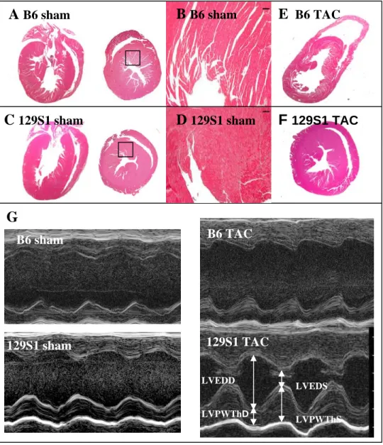
to eight weeks post-TAC in male B6, 129S1, and B6129S1F1 (F1) mice. B6 mice had earlier
onset and more pronounced impairment in contractile function, with corresponding LV and RV
dilatation, fibrosis, change in expression of hypertrophy marker, and increased liver weights at
five weeks post-TAC. These observations suggest that B6 mice had eccentric hypertrophy with
systolic dysfunction and right-sided heart failure. By contrast, we found that 129S1 and F1 mice
delayed transition to decompensated heart failure, with 129S1 mice exhibiting preserved systolic
function until eight weeks post-TAC, and relatively mild alterations in histology and markers of
hypertrophy at five weeks post-surgery. Consistent with concentric hypertrophy, our results