ROLE OF EPIDERMAL GROWTH FACTOR RECEPTOR ON CARDIAC FUNCTION
Yuying Xie
A dissertation submitted to the faculty of the University of North Carolina at Chapel Hill in partial fulfillment of the requirements for the degree of Doctor of Philosophy in the Curriculum
of Genetics and Molecular Biology in the School of Medicine.
Chapel Hill 2015
Approved by: David Threadgill
Fernando Pardo-Manuel de Villena Tim Wiltshire
©2010 Yuying Xie
ABSTRACT
YUYING XIE: Role of Epidermal Growth Factor Receptor on Cardiac Function (Under the direction of David Threadgill)
The epidermal growth factor receptor (EGFR/ERBB1) was the first discovered member of the ERBB family of tyrosine kinas receptors that includes ERBB2, ERBB3 and ERBB4. After binding by EGF-related ligands, EGFR is activated to induce homodimerization or heterodimerization with other ERBB receptors, resulting in tyrosine kinase activity. Subsequently, autophosphorylation or transphosphorylation of tyrosine residues in the C-terminal tail of the receptor allows the binding of adaptor proteins to trigger intracellular signaling cascades that can lead to proliferation, survival, and anti-apoptosis. As EGFR is expressed in the majority of developing and adult tissues including heart, dysfunction of EGFR activity can cause severe damage in different tissues and even initiate cancers. Several anti-EGFR drugs are already available in the clinic for late stage cancer patients with activated anti-EGFR activity. However, in cancer therapy using anti-ERBB2 drugs, severe cardiotoxicity among patients has been reported, emphasizing the importance of ERBB signaling in cardiac
homeostasis. Also with the increases in life expectancy of patients, some types of cancers tend to be treated as a chronic disease. Therefore it is importance to understanding possible cardiac toxicity under chronic suppression of EGFR pathway.
We propose the use of conditional knockout mice lacking EGFR activity in
demonstrated that chronic repression of EGFR pathway would cause severe cardiac dysfunction with chamber dilations, left ventricular wall thinning and depressed cardiac function.
Left ventricular hypertrophy (LVH) is associated with many cardiovascular diseases and is a risk factor for cardiac related morbidity and mortality. Mice homozygous with EGFR
ACKNOWLEDGEMENTS
I would never have been able to complete this work without the guidance of my
committee members, courage from friends, and support from my wife. I would not be here at the University of North Carolina at Chapel Hill if it was not for my grandma, Chunying Huang, who had spent an enormous of effort and patient to educate me and protect me in the earliest stage of my life.
First of all, I would like to express my deepest gratitude to my mentor, Dr. David Threadgill for supporting me during the past five years. David is someone you will never forget when you ever meet him. He is the most supportive advisor and one of the smartest person I know. He taught me how to pick important problems and provided me an extraordinary amount of freedom to create my own research agenda. During the hard day of my research, he was always optimistic and spent a huge amount of time and effort to guide me through the dark. After my training in Threadgill’s Lab, I become not only a better scientist but also a better person.
I would also like to thank Delia Barrick who has done pioneering work on this project and taught me all the echography techniques. I am also thankful to her for reading my thesis, commenting on my project and helping me understand my idea.
I would like to thank all current and former Threadgillians for their help and support, especially for Jill Steigerwalt who helped me breeding mice and gave me supports; Ming, Yu, who
introduced me to the lab and taught me experiments, Ryan, Dave, Emily, Chevonne and Jenn for numerous discussions.
TABLE OF CONTENTS
LIST OF TABLES... xi
LIST OF FIGURES... xii
LIST OF ABBREVATIONS AND SYMBOLS...xiv
Chapter I: ROLE OF ERBB SIGNALING IN CARDIAC DEVELOPMENT AND HOMESTASIS... 1
I.1: Introduction to cardiomyopathy... 1
I.1.1: Dilated cardiomyopathy... 1
I.1.2: Hypertrophic cardiomy... 3
I.2: Brief outline of heart development... 6
I.2.1: Cardiac development... 6
I.2.2: Valve development... 7
I.3: Role of ERBB family signaling in cardiac development and function………. 8
I.3.2: ERBB and ligand knockouts... 10
I.3.3: ERBB signaling in cardiac development... 10
I.3.4: ERBB signaling in maintenance of mature heart function... 12
I.3.5: Genetic modifiers for EGFR signaling... 15
References... 22
Chapter II: EGFR IS ESSENTIAL IN THE PREVENTION OF DILATED CARDIOMYOPATHY... 31
II.1: Overview ... 31
II.2: Introduction……... 32
II.3: Materials and Methods... 34
II.4: Results... 37
II.5: Discussion... 43
References... 57
Chapter III: GENETIC MODIFIER LOCUS AFFECTING LEFT VENTRICULAR HYPERTROPHY IN THE EGFRWA2 MOUSE MODEL OF AORTIC STENOSIS... 60
III.1: Overview ... 60
III.3: Materials and Methods... 62
III.4: Results... 64
III.5: Discussion... 68
References... 79
Chapter IV: CONCLUSIONS AND FUTURE DIRECTIONS... 82
LIST OF TABLES
Table 1.1: Causes of cardiomyopathy... 17
Table 1.2: Phenotype summary from genetic ablation of ERBB receptors, ERBB ligands... 18
Table 3.1: Representative echocardiographic parameters and
LIST OF FIGURES
Figure 1.1: A schematic figure of the dilated cardiomyopathy (DCM) causing mechanism... 18
Figure 1.2: The principal stages of cardiac development... 19
Figure 1.3: Ligand binding specificity for ERBB family... 20
Figure 2.1: Cre expression in MHCcre/+mice... 47
Figure 2.2: Cre-mediated mutation of Egfr in cardiomyocyte... 48
Figure 2.3: Egfr CKO mice develop marked deficits in heart function... 49
Figure 2.4: Ventricular dilation and myofiber hypertrophy in Egfr-CKO mice... 50
Figure 2.5: Comparison of relative expression of cardiac hypertrophy markers in LV... 51
Figure 2.6: Correlation between FS% and Egfrwt%... 52
Figure 2.7: Comparison of aortic valve function and morphology between Egfr-CKO and wildtype mice... 53
Figure 2.8: Cardiac pathology progression in Egfr-CKO mice... 54
Figure 2.9: Apoptosis in the hearts of Egfr CKO and wildtype mice... 55
Figure 2.10: Deterioration of cardiac function in Egfr-CKO mice following TAC treatment.... 56
Figure 3.2: Distribution of heart weight and body weight... 71
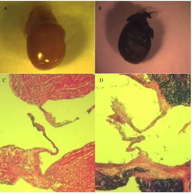
Figure 3.3: Representative histological sections from F2 Egfrwa2/wa2 three-month-old littermates………...…. 73
Figure 3.4: Whole genome scan performed for HW on F2 progeny, using sex as a covariate... 74
Figure 3.5: Linkage analysis on F2 male mice... 75
Figure 3.6: Linkage analysis on F2 female mice... 76
Figure 3.7: Test with increased samples size... 77
LIST OF ABBREVATIONS AND SYMBOLS
Actb beta-actin
ADAM12 a disintegrin and metalloproteinase domain 12
Adam19 a disintegrin and metalloproteinase domain 19
ANG-II angiotensin II
AR Amphiregulin
AREG amphiregulin
AS aortic stenosis
ATP adenosine triphosphate
AV atrial ventricular
Bax BCL2-associated X protein
BTC beta-cellulin
BW body weight
cAMP cyclic adenosine monophosphate
cDNA Complementary DNA
CK creatine kinase
DCM dilated cardiomyopathy
E7.0 embryonic day 7.0
EGF epidermal growth factor
EGFR epidermal growth factor receptor
Egfr CKO Egfr conditional knockout
Egfrwa2/wa2 homozygous Egfr mutant mice
Egfrwt % = 1- recombination efficiency
EgfrΔ Egfr with deletion
EH essential hypertension
EMT epithelial-to-mesenchymal transition
EPG epigen
EREG epiregulin
ERK1 Extracellular signal-regulated kinase 1
ET-1 endotelin-1
FS% fractional shortening percent
Gapdh Glyceraldehyde 3-phosphate dehydrogenase
GPCR G-protein coupled receptor
HB-EGF heparin-binding EGF-like growth factor
HCM human leukocyte antigen
hEGFRKI/KI homozygous human EGFR knockin mice
HER2 ErbB-2
HLA human leukocyte antigen
HW heart weight
HW/BW heart-to-body weigh ratio
IF intermale fighting
Kirrel3 kin of IRRE like 3
KO knockout
LDH lactate dehydrogenase
LOD logarithm (base 10) of odds
LV left ventricule
LVH left ventricular hypertrophy
LVID,d left ventricular internal dimension, diastole
LVID,s left ventricular internal dimension, systole
LVPW,d left ventricular posterior wall, diastole
LVPW,s left ventricular posterior wall, systole
mAbs monoclonal antibodies
M-mode motion mode
MPD mouse phenome database
MT masson’s trichrome
Mybpc myosin binding protein-C
MYBPC3 cardiac myosin-binding protein C
Nppa atrial nautriuretic peptide
Nppb brain nautriuretic peptide
NRG1 neuregulin 1
NRG2 neuregulin 2
NRG3 neuregulin 3
NRG4 neuregulin 4
p p value
PAF platelet-activating factor
PAS-H periodic acid-schiff counterstained with hematoxylin
PBS Phosphate buffered saline
PE phenylephrine
PI3K phosphatidylinositol 3-kinase
PRKAG2 γ-2-regulatory subunit of AMP-activated protein kinase
PTB phosphotyrosine binding domain
Q2 quantitative trait locus 2
QTL quantitative trait locus
R26R Rosa26 reporter
RCE restraint and cold (4˚C) exposure
RT-PCR reverse transcription polymerase chain reaction
SEM standard error of the mean
SH2 Src Homology 2 domain
SNP single-nucleotide polymorphism
St3gal4 ST3 beta-galactoside alpha-2,3-sialyltransferase 4
STD standard deviation
TAC transverse aortic constriction
Tace TNF-alpha converting enzyme
TGFA or TGF-α transforming growth factor-α
TK tyrosine kinase
TNNI3 cardiac troponin I
TNNT2 cardiac troponin T
Tnt troponin-T
TPM1 α-tropomyosin
TUNEL Terminal deoxynucleotidyl transferase mediated dUTP Nick End Labeling assay
Chapter I: ROLE OF ERBB SIGNALING IN CARDIAC DEVELOPMENT AND HOMESTASIS
I.1 Introduction to cardiomyopathy
Cardiomyopathies, diseases of the myocardial tissue, are strongly linked to cardiac dysfunction.1 In early stages, cardiomypothies may be asymptomatic but as disease progresses typical heart failure-like symptoms present, such as shortness of breath, orthopnea, paroxysmal nocturnal dyspnea and edema. More importantly, cardiomyopathies are often high risk signs for arrhythmia or sudden cardiac death.1
The four major types of cardiomyopathies are dilated cardiomyopathy, hypertrophic cardiomyopathy, restrictive cardiomyopathy, and arrhythmogenic right ventricular
cardiomyopathy. The cause for cardiomyopathy is still unclear. The possible etiologies include hypertension, coronary artery disease, metabolic disorders, nutritional deficiencies, heart tissue damage, viral myocarditis, chronic rapid heart rate, use of cocaine, pregnancy and genetic defects.2(Table 1-1)
I.1.1 Dilated cardiomyopathy
Dilated cardiomyopathy (DCM), the most common form of non-ischemic
Epidemiology
The estimated incidence of DCM in the United States is five per 100,000 adults and 0.57 per 100,000 children and the incidence has been increasing, most likely due to better diagnostic methods.3,4 The mortality rates are approximately 25 percent at one year and 50 percent at five years post-diagnosis.5,6
Pathological features
Dilatation of both ventricles is the chief pathological feature of DCM. Frequently, mural thrombi present in the left ventricle and occasionally in both atria, which are also usually
dilated.7 Both DCM and left ventricular hypertrophy (LVH) present with electrocardiographic changes (increased voltage) and elevated heart weight. However, the thicknesses of the left ventricular free wall and septum are typically normal or thinner with DCM.7 In addition, secondary dilatations in mitral and tricuspid annuli are frequently present, and microscopical features show hypertrophy and degeneration of myocyte and interstitial fibrosis with presentation of DCM.7
Etiology
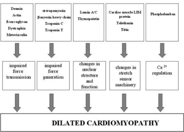
generation or force transmission is thought to be the mechanism for sarcomere and cytoskeletal protein genes (actin, troponin C, and troponin T). Additional mechanisms include impaired energy production for mitochondrial mutations, disturbed Ca2+ metabolism (phospholamban), impaired stretch sensor machinery (titin, telethonin), and defects in nuclear envelope (lamin A/C and tafazzin) (Fig 1-1).10 Recently, several groups have found DCM causing mutations linked with troponin T, which is part of the thin filament.11,12 Other groups have mdeled this mutation in mice to study its effect on muscle fibers. Troponin T mutations result in significantly lower Ca2+ sensitivity in force generation in the sarcomere, which would be a possible mechanism for the pathogenesis of DCM. Additionally, TnT-/- mice develop severe DCM, which recapitulates the phenotype of patients.12
Genetic diseases can be classified as Mendelian diseases (monogenic diseases) or
multifactorial diseases.13 Monogenic diseases are rare and are caused by single gene mutations. Although the gene mutations described above are monogenetic with high penetrance, they only explain DCM in rare familial cases. Multifactorial diseases are more common and complex because they may be due to additive or interactive effects of multiple genes. Genes responsible for multifactorial diseases tend to have low penetrance, and to have interactions with
environmental factors. Genetic association studies have identified several low-penetrance polymorphisms (susceptibility genes) contributing to DCM including the G994T mutation in
plasma platelet-activatingfactor (PAF) acetylhydrolase, and a 14-bp deletion polymorphism in human leukocyte antigen (HLA).14-16
I.1.2 Hypertrophic cardiomyopathy
interventricular septum.1 Typically in patients with HCM, the left ventricular volume is normal or reduced and the systolic gradients are normal. HCM is the leading cause of sudden cardiac death in young athletes.2,17 However, it can occur patients of any age with disabling cardiac symptoms or sudden unexpected cardiac death.
Epidemiology
Hypertrophic cardiomyopathy is likely the most frequently occurring cardiomyopathy with an incidence of 0.2% in the general population, affecting around 600,000 people in the United States.2 Previous studies have shown that annual mortality for HCM is about 1.4%, and this mortality can be stratified to sudden death (0.7%), progressive heart failure (0.5%) or stroke-related death (0.2%).18 In young patients sudden death is the major outcome, while progressive heart failure and stroke related death is most common in patients after mid-age.18 Most patients with HCM have little or no disability and can have normal life expectancy indicating additional factors contribute to sudden death or progressive disease.
Pathological features
Left Ventricular Hypertrophy (LVH) is the most common feature of HCM. However, with the heterogeneity of HCM, no single pattern of LVH is dominant and can include
electrophysiological characteristic, ultimately leading to arrhythmias and sudden death in HCM.20
Because of the heterogeneity of HCM, the clinical course for individual patients can be classified into one of subgroups: 1) no or mild symptoms, 2) high risk for sudden cardiac death, 3) progressive heart failure with exertional dyspnea and functional disability, and 4) atrial fibrillation often accompanied by embolic stroke.19 Although sudden death can occur at a wide range of ages, progressive heart failure and stroke occur more frequently in mid-age and older.21 Risk factors for sudden death include: 1) patients with a prior cardiac arrest or sustained
ventricular tachycardia, 2) family history of sudden death due to HCM, 3) syncope or near syncope particularly when it is related to physical activity or when it occurs several times, 4) repeatedly ventricular tachycardia on serial ECG recording, 5) failure of blood pressure to respond to exercise, especially for people younger than 50, and 6) extreme LVH with wall thickness > 30 mm.19,22 Additional risk factors that have been suggested include atrial
fibrillation, myocardial ischemia, alcohol septal ablation surgery, LV outflow obstruction and bridged left anterior descending coronary artery.19,22
Etiology
Approximately 50% of hypertrophic cardiomyopathy cases are familial with most inherited as a Mendelian autosomal dominant trait and caused by mutations in one of a number of genes that encode proteins of the cardiac sarcomere (thick or thin filaments).19,23 Genes responsible for HCM include β–cardiac myosin heavy chain (MYH7), cardiac myosi-binding protein C (MYBPC3), cardiac troponin T (TNNT2), cardiac troponin I(TNNI3), and
protein mutations, γ-2-regulatory subunit of AMP-activated protein kinase (PRKAG2) and
lysosome-associated membrane protein2 (LAMP-2) have been linked to HCM.2 However, the molecular defects and mechanisms responsible for HCM are usually different in unrelated patients, and many additional mutations responsible for HCM remain to be identified. Possible mechanisms for HCM are impaired ATPase activity in cardiomyocytes leading to improper systolic and diastolic pressures, reduced contractile function leading to hypertrophy in
cardiomyoctes, and disorganized sarcomere structure leading to stimulation of growth factors which would cause hypertrophy and fibrosis.25
In recent years, an increasing number of mouse models have been created to model human HCM, such as missense mutations in α-MHC gene, transgenic mice expressing a
troponin-T (Tnt) missense mutation, and transgenic mice with mutant myosin binding protein-C (Mybpc) lacking binding domains.26-28
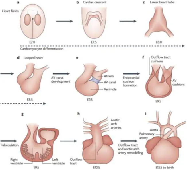
I.2 Brief outline of heart development I.2.1 Cardiac development
series of step including formation of the atrioventricular canal (Figure 1.2 e), formation of the endocardial cushion that will develop to four major heart valves (Figure 1.2 f), and formation of trabeculae within walls of left and right ventricle. (Figure 1.2 g)29 After spetation of the atria and ventricle and remodeling of the outflow tract, cardiac maturation is complete. The heart is the first definitive organ that forms during embryogenesis. At embryonic day 7.0 (E7.0), cardiac progenitors are located in the primary heart field, a region of mesoderm that migrates and converges at the midline of the embryo to form the cardiac crescent (Figure 1.3 a,b).29 By E8.0, the halves of the cardiac crescent have fused to become a heart tube that consists of an
endothelial tube surrounded by a layer of myocardial cells (Figure 1.3 c).29 A crucial remodeling process known as cardiac looping then starts from E8.5 to E10.5 (Figure 1.3 d-h). During
cardiac looping, the tube elongates and adopts a pronounced rightward curvature. The cardiac tube is then transformed into a heart structure with four distinct chambers through a series of step including formation of the atrioventricular canal (Figure 1.3 e), formation of the endocardial cushion that will develop to four major heart valves (Figure 1.3 f), and formation of trabeculae within walls of left and right ventricle (Figure 1.3 g).29 After septation of the atria and ventricle and remodeling of the outflow tract, cardiac maturation is complete.
I.2.2 Valve development
There are two steps for cardiac valve formation: cardiac cushion formation and valve remodeling. Following cardiac looping, the extracellular matrix, known as cardiac jelly, expands to form the cardiac cushion in the atrioventricular canal and the distal portion of the outflow tract, which are precursors of tricuspid and mitral valves and the aortic and pulmonic valves,
respectively. Before onset of cushion formation, the cardiac jelly is surrounded by the
epithelial-to-mesenchymal transition (EMT) following activation from adjacent myocardium.29 This transition involves down-regulation of the cell-adhesion molecule vascular endothelial cadherin (Cdh5), which enables a subset of endocardial cells to delaminate and invade the cardiac jelly.29 These cells differentiate to mesenchymal cells and proliferate to form the cardiac cushions. Endocardial cushion formation is complete by E12.5, and then cushions undergo a valve remodeling process in which cell proliferation is decreased and apoptosis increased changing the cushion into a slender valve leaflet by E15.5.
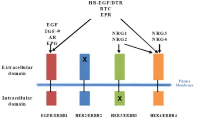
I.3 Role of ERBB family signaling in cardiac development and function I.3.1 EGFR and ERBB family
The epidermal growth factor receptor (EGFR/ERBB1) was the first discovered member of the ERBB family of tyrosine kinas receptors that includes ERBB2, ERBB3 and ERBB4.30 Members of ERBB family are membrane bound glycoproteins whose basic function is to transmit extracellular signals into cellular responses. EGFR as well as other members of the ERBB family have a conserved protein structure containing an extracellular cysteine-rich ligand binding domain (except for ERBB2/HER2), a single alpha-helix transmembrane domain, an intracellular tyrosine kinase (TK) domain (except for ERBB3/HER3) and a C-terminal tail with several tyrosine residues that can serve as docking sites for adaptor proteins after
phosphorylation. The ERBB receptor is activated by binding EGF-related ligands in an
on the ligand, biological output of signaling cascade can be diverse including proliferation, migration, adhesion, motility and survival.32 ERBB2 is an orphan receptor with no recognized ligand, while ERBB3 lacks intrinsic kinase activity. Therefore, ERBB2 and ERBB3 can only function through heterodimerization with other ERBB receptors.33,34
One of the complexities of ERBB signaling stems from the diversity of ligands. In addition to epidermal growth factor (EGF), there are ten known ligands for ERBB receptors including amphiregulin (AREG), transforming growth factor-α (TGFA), heparin-binding EGF-like growth factor (HB-EGF), betacellulin (BTC), epiregulin (EGEG), epigen (EPG), and
neuregulins (NRG 1-NRG4).35-43 EGF, TGFA, AREG and EREG are specific for EGFR/ERBB1, while HB-EGF, BTC and EPR can activate both EGFR/ERBB1 and ERBB4. (Figure 1.2) NRG1 and NRG2 are ligands for both ERBB3 and ERBB4, but NRG3 and NRG4 have higher
preference for ERBB4.44 After ligand binding, the activated receptors recruit and phosphorylate downstream effecter proteins to activate a cascade of intracellular signaling pathways. The mitogen-activated protein kinase pathway, which is a target of all ERBB receptors, and the phosphatidylinositol 3-kinase (PI3K)-AKT pathway, which are target for a subset of ERBB dimmers, are the most well-studied. The activated receptors are subsequently endocytosed and either degraded in the endosome or recycled to the plasma membrane. Signal termination is an additional control for the biological response of ERBB signaling.
To add another layer of complexity, the ERBB family is also involved in other signaling networks through cross-activation with other receptor classes. For example, members of the G-protein coupled receptor G-protein (GPCR) family can transactivate EGFR by a so called “triple membranepassing signaling” paradigm, which involve the activation of metalloproteases and
proposed the mechanism that GPCR agonists, such as angiotensin II(ANG-II) and endotelin-1 (ET-1), triggers cardiomyocyte hypertrophy.47
I.3.2 ERBB and ligand knockouts
Evidences from knockouts in genes coding for ERBB receptors and their ligands have suggested the vital role for ERBB signaling in cardiac pathologies (Table 1-2). Erbb2, Erbb4
and Nrg1 null mice all die around E10.5 with severe defects in cardiac trabeculae formation.48-50 Although mice deficient for Erbb3 have normal cardiac trabeculae, they have defective valve formation resulting embryonic lethal at E13.5.51,52 Egfr null mice show various timing of
lethality ranging from pre-implantation to two to three weeks after birth depending on the genetic background.53-55 Surviving Egfr null mice on a mixed CD-1 background exhibit severe
semilunar valve enlargement.48 Compared with the critical role of ERBB receptors on cardiac development, cardiac phenotype in mice lacking ERBB ligands are modest. Mutations in the majority of the ten known ligands including Egf, Areg, Tgfa, Btc, Ereg and Nrg-2 show no cardiovascular defects indicating that there is considerable redundancy or functional overlap in the action of specific ligands.44 However, genetic ablation of Hb-egf exhibits severe defects in heart chamber and valve formation.56 Moreover, Nrg-1 null mice also have cardiac traveculae defect.50 In sum, Erbb and ligand knock-out models suggest distinct roles for ERBB receptors and ligands during cardiovascular development with ERBB2, ERBB4 and NRG1 signaling being involved in trabeculae formiation, while HB-EGF and EGFR being central to cardiac valve development.
I.3.3 ERBB signaling in cardiac development
ERBB signaling is required for the mid to late gestational cardiac development,
formation both require reciprocal signaling between endocardium and myocardium to regulate cell proliferation, differentiation, and cell invasion to cardiac jelly. In wild-type mice, between E9.5 to 10.5, the expression of ERBB2 and ERRB4 are restricted to cardiac myocardial cells, and NRG-1 is specifically expressed in endocardial cells. From gene targeting studies, mice lacking Erbb2, Erbb4 and Nrg1 all die around E10.5 due to the trabeculae defects that develop into arrhythmia, enlarged ventricule, and reduced blood flow.48-50 Moreover, other studies have shown expression of ERBB2 and ERBB4 under the control of cardiac specific promoters in
Erbb2 and Erbb4 null mice, respectively, can rescue the trabeculae defects.57,58 Because Egfr
and Erbb3 null mice show no trabecular defects and neither Erbb2 nor Erbb4 can compensate for the loss of the other, cardiac trabeculae formation seems to require NRG-1 signaling through ERBB2/ERBB4 heterodimers.
While ERBB2 and ERBB4 are particularly important in trabeculation, ERBB3 and EGFR have vital roles in cardiac valve formation. During cardiac development, the expression of ERBB3 is restricted to endocardial cushion mesenchyme, and Erbb3 null mice die at E13.5 due to defective cardiac cushions completely lacking mesenchymal cells.50,51 Although ERBB3 lacks tyrosine kinase activity, NRG-1 can induce the heterodimerization between ERBB2 and ERBB3, which can trigger downstream signaling by the tyrosine kinase activity on ERBB2. Moreover, close inspection of endocardial cushions in Erbb2 and Nrg-1 null embryos shows
underdeveloped cushion at E10.5.48,50 This result suggests that NRG-1 signaling to– ERBB2/ERBB3 heterodimers is required at the early stages of cellular proliferation and differentiation in endocardial cushion formation.
which is a later stage during valve formation. Mice homozygous for the Egfr hypomorphic
waved-2 mutation (Egfrwa2/wa2) have enlarged aortic and pulmonic valves, and another study using Egfr null mice showsed hyperplastic semilunar (aortic and pulmonic) and AV (mitral and tricuspid) valves.48,59 The role of EGFR signaling during cardiac development is also observed in zebrafish.60 Addition of an EGFR kinase inhibitor or the transient knockdown of EGFR expression also results in decreased circulation and a narrowed outflow tract suggesting that EGFR signaling function in cardiac development is conserved across species.56,59,60 Consistent with valve defects in Egfr null mice, mice lacking its ligand Hb-egf or the convertases Tace
(TNF-alpha converting enzyme) or Adam19 (a disintengrin and metalloproteinase domain 19) also show cardiac valve abnormalities in semilunar and AV valve.59,61 During embryonic heart valve development the expression of HB-EGF is restricted exclusively to endocardial cells, and is not detected in differentiated mesenchymal cells.59 Analysis of Hb-egf null mice suggests that HB-EGF-EGFR signaling might regulate valve remodeling rather than cushion formation
because 1) EMT is normal in Hb-egf null mice, 2) there is no change in apoptosis rate in Hb-egf
null mice, and 3) excessive proliferation of mesenchymal cells is detected in Hb-egf null mice.59 Available data suggests a paracrine model for EGFR signaling in valve formation where HB-EGF is released from the plasma membrane of the endocardial cells and diffuses to activate EGFR on mesenchymal cells to activate downstream signals that suppresses mesenchymal cell proliferation.
I.3.4 ERBB signaling in maintenance of mature heart function
ERBB4 as the most prevalent receptor.48 In vitro studies have provided evidence that NRG-1, ERBB2, and ERBB4 are implicated in both hypertrophic and survival signaling pathways in adult cardiomyocyte.48 Several clinical studies suggest an association between heart failure and decreased ERBB2 and ERBB4 protein level.62,63 Consistent with these clinical finding, ERBB2 and ERBB4 expression is upregulated in heart failure patients whose cardiac function was improving after mechanical ventricular unloading.62 Moreover, downregulation of ERBB2 activity by using anti-breast cancer drug Trastuzmab, a humanized monoclonal antibody designed to block ERBB2’s ligand binding site, resulted in cardiac dysfunction in some patients.64,65 These findings were recapitulated in Erbb2-deficient conditional mutant mice (Erbb2 CKO mice) which develop severe heart failure with dilated ventricles and decreased contractility by three months of age. 48,66 Erbb4 conditional knock-out mice with 80% reduction in ventricular ERBB4 protein levels also develop a cardiac phenotype similar to Erbb2 CKO mice, suggesting that ERBB2 may partner with ERBB4 to form a heterodimer required for maintenance of normal cardiac function.67
Ligands for ERBB receptors, such as HB-EGF and NRG1, have also been shown to play important role in postnatal heart. Adult cardiomyocyte strongly express HB-EGF, and the constitutive tyrosine phosphorylation levels of ERBB2 and ERBB4 are significantly reduce in cardiomyocyte from HB-EGF knock out mice.44 These mice also developed a dilated heart with enlarged cardiomyocytes and depressed cardiac function. Moreover, in mice with hypertrophy induced by pressure overload or GPCR agonists, inhibition of HB-EGF shedding by adding DAM12 inhibitor attenuated hypertrophic changes and improved cardiac function.68
cardiomyocyte death.69 In this model, the protein level of phosphorylated ERBB2, AKT and ERK1/2 are significantly reduced in Nrg1+/- compared with wild-type control indicating the involvement of survival pathway.69 Consistent with data above, short term intravenous administration of NRG1 improved cardiac performance in ischemic, drug-induced
cardiomyopathy, myocaritis and chronic rapid pacing model in rodent and canine models.70 Interestingly, the survival benefit from NRG1 is additive to angiotensin-converting enzyme inhibitor therapy in ischemic models.70
Similar as other ERBB members, EGFR signaling also has vital role in maintenance of mature heart function. Exogenous EGF increases contractility and heart rate by elevating cyclic adenosine monophosphate (cAMP) levels in cardiac myocytes.71 The increase in cAMP occurs through activation of adenylyl cyclase by EGFR-mediated activation of Gs protein.71 Moreover,
in vitro studies using isolated cardiomyocytes showed that exposure to HB-EGF or EGF induce hypertrophy.72 By using a conditional knock-in approach using the human EGFR cDNA,
shortening were observed in female mice.53 Taken together, these data suggest that EGFR signaling is required to maintain normal function.
EGFR signaling may protect the heart against stress-induced injury. Using a restraint-and-cold (4˚C)-exposure (RCE) mice model, Miguel Pareja et al. found that heart injury biomarker, including plasma lactate dehydrogenase (LDH) and creatine kinase (CK), were increased in a time-dependent manner.75 By contrast, in another common used stress model, intermale fighting (IF), only LDH activity is raised.75 One difference between these two models is that with IF, but not RCE, plasma EGF concentration is strongly elevated. When mice were exposed to the EGFR inhibitor, AG-1478, immediately before IF, plasma levels of both
biomarkers was increased.75
Conversely, injecting Egf prior to RCE exposure significantly reduce LAH and CK activity. Together, this data supports a cardiac-protective role of EGFR signaling on stress-induced injury.
EGFR signaling also contributes to cardiac hypertrophy induced by activation of the G-protein-coupled receptor (GPCR) pathway.76 GPCR agonists, such as phenylephrine (PE), angiotensin II (AngII) and endothelin-1 (Et-1), are well-know inducer of cardiomyocyte
hypertrophy.77-79 All of these molecules transactivate EGFR by increasing shedding ofHB-EGF caused by activation of a specific metalloproteinase.45 Consistent with this model, blocking the cross-talk between EGFR and GPCR signaling through metalloproteinase or EGFR inhibitor attenuates hypertrophic phenotype.47,68
I.3.5 Genetic modifiers for EGFR signaling
which results in peri-implantation death.55 Contrastingly, on a CD-1 background, Egfr mutant mice can live up to three weeks after birth with abnormalities in skin, kidney and other
organs.53,55 Additionally, mice homozygous for Egfrwa2 have abnormalities in aortic valves and development left ventricular hypertrophy, phenotypes that are also dependent on genetic
background.53 Egfrwa2/wa2 mice on a C57BL/6J (B6) background have thicker aortic cusps, higher incidence of heart failure, and shorter lifespan compared with Egfrwa2/wa2 mice on
Figure 1.1: A schematic figure of the dilated cardiomyopathy (DCM) causing mechanism.
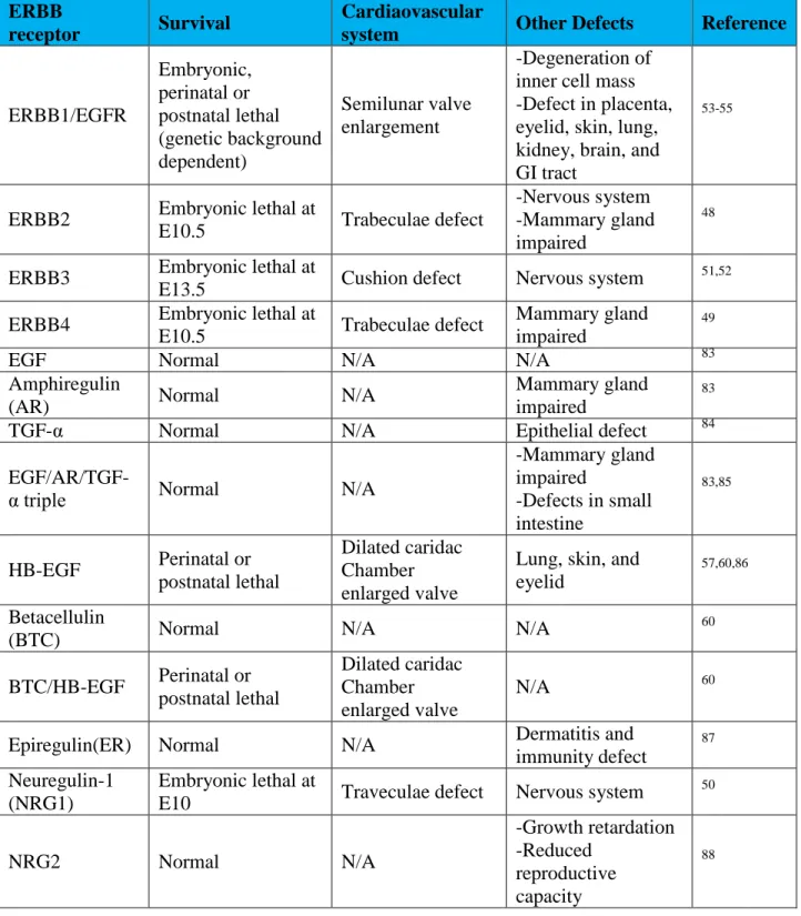
Table 1.2: Phenotype summary from genetic ablation of ERBB receptors, ERBB ligands. ERBB
receptor Survival
Cardiaovascular
system Other Defects Reference
ERBB1/EGFR Embryonic, perinatal or postnatal lethal (genetic background dependent) Semilunar valve enlargement -Degeneration of inner cell mass -Defect in placenta, eyelid, skin, lung, kidney, brain, and GI tract
53-55
ERBB2 Embryonic lethal at
E10.5 Trabeculae defect
-Nervous system -Mammary gland impaired
48
ERBB3 Embryonic lethal at
E13.5 Cushion defect Nervous system
51,52
ERBB4 Embryonic lethal at
E10.5 Trabeculae defect
Mammary gland impaired
49
EGF Normal N/A N/A 83
Amphiregulin
(AR) Normal N/A
Mammary gland impaired
83
TGF-α Normal N/A Epithelial defect 84
EGF/AR/TGF-α triple Normal N/A
-Mammary gland impaired
-Defects in small intestine
83,85
HB-EGF Perinatal or postnatal lethal
Dilated caridac Chamber enlarged valve
Lung, skin, and eyelid
57,60,86
Betacellulin
(BTC) Normal N/A N/A
60
BTC/HB-EGF Perinatal or postnatal lethal
Dilated caridac Chamber enlarged valve
N/A 60
Epiregulin(ER) Normal N/A Dermatitis and
immunity defect
87
Neuregulin-1 (NRG1)
Embryonic lethal at
E10 Traveculae defect Nervous system
50
NRG2 Normal N/A
-Growth retardation -Reduced
reproductive capacity
REFERENCES
1. Richardson, P. et al. Report of the 1995 World Health Organization/International Society and Federation of Cardiology Task Force on the Definition and Classification of cardiomyopathies. Circulation 93, 841-2 (1996).
2. Maron, B.J. et al. Contemporary definitions and classification of the cardiomyopathies: an American Heart Association Scientific Statement from the Council on Clinical Cardiology, Heart Failure and Transplantation Committee; Quality of Care and Outcomes Research and Functional Genomics and Translational Biology Interdisciplinary Working Groups; and Council on Epidemiology and Prevention. Circulation 113, 1807-16 (2006).
3. Dec, G.W. & Fuster, V. Idiopathic dilated cardiomyopathy. N Engl J Med 331, 1564-75 (1994).
4. Towbin, J.A. et al. Incidence, causes, and outcomes of dilated cardiomyopathy in children. Jama 296, 1867-76 (2006).
5. Diaz, R.A., Obasohan, A. & Oakley, C.M. Prediction of outcome in dilated cardiomyopathy. Br Heart J 58, 393-9 (1987).
6. Fuster, V. et al. The natural history of idiopathic dilated cardiomyopathy. Am J Cardiol
47, 525-31 (1981).
7. Roberts, W.C., Siegel, R.J. & McManus, B.M. Idiopathic dilated cardiomyopathy: analysis of 152 necropsy patients. Am J Cardiol 60, 1340-55 (1987).
8. Grunig, E. et al. Frequency and phenotypes of familial dilated cardiomyopathy. J Am Coll Cardiol 31, 186-94 (1998).
10. Karkkainen, S. & Peuhkurinen, K. Genetics of dilated cardiomyopathy. Ann Med 39, 91-107 (2007).
11. Hanson, E.L. et al. Cardiac troponin T lysine 210 deletion in a family with dilated cardiomyopathy. J Card Fail 8, 28-32 (2002).
12. Kamisago, M. et al. Mutations in sarcomere protein genes as a cause of dilated cardiomyopathy. N Engl J Med 343, 1688-96 (2000).
13. Colombo, M.G., Botto, N., Vittorini, S., Paradossi, U. & Andreassi, M.G. Clinical utility of genetic tests for inherited hypertrophic and dilated cardiomyopathies. Cardiovasc Ultrasound 6, 62 (2008).
14. Ichihara, S., Yamada, Y. & Yokota, M. Association of a G994-->T missense mutation in the plasma platelet-activating factor acetylhydrolase gene with genetic susceptibility to nonfamilial dilated cardiomyopathy in Japanese. Circulation 98, 1881-5 (1998).
15. Lin, A. et al. 14 bp deletion polymorphism in the HLA-G gene is a risk factor for idiopathic dilated cardiomyopathy in a Chinese Han population. Tissue Antigens 70, 427-31 (2007).
16. Small, K.M., Wagoner, L.E., Levin, A.M., Kardia, S.L. & Liggett, S.B. Synergistic polymorphisms of beta1- and alpha2C-adrenergic receptors and the risk of congestive heart failure. N Engl J Med 347, 1135-42 (2002).
17. Rosamond, W. et al. Heart disease and stroke statistics--2008 update: a report from the American Heart Association Statistics Committee and Stroke Statistics Subcommittee.
Circulation 117, e25-146 (2008).
18. Maron, B.J. et al. Epidemiology of hypertrophic cardiomyopathy-related death: revisited in a large non-referral-based patient population. Circulation 102, 858-64 (2000).
20. Maron, B.J. Contemporary insights and strategies for risk stratification and prevention of sudden death in hypertrophic cardiomyopathy. Circulation 121, 445-56.
21. Kubo, T., Kitaoka, H., Okawa, M., Nishinaga, M. & Doi, Y.L. Hypertrophic cardiomyopathy in the elderly. Geriatr Gerontol Int 10, 9-16.
22. Maron, B.J. Cardiology patient pages. Hypertrophic cardiomyopathy. Circulation 106, 2419-21 (2002).
23. Spirito, P., Seidman, C.E., McKenna, W.J. & Maron, B.J. The management of hypertrophic cardiomyopathy. N Engl J Med 336, 775-85 (1997).
24. Bonne, G., Carrier, L., Richard, P., Hainque, B. & Schwartz, K. Familial hypertrophic cardiomyopathy: from mutations to functional defects. Circ Res 83, 580-93 (1998).
25. Jurynec, J. Hypertrophic cardiomyopathy: a review of etiology and treatment. J Cardiovasc Nurs 22, 65-73; quiz 74-5 (2007).
26. Vikstrom, K.L., Factor, S.M. & Leinwand, L.A. Mice expressing mutant myosin heavy chains are a model for familial hypertrophic cardiomyopathy. Mol Med 2, 556-67 (1996).
27. Tardiff, J.C. et al. Cardiac troponin T mutations result in allele-specific phenotypes in a mouse model for hypertrophic cardiomyopathy. J Clin Invest 104, 469-81 (1999).
28. Yang, Q. et al. In vivo modeling of myosin binding protein C familial hypertrophic cardiomyopathy. Circ Res 85, 841-7 (1999).
29. High, F.A. & Epstein, J.A. The multifaceted role of Notch in cardiac development and disease. Nat Rev Genet 9, 49-61 (2008).
30. Gullick, W.J. Type I growth factor receptors: current status and future work. Biochem Soc Symp 63, 193-8 (1998).
32. Wells, A. EGF receptor. Int J Biochem Cell Biol 31, 637-43 (1999).
33. Klapper, L.N. et al. The ErbB-2/HER2 oncoprotein of human carcinomas may function solely as a shared coreceptor for multiple stroma-derived growth factors. Proc Natl Acad Sci U S A 96, 4995-5000 (1999).
34. Horan, T. et al. Binding of Neu differentiation factor with the extracellular domain of Her2 and Her3. J Biol Chem 270, 24604-8 (1995).
35. Marquardt, H., Hunkapiller, M.W., Hood, L.E. & Todaro, G.J. Rat transforming growth factor type 1: structure and relation to epidermal growth factor. Science 223, 1079-82 (1984).
36. Ciardiello, F., Dono, R., Kim, N., Persico, M.G. & Salomon, D.S. Expression of cripto, a novel gene of the epidermal growth factor gene family, leads to in vitro transformation of a normal mouse mammary epithelial cell line. Cancer Res 51, 1051-4 (1991).
37. Ciardiello, F. et al. Differential expression of epidermal growth factor-related proteins in human colorectal tumors. Proc Natl Acad Sci U S A 88, 7792-6 (1991).
38. Higashiyama, S., Abraham, J.A., Miller, J., Fiddes, J.C. & Klagsbrun, M. A heparin-binding growth factor secreted by macrophage-like cells that is related to EGF. Science
251, 936-9 (1991).
39. Barnard, J. Betacellulin: newest addition to the epidermal growth factor family. J Pediatr Gastroenterol Nutr 17, 343-4 (1993).
40. Toyoda, H. et al. Epiregulin. A novel epidermal growth factor with mitogenic activity for rat primary hepatocytes. J Biol Chem 270, 7495-500 (1995).
42. Carraway, K.L., 3rd et al. Neuregulin-2, a new ligand of ErbB3/ErbB4-receptor tyrosine kinases. Nature 387, 512-6 (1997).
43. Harari, D. et al. Neuregulin-4: a novel growth factor that acts through the ErbB-4 receptor tyrosine kinase. Oncogene 18, 2681-9 (1999).
44. Iwamoto, R. & Mekada, E. ErbB and HB-EGF signaling in heart development and function. Cell Struct Funct 31, 1-14 (2006).
45. Prenzel, N. et al. EGF receptor transactivation by G-protein-coupled receptors requires metalloproteinase cleavage of proHB-EGF. Nature 402, 884-8 (1999).
46. Daub, H., Weiss, F.U., Wallasch, C. & Ullrich, A. Role of transactivation of the EGF receptor in signalling by G-protein-coupled receptors. Nature 379, 557-60 (1996).
47. Thomas, W.G. et al. Adenoviral-directed expression of the type 1A angiotensin receptor promotes cardiomyocyte hypertrophy via transactivation of the epidermal growth factor receptor. Circ Res 90, 135-42 (2002).
48. Crone, S.A. et al. ErbB2 is essential in the prevention of dilated cardiomyopathy. Nat Med 8, 459-65 (2002).
49. Gassmann, M. et al. Aberrant neural and cardiac development in mice lacking the ErbB4 neuregulin receptor. Nature 378, 390-4 (1995).
50. Meyer, D. & Birchmeier, C. Multiple essential functions of neuregulin in development.
Nature 378, 386-90 (1995).
51. Erickson, S.L. et al. ErbB3 is required for normal cerebellar and cardiac development: a comparison with ErbB2-and heregulin-deficient mice. Development 124, 4999-5011 (1997).
53. Barrick, C.J., Yu, M., Chao, H.H. & Threadgill, D.W. Chronic pharmacologic inhibition of EGFR leads to cardiac dysfunction in C57BL/6J mice. Toxicol Appl Pharmacol 228, 315-25 (2008).
54. Miettinen, P.J. et al. Epithelial immaturity and multiorgan failure in mice lacking epidermal growth factor receptor. Nature 376, 337-41 (1995).
55. Sibilia, M. & Wagner, E.F. Strain-dependent epithelial defects in mice lacking the EGF receptor. Science 269, 234-8 (1995).
56. Iwamoto, R. et al. Heparin-binding EGF-like growth factor and ErbB signaling is essential for heart function. Proc Natl Acad Sci U S A 100, 3221-6 (2003).
57. Morris, J.K. et al. Rescue of the cardiac defect in ErbB2 mutant mice reveals essential roles of ErbB2 in peripheral nervous system development. Neuron 23, 273-83 (1999).
58. Tidcombe, H. et al. Neural and mammary gland defects in ErbB4 knockout mice genetically rescued from embryonic lethality. Proc Natl Acad Sci U S A 100, 8281-6 (2003).
59. Jackson, L.F. et al. Defective valvulogenesis in HB-EGF and TACE-null mice is associated with aberrant BMP signaling. Embo J 22, 2704-16 (2003).
60. Goishi, K. et al. Inhibition of zebrafish epidermal growth factor receptor activity results in cardiovascular defects. Mech Dev 120, 811-22 (2003).
61. Zhou, B., Rao, L., Peng, Y., Zhang, Q. & Zhang, L. Epidermal growth factor receptor gene polymorphisms, R497K, but not (CA)n repeat, is associated with dilated cardiomyopathy. Clin Chim Acta 403, 184-7 (2009).
62. Uray, I.P. et al. Left ventricular unloading alters receptor tyrosine kinase expression in the failing human heart. J Heart Lung Transplant 21, 771-82 (2002).
63. Rohrbach, S., Niemann, B., Silber, R.E. & Holtz, J. Neuregulin receptors erbB2 and erbB4 in failing human myocardium -- depressed expression and attenuated activation.
64. Schneider, J.W., Chang, A.Y. & Rocco, T.P. Cardiotoxicity in signal transduction therapeutics: erbB2 antibodies and the heart. Semin Oncol 28, 18-26 (2001).
65. Schneider, J.W., Chang, A.Y. & Garratt, A. Trastuzumab cardiotoxicity: Speculations regarding pathophysiology and targets for further study. Semin Oncol 29, 22-8 (2002).
66. Ozcelik, C. et al. Conditional mutation of the ErbB2 (HER2) receptor in cardiomyocytes leads to dilated cardiomyopathy. Proc Natl Acad Sci U S A 99, 8880-5 (2002).
67. Garcia-Rivello, H. et al. Dilated cardiomyopathy in Erb-b4-deficient ventricular muscle.
Am J Physiol Heart Circ Physiol 289, H1153-60 (2005).
68. Asakura, M. et al. Cardiac hypertrophy is inhibited by antagonism of ADAM12 processing of HB-EGF: metalloproteinase inhibitors as a new therapy. Nat Med 8, 35-40 (2002).
69. Liu, F.F. et al. Heterozygous knockout of neuregulin-1 gene in mice exacerbates doxorubicin-induced heart failure. Am J Physiol Heart Circ Physiol 289, H660-6 (2005).
70. Liu, X. et al. Neuregulin-1/erbB-activation improves cardiac function and survival in models of ischemic, dilated, and viral cardiomyopathy. J Am Coll Cardiol 48, 1438-47 (2006).
71. Nair, B.G., Rashed, H.M. & Patel, T.B. Epidermal growth factor produces inotropic and chronotropic effects in rat hearts by increasing cyclic AMP accumulation. Growth Factors 8, 41-8 (1993).
72. Clerk, A., Aggeli, I.K., Stathopoulou, K. & Sugden, P.H. Peptide growth factors signal differentially through protein kinase C to extracellular signal-regulated kinases in neonatal cardiomyocytes. Cell Signal 18, 225-35 (2006).
74. Rajagopalan, V., Zucker, I.H., Jones, J.A., Carlson, M. & Ma, Y.J. Cardiac ErbB-1/ErbB-2 mutant expression in young adult mice leads to cardiac dysfunction. Am J Physiol Heart Circ Physiol 295, H543-54 (2008).
75. Pareja, M., Sanchez, O., Lorita, J., Soley, M. & Ramirez, I. Activated epidermal growth factor receptor (ErbB1) protects the heart against stress-induced injury in mice. Am J Physiol Regul Integr Comp Physiol 285, R455-62 (2003).
76. Saito, Y. & Berk, B.C. Transactivation: a novel signaling pathway from angiotensin II to tyrosine kinase receptors. J Mol Cell Cardiol 33, 3-7 (2001).
77. Simpson, P., McGrath, A. & Savion, S. Myocyte hypertrophy in neonatal rat heart cultures and its regulation by serum and by catecholamines. Circ Res 51, 787-801 (1982).
78. Ito, H. et al. Endothelin-1 induces hypertrophy with enhanced expression of muscle-specific genes in cultured neonatal rat cardiomyocytes. Circ Res 69, 209-15 (1991).
79. Sadoshima, J., Xu, Y., Slayter, H.S. & Izumo, S. Autocrine release of angiotensin II mediates stretch-induced hypertrophy of cardiac myocytes in vitro. Cell 75, 977-84 (1993).
80. Luetteke, N.C. et al. Targeted inactivation of the EGF and amphiregulin genes reveals distinct roles for EGF receptor ligands in mouse mammary gland development.
Development 126, 2739-50 (1999).
81. Luetteke, N.C. et al. TGF alpha deficiency results in hair follicle and eye abnormalities in targeted and waved-1 mice. Cell 73, 263-78 (1993).
82. Troyer, K.L. et al. Growth retardation, duodenal lesions, and aberrant ileum architecture in triple null mice lacking EGF, amphiregulin, and TGF-alpha. Gastroenterology 121, 68-78 (2001).
Chapter II: EGFR IS ESSENTIAL IN THE PREVENTION OF DILATED CARDIOMYOPATHY
II.1 Overview
Approximately one third of all human cancer has increased activity in EGFR/ERBB1 signaling, which is associated with poor prognosis. EGFR targeted therapy, which includes monoclonal antibodies (mAbs) and tyrosine kinase inhibitors (TKI), has been widely used on non-small-cell lung cancer, pancreatic cancer, and colorectal cancer, and has improved patient survival significantly. With the improvement in cancer treatment regimen, patients tend to have a longer prognosis, which leads to a prolonged exposure to anti-EGFR drugs. EGFR is known to protect the heart against acute stress, and its ligand, heparin-binding EGF is required to maintain homeostasis of the heart. However, the role of EGFR in a normal heart is not clear. Therefore, the chronic suppression of EGFR signaling from anti-EGFR drugs may lead to unexpected cardiac-toxicity similar to trastuzumab, which is a mAb for ERBB2. To investigate the
physiological role of EGFR signaling in the adult heart, we created mice with a cardiomyocyte specific deletion of Egfr. These Egfr conditional mutant mice were viable and displayed a normal phenotype. However, physiological analysis revealed a progressively dilated
II.2 Introduction
The epidermal growth factor receptor (EGFR/ERBB1) was the first discovered member of the ERBB family of tyrosine kinase receptors that includes ERBB2, ERBB3 and ERBB4.1 EGFR as well as other members of the ERBB family has a conserved protein structure
containing an extracellular cysteine-rich ligand binding domain (except for ERBB2/HER2), a single alpha-helix transmembrane domain, an intracellular tyrosine kinase (TK) domain (except for ERBB3/HER3), and a C-terminal tail with several tyrosine residues that can serve as docking sites for adaptor proteins after phosphorylation.2 The EGFR receptor is activated by binding EGF-related ligands, which induce homodimerization or heterodimerization with other ERBB receptors, resulting in tyrosine kinase activity.3 Subsequently, autophosphorylation or
transphosphorylation of tyrosine residues in the C-terminal tail allows the binding of adaptor protein to trigger intracellular signaling cascades, including proliferation, survival, and anti-apoptosis.4
Mutant EGFR is the main etiology for non-small-cell lung cancer, which accounts for more than 80% of patients.5 EGFR is also mutated or overexpressed in many other types of tumors including lung cancer, colorectal cancer, and kidney cancer. Tumors with mutant EGFR normally have poor prognosis, including chemotherapy resistance and decreased
ligand binding (Getuximab and Panitumumab) or competing with adenosine triphosphate (ATP) for binding to kinase pockets (Gefitinib and Erlotinib).11 With the advance in cancer therapy and the resultant increases in life expectancy among patients, some types of cancers are now treated as a chronic disease. The use of anti-EGFR drugs on a long-term basis emphasizes the
importance of knowing the possible cardiac toxicity under chronic suppression of EGFR pathway.
The ERBB signaling pathway is known to be essential for maintaining cardiac
homeostasis. The expressions of EGFR, ERBB2 and ERBB4 are all detectable in adult human and rodent hearts. The expression and activation of ERBB2 and ERBB4 are inhibited in failing hearts.12-14 Moreover, it has been shown that breast cancer therapy using ERBB2 antibody trastuzumab combined with chemotherapy results in unexpected cardiac dysfunction in some patients.15-17 Mice lacking ERBB2 and ERBB4 in the cardiac ventricles also showed to have dilated cardiomyopathy.11,18,19 EGFR signaling is involved in promoting cardiachypertrophy through transactivation by the G protein couple receptor (GPCR) pathway.20,21 Several studies have also shown that ERGF signaling protect the heart against stress-induced injury.22 Previous studies by our laboratory have shown that mice homozygous for Egfrwa2, which is a hypomorphic mutation of Egfr, develop cardiac hypotrophy in a C57BL/6J background.23 However, Egfrwa2
analysis revealed a progressive onset of multiple independent parameters of dilated
cardiomyopathy. In addition, we found that Egfr deficient mice were also more susceptible to stress-induced cardiac dysfunction.
II.3 Materials and Methods Animals
The generation and genotyping of Egfr floxed (Egfrf/f) mice and a MHC-cre (MHCcre/+) mice have been described.11,25 The Rosa26 reporter (R26R) was a gift from Dr. Terry Van Dyke (University of North Carolina at Chapel Hill).
In vivo pressure overload
In vivo pressure overload was induced by transverse aortic constriction (TAC) on 10 Egfr
conditional knockout (Egfr CKO) mice and 6 wildtype littermates with compatible cardiac function, as described previously.26 The aorta was constricted between the innominate and left common carotid arteries by tying a silk thread, which produced a stenosis of the vessel. The load produced by TAC was measured using Doppler in the right and left carotid arteries before and after silk ligation. Three mice of Egfr CKO and control group received a sham operation in which the aortic arch was isolated but not ligated.
Echocardiography
wall, diastole (LVPW,d), left ventricular posterior wall, systole (LVPW,s) were measure from long-axis motion mode (M-mode) tracing. Fractional shortening (FS), a measure of the pumping function of the heart, is calculated by (LVID,d-LVID,s)/LVID,d. All measurement were done from leading edge to leading edge following the American Society of Echocardiography
guildlines.27 Areas of increased velocities in outflow tract are identified by color flow Doppler. Pulsed Doppler was then used to quantify these velocities.
Tissue collection
After mice were weighted, hearts, kidneys and livers were removed from the mice, rinsed in PBS and weighted. Hearts were cut in half at the level of papillary muscle. The top half of heart was fxied in 10% neutral buffered formalin at 4˚C overnight and embedded in paraffin for
histological analysis, and the bottom half of heart was snap-frozen for use in cryo-sectioning and RNA extraction.
Histology
The sections were stained with hematoxylin-eosin for examination of gross appearance; while masson’s trichrome (MT) or Periodic Acid-Schiff counterstained with hematoxylin
X-gal staining
Cryo-sections of heart samples were fixed with 2% paraformaldehyde on ice for 10 min, and washed in rinse buffer (2 mM MgCl2, 1 X PBS, pH 7.2) for 10 min. Then slides were
washed in detergent rinse buffer (2 mM MgCl2, 0.02% Igepal CA 630, 0.01% sodium
deoxycholate, 1X PBS, pH 7.2) for 10 min, and followed by incubating in x-gal staining solution for 4 hours at 37 ˚C in a humidified box. Sections were counterstained with hematoxylin and
eosin.
Real-Time RT-PCR
Total RNA was extracted from the lower half of the LV using TRIZOL (Invitrogen, Carlsbad, California) according to the manufacturer’s protocol. After DNAse treatment, total
RNA was reverse transcribed using the High Capacity cDNA Archive Kit (Applied Biosystems, Foster City, California). Taqman primers and probes for mouse Glyceraldehyde 3-phosphate dehydrogenase (Gapdh), Egfr, atrial nautriuretic peptide (Nppa), brain nautriuretic peptide (Nppb), BCL2-associated X protein (Bax) and beta-actin (Actb) were purchased from Applied Biosystem. Real-time quantitative PCR was carried out using Stratagene MX3000P. The threshold count values were normalized to either GAPDH or ACTB.
TUNEL assay
TUNEL was performed using ApopTag Fluorescein In Situ apoptosis detection kit (Chemicon) according to the manufacturer’s protocol.
Statistical analysis
-test or nonparametric Mann-Whitney -test; while one way ANOVA was used to determine statistical significance between three groups. A p-value of less than 0.05 was considered significant.
II.4 Results
Generation of cardiomyocyte-restricted deletion of Egfr
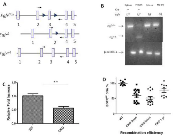
Egfrwa2/wa2 and Egfr null mice develop enlarged aortic valves, showing that EGRF signaling is required for normal valve remodeling processes. However, the enlarged valves complicate analysis of associated cardiac phenotypes that subsequently occur.24 To overcome this limitation, Egfr was deleted exclusively in cardiomyocyte by crossing mice carrying the conditional Egfrf allele with mice carrying the MHCcre allele, which drives high-efficiency cardiomyocyte specific Cre expression. To assess distribution of Cre expression in MHCcre/+
mice, MHCcre/+ mice were crossed with R26R mice to obtain MHCcre/+/R26R mice. After staining with x-gal, hearts from MHCcre/+/R26R mice showed β-galactosidase activity in the myocardium, but not in the valves. (Figure 2.1) Although MHCcre introduced recombination of the Egfrf allele in cardiomyocytes with high efficiency as determined by PCR of left ventricle DNA, some non-recombined Egfrf alleles remained in the left ventricle resulting in mosaicism of the heart muscle. (Figure 2.2 B) Recombination was not detected in other tissues. Quantitative RT-PCR analysis using total RNA prepared from left ventricles of three-month old mice
revealed that the expression of Egfr was significantly lower in Egfr CKO mice compared to their wild-type littermates. (Figure 2.2 C) Analysis of genotypes of offspring showed that Egfr CKO mice were present at the expected Mendelian ratios, indicating that loss of EGFR in
recombination efficiencies, we designed a recombination specific Taqman assay, Egfr KO, with primer and the probes located Intron 2, which is deleted in EgfrΔ. Quantitative RT-PCR using this assay in DNA extracted from LV showed various recombination efficiencies in different age groups with the lowest efficiency in one-year old mice, indicating that some mice with high recombination efficiency likely died before one year of age. (Figure 2.2 D) Consistent with this result, survival analysis showed that only 40% of Egfr CKO mice live to one year (n=35) compared with 76.9% for wild-type controls (n=26), and 75% of Egfr CKO mice live to six months of age (n=12) compared with 100% of controls (n=8). (Data not shown)
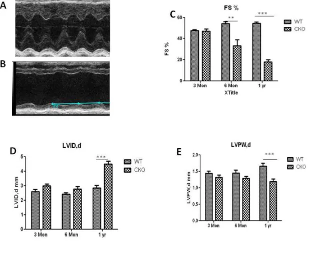
Egfr CKOmice develop severe heart dysfunction
Although Egfr CKOmice were born at Mendelian frequencies with normal cardiac morphogenesis at birth, and can live to adulthood. The life-span for the majority of Egfr CKO mice are less than one year because of sudden death. Physiological examination of the hearts from adult Egfr CKOmice using echocardiography showed an age-related dilated
echocardiographic examinations, which is a common symptom for dilated cardiomyopathy patients.
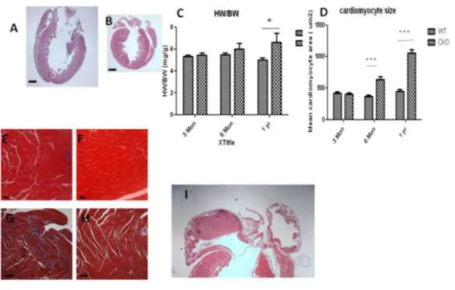
Histology of adult Egfr CKO hearts revealed several abnormal features that are common with heart from dilated cardiomyopathy patients. Ventricular chamber dilation was evident in
Egfr CKO mice at one year of age, as manifested by the significant reduction in LV thickness and the increased LV diameter. (Figure 2.4 A-B) Moreover, heart-to-body weight ratios in mice at one year of age were higher in Egfr CKO than control mice despite having similar body weights (41.83±2.6 g for CKO mice vs 39.7±1.8 g for control mice), indicating a hypertrophic growth in the heart. (Figure 2.4 C) Consistent with the increased heart-to-body weight ratio, the size of cardiomyocytes from CKO was significantly enlarged at six months and one year of age but not at three months. (Figure 2.4 D-F) Additionally, extensive interstitial fibrosis and atrial thrombus were also observed in the histological section from one-year-old Egfr CKOmice. (Figure 2.4 G,I)
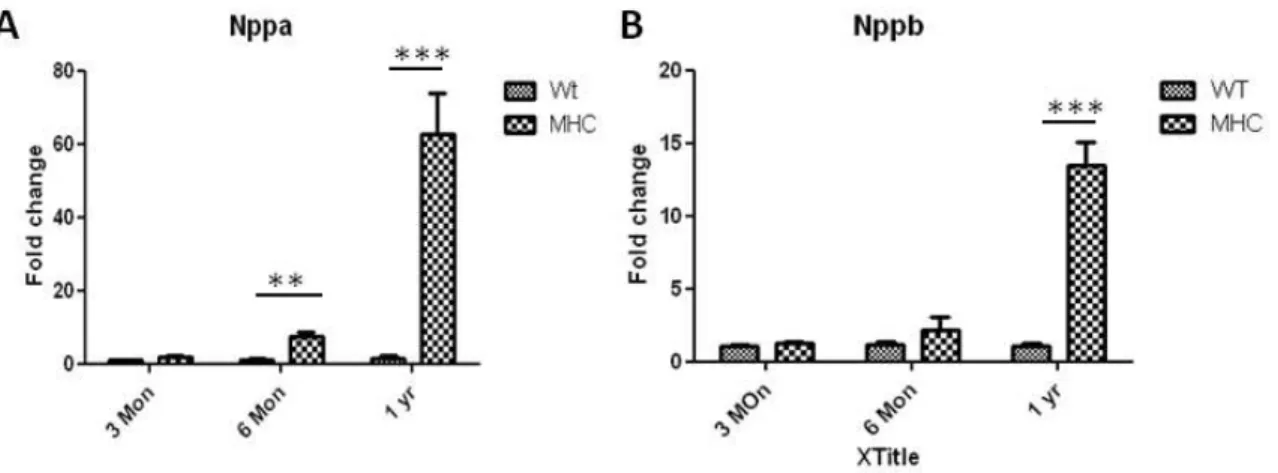
The expression of several hypertrophy-related genes, including atrial nautriuretic peptide (Nppa) and brain nautriuretic peptide (Nppb) have been used as sensitive and consistent
biomarkers for cardiac hypertrophy in humans and mice.28 The re-expression of ventricular NPPA and NPPB is known as a marker for the induction of the embryonic gene program in cardiomyocyte hypertrophy. The expression of NPPA and NPPB were not different between
compared to controls. (Figure 2.5 A,B) This expression pattern across age was correlated with cardiac function and cardiomyocyte cell size.
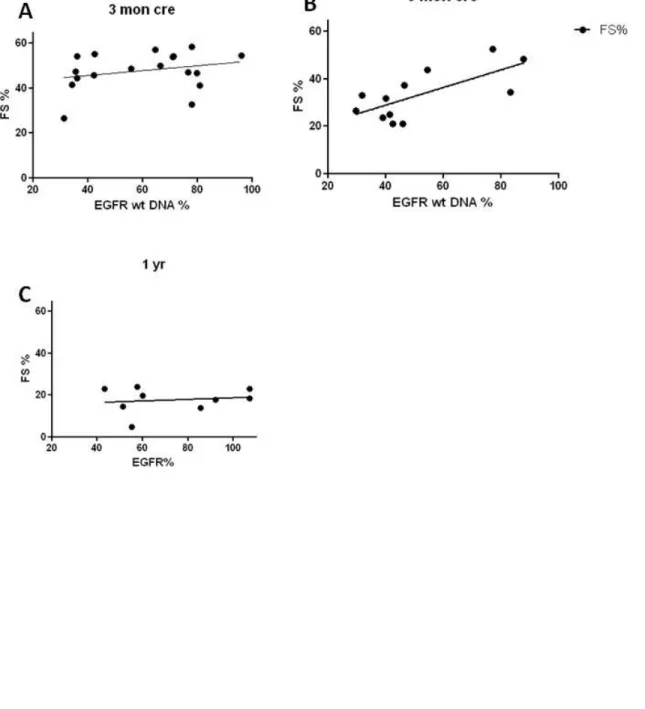
Severity of cardiac dysfunction in Egfr CKO mice associates with recombination efficiency The recombination efficiency varied among mice from 5-70%. (Figure 2.2 D) Because the heart is a mixture of different cell types including cells other than cardiomyocytes, the recombination efficiency for MHC-cre mice in total LV cannot be 100%. Since fractional shortening (FS%), a measurement of the pumping function of the heart, is the most direct and sensitive parameter for cardiac dysfunction, correlation between the recombination efficiency and cardiac dysfunction using linear regression analysis for Egfrwt % ( = 1- recombination efficiency) vs FS% for Egfr CKO mice at three months, six months and one year of age. (Figure 2.6 A-C)
Although the correlation between Egfrwt % and FS% was not significant at three months and one year of age groups, Egfrwt% showed a positive correlation with FS% in the six months of age group (p value < 0.0107; R2 = 0.4951). (Figure 2.6 B) The onset for cardiac dysfunction in
Egfr CKO mice occurs after three months of age, which is why no correlation was observed at young ages. Moreover, because only 40% of Egfr CKO mice live to one year, the correlation between recombination efficiency and FS% was likely weakened by loss of mice with high recombination levels die before one year of age.
Normal aortic valves in Egfr CKOmice
EGFR has vital roles in cardiac valve formation. Mice homozygous for Egfrwa2, a hypormorphic mutation in Egfr, develop semi-lunar valve thickening and aortic stenosis (AS). In order to rule out the possibility that cardiac dysfunction observed in Egfr CKO mice is due to AS, the morphology and function of aortic valves was assessed. Increased peak velocity across the aortic valve is a feature of aortic stenosis. Doppler tracing taken at the level of the aortic root revealed no peak velocity differences between Egfr CKO mice and littermate controls indicating normal aortic valve function in Egfr CKOmice. (Figure 2.7 A) Aortic valve diameter was also measured from H&E stained sections. Egfr CKOmice had similar aortic valve morphology and thickness compared with wild-type controls. (Figure 2.7 B)
then progresses to a late stage dilated cardiomyopathy with reduce FS% and high HW/BW. This pathological progression of Egfr CKO mice suggests that AS is not the cause of cardiac
dysfunction in Egfr CKO mice.
Apoptosis rates for cardiomyocytes is unchanged in Egfr CKOmice
Loss of cardiomyocytes can increase cardiac stress and can cause cardiac dysfunction and cardiomyocyte hypertrophy. Because activation of EGFR is known to confer cardio-protection by activating cell survival signaling pathways22, Quantitative RT-PCR and TUNEL assays were performed to investigate whether apoptosis rates are increased in Egfr CKO mice.29 To minimize effects on apoptosis caused by impaired cardiac function, we focus on the Egfr CKO mice at three months of age when they display no overt signs of heart dysfunction. Our results show there was no significant difference in TUNEL-positive cardiac cells between Egfr CKO and wildtype controls mice (Figure 2.9 A). Consistent with results from the TUNEL assay, expression of the pro-apoptotic gene Bax was also not significantly altered in Egfr CKO compared with control mice (Figure 2.9B).
Effect of Egfr deletion on gene expression in the hearts of conditional knockout mice To identify target genes of EGFR signaling in the heart, RNAs from the LVs of Egfr
CKO and wildtype mice were isolated and analyzed using Agilent microarrays. To avoid effects on gene expression caused by secondary changes such as cardiac hypertrophy, three-month-old mice were used (n = 6 for Egfr CKO, n = 6 for wildtype controls). The microarrays confirmed down-regulation of Egfr in the LV of Egfr CKO mice. No other ERBBfamily members showed altered expression between the Egfr CKO and wildtype mice. Although the expression of ACTA, a cardiac hypertrophy biomarker, was significantly up-regulated, expressions of other
show enrichment for the apoptosis pathway, consistent with TUNEL results, but did show over-representation of genes associated with the adhesions junction (p value = 0.02), including Ep300,
Actb, catenin and Snai2.
Egfr CKOmice are more sensitive to pressure overload
To determine the effect of cardiac-specific Egfr inactivation on the heart’s response to stress, pressure overload was induced by transverse aortic constriction (TAC) in three month-old wild-type and CKO male mice. Cardiac function was assessed in TAC mice and sham controls of each genotype by echocardiography. By two weeks post-surgery, TAC control mice developed compensated hypertrophy, reflected by increased contractile function, decreased LVEDD and LVEDS, and increased wall thickness (Figure 2.10). However, hearts from Egfr CKO mice showed reduced FS% without compensating with increased pressure in the LV after aortic banding. Interestingly, a subset of Egfr CKO mice developed dilated cardiamyopathy (20%, n = 10) two weeks post aortic binding, but none of the wild-type mice showed dilated heart even at six weeks post surgery (n = 8).
II.5 Discussion
Here a mouse model is described in which cardiomyocyte-restricted deletion of Egfr
leads to dilated cariomyopathy. Although EGFR is known to have an essential role in aortic valve development, Egfr CKO mice had normal valve function ruling out the possibility that observed cardiac dysfunction was secondary to valve abnormalities.24 The data reported here shows that EGFR is also important for maintaining cardiac function in adult hearts. Most Egfr