PHARMACOLOGICAL AND IMMUNOLOGICAL CONTROL OF ZIKA VIRUS IN MICE DEFICIENT IN ADAPTIVE IMMUNE RESPONSES
Nathaniel Schramm
A thesis submitted to the faculty at the University of North Carolina at Chapel Hill in partial fulfillment of the requirements for the degree of Maters of Science in the Department of Microbiology and Immunology
in the School of Medicine.
Chapel Hill 2019
Approved by:
J. Victor Garcia-Martinez Ralph Baric
iii
ABSTRACTNathaniel Schramm: Pharmacological and immunological control of Zika virus replication in mice deficient in adaptive immune responses.
(Under the direction of J. Victor Garcia-Martinez)
Zika virus (ZIKV) has recently demonstrated epidemic potential with prolonged infection, sexual and mother to fetus transmission, severe clinical manifestation of fetal microcephaly and congenital malformations and Guillain-Barré syndrome in adults. Existing small animal models for ZIKV infection
v
ACKNOWLEDGMENTS
vi
TABLE OF CONTENTS
LIST OF TABLES ……….… viii
LIST OF FIGURES ………...ix
LIST OF ABBREVIATIONS ……….……….…..x
CHAPTER 1: Investigating the susceptibility of immune competent and immune deficient mouse models to Zika virus infection ……….……….1
Introduction ………1
Section 1.1 Results ………...5
Immune competent BALB/c mice maintain detectable levels of Zika virus RNA in multiple tissues up to six months post exposure …….……….5
BALB/c mice do not completely clear ZIKV-RNA from the periphery when CD4+ and CD8+ T cells are depleted ………..6
Analysis of replication and long-term persistence of ZIKV in immune competent mice ………6
Immune competent BALB/c mice lose coordination and balance following intranasal ZIKV exposure ………7
Section 1.2 Discussion ………...9
Section 1.3 Figures and Tables ….………11
Section 1.4 Methods ………...17
Section 1.5 References ………..20
CHAPTER 2: Pharmacological and immunological control of Zika Virus replication in mice deficient in adaptive immune responses……….………24
Introduction ………..24
Section 2.1 Results ……….27
Immune deficient mice lacking T cells, B cells, and NK cells maintain high levels of ZIKV-RNA in the periphery ………27
vii
Systemic replication of ZIKV in tissues of immune deficient mice ………..29
ZIKV replication in the male and female reproductive tracts ………...29
7-Deaza-7-fluoro-2’-C-methyl-adenoside (DFMA) reduces viral burden and improves survival after ZIKV infection ………..…30
Pretreatment with C10, a neutralizing anti-ZIKV antibody, markedly reduces virus replication, shedding and overall plasma viral burden ………..30
A single dose of C10 greatly reduces ZIKV replication in tissues ……….31
C10 efficiently suppresses ZIKV replication in the male and female reproductive tracts ………32
Section 2.2 Discussion ………...33
Section 2.3 Author contributions and declaration of interests ………...……36
Section 2.4 Figures and tables …....……….37
Section 2.5 Methods ………...50
viii
LIST OF TABLESTable 1.1. Detectable Zika virus RNA in tissues of immune
ix
LIST OF FIGURES
Figure 1.1. Zika virus RNA in immune competent BALB/c mice ………...11 Figure 1.2. Zika virus RNA in the periphery of CD4 and CD8 depleted BALB/c mice ………..13 Figure 1.3. Control of ZIKV replication and persistence in BALB/c mice ……….14
Figure 1.4. Intranasal exposure to Zika virus induces neurological
degeneration in immune competent BALB/c mice ………..16
Figure 2.1. Immune deficient NSG mice are permissive to Zika virus
infection at a range of inoculum doses ………..37 Figure 2.2. Sustained viremia and viral shedding in the saliva of
ZIKV-infected immune deficient mice ………38 Figure 2.3. Analysis of systemic infection in immune deficient mice exposed to ZIKV ……….39
Figure 2.4 Treatment of ZIKV infected mice with DFMA reduces
viremia and improves survival ………41 Figure 2.5. C10 neutralizing antibody administration dramatically
reduces ZIKV replication and prevents viral shedding ………42 Figure 2.6. C10 antibody administration effectively reduces ZIKV
replication in tissues ……….44 Supplementary figure 2.1. Male and female mice were similar in mean
levels of peripheral ZIKV-RNA and survival ……….46
Supplementary figure 2.2. ZIKV isolated from infected mice efficiently
x
LIST OF ABBREVIATIONS
BM Bone marrow
CVL Cervical vaginal lavage
DENV Dengue virus
DFMA 7-Deaza-7-fluoro-2’-C-methyl-adenoside FRT Female reproductive tract
IEL Intraepithelial layer of the gastrointestinal tract
IFN Interferon
IP Intraperitoneal
IV Intravenous
LPL Lamina propria layer of the gastrointestinal tract NOD/SCID NOD.CB17-Prkdcscid/J
NOG NOD.Cg-Prkdcscid Il2rgtm15ug/JicTac
NSG NOD.Cg-Prkdcscid Il2rgtrm1Wjl/SzJ
1
CHAPTER 1: INVESTIGATING THE SUSCEPTIBILITY OF IMMUNE COMPETENT MOUSE MODELS TO ZIKA VIRUS INFECTION
Introduction
The flavivirus ZIKV envelope is highly dynamic and sensitive to pH changes, providing variable interaction sites with cellular receptors based on the degree of maturation of the virion (1, 2). This results in a large number of cell types being susceptible to ZIKV infection and wide tissue tropism (1, 3).
One of the most critical factors in replication of the ZIKV and production of mature virus particles is the pH changes from one intracellular compartment to the next. The structure of the ZIKV envelope protein, E, is particularly sensitive to changes in pH. After an endosome forms around the virus during cell entry, the low pH of 6.0 in the endosome causes structural rearrangements in the E glycoprotein, leading to the disassembly of the viral shell and fusion of the viral capsid (composed of C proteins) with the endosome (4). This fusion leads to the cytoplasmic release of the viral RNA (5).
The subgenomic ZIKV-RNA is then translated as a single polyprotein by cellular mechanisms. This polyprotein is cleaved into 3 structural (C, prM, and E) and 7 nonstructural proteins (NS1, NS2A, NS2B, NS3, NS4A, NS4B, and NS5) (6). The first of these cleavages is performed by NS3, which functions as a serine protease to auto-cleave the ZIKV polyprotein while associated with cofactor NS2B (7).
Genome replication, mRNA production, and initial assembly of the immature virion is initiated at the ER membrane (8). After cleavage, ZIKV NS1 localizes to the ER membrane, where it recruits
2
Newly replicated (+) sense ssRNA genomes generated at the replication factories associated with the ER membrane will associate with capsid C protein and become enveloped by glycoproteins prM and E, along with cellular lipids from the ER membrane. ZIKV NS2A also plays a critical role here facilitating the incorporation of ER-derived membrane lipids into budding virions (11).
This culminates in budding of the immature virion into the ER. From there, the virion is transported to the cis-golgi along the secretory pathway. There, cellular furin cleaves prM into the pr peptide and the membrane protein M (12). Critically, this results in further structural rearrangement of the E protein as the virion approaches full maturation. Normally, the low pH (6.7 at cis-golgi – 6.0 at trans-golgi) in the golgi compartments would induce changes in the E protein leading to virion disassembly as happens in the endosome of a newly infected cell, but the pr peptide remains associated with the E protein at this low pH to prevent premature fusion (12). The secretory pathway continues and culminates in the release of the virion outside of the cell. Once outside of a golgi-derived vesicle, the higher pH outside of the cell allows for the dissociation of the pr peptide from the virion shell and the final maturation of the glycoprotein shell into a mature ZIKV protected by self-dimerized E proteins (12).
The flavivirus envelope maturation states have been well characterized in DENV, and a recent, comprehensive review proposes that based on sequence similarity and characterization, the ZIKV envelope shares these traits (1). Typically, the flavivirus particle is surrounded by M and E protein homodimers which are responsible for interacting with cell receptors for entry (13). However, prM cleavage is inefficient, resulting in a heterogeneic viral envelope (14). These viruses are still infection-competent, even when the shell is not in the fully matured state (1). In those cases, alternating conformations of the E proteins, the pr peptide, and the M protein can each be exposed for interacting with cell receptors (1). For example, a neutral pH ER during replication can cause final presentation of trimeric pr-M projections (2).
clatherin-3
mediated endocytosis begins (16, 17). Of course, ZIKV has also been strongly linked to congenital malformation and Guillain-Barré syndrome as a result of its ability to directly infect neural progenitor cells and impair their proliferation (18). So, depending on which cell types are infected, different disease outcomes will be produced.
One chronic ZIKV-associated condition is microcephaly. ZIKV-associated microcephaly occurs with the highest incidence when mothers are infected with ZIKV during the first trimester of pregnancy (19). ZIKV is unique among flaviviruses in that it infects placental tissue and new virions are secreted from there directly into fetal capillaries, allowing access to the immune privileged fetal tissue (19). This trimester is when critical neural development would normally occur but can be impaired by ZIKV infection (20). One possible cause of the neurodevelopmental impairment is the ability of ZIKV NS2A to disrupt cortical neurogenesis via degradation of the adherens junction complex (18). A case study compiled using Brazilian patient data proposes that pregnant women infected with ZIKV are 8.6 times more likely to have a child who developed microcephaly compared to non-infected women (21).
In adults, ZIKV infection can sometimes lead to Guillain-Barré syndrome. Guillain-Barré is an autoimmune disorder which occurs infrequently in adults following infection or vaccination. However, South American case studies have reported higher incidence of Guillain-Barré syndrome during ZIKV epidemics, with increased severity and morbidity compared to patients who did not present with ZIKV infection, though the retroactive nature of the studies has made specific statistical estimates imprecise (22, 23). Although the precise mechanism of ZIKV-associated Guillain-Barré remains unidentified, it is likely related to the potential for ZIKV to broadly target C-type lectin receptors which are highly expressed in immune cells (15). Additionally, patients previously infected with DENV are at risk for an antibody dependent enhancement of ZIKV, which can greatly increase the risk of severe disease complications (24, 25).
4
27). We sought to characterize a mouse model with a Zika virus pathology that better mimics the nonlethal, persistent nature of human infection.
5
Results
Immune competent BALB/c mice maintain detectable levels of Zika virus RNA in multiple
tissues up to six months post exposure. Due to the capacity for mouse IFN to effectively restrict ZIKV,
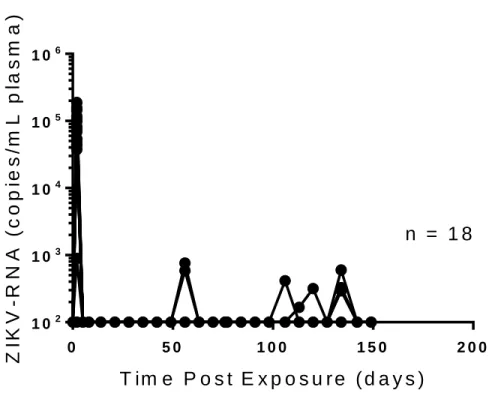
the virus is rapidly cleared in the periphery of immune competent mice. We exposed a group of male BALB/c mice (n=18) intravenously to H/PF/2013 (5.0 x 10⁵ FFU).
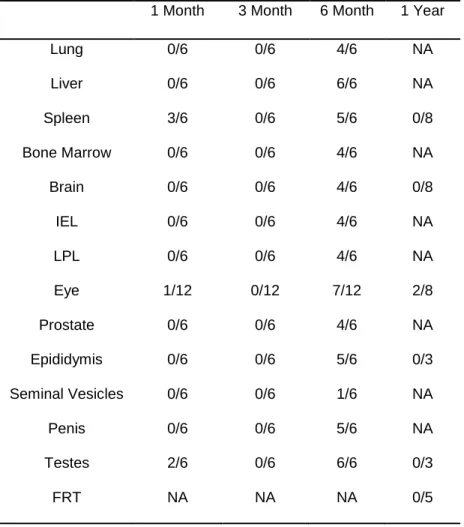
At one, three, and six months post exposure, tissues were harvested from six mice. The tissues we assessed for localized ZIKV-RNA from each mouse were lung, liver, spleen, bone marrow, brain, intraepithelial layer of the gastrointestinal tract (IEL), lamina propria layer of the gastrointestinal tract (LPL), eye, prostate, epididymis, seminal vesicles, prostate, penis, and testes (Table 1.1) At one month post exposure, low levels of ZIKV-RNA were detected in 4/6 mice. Virus was found in 3/6 spleens, 2/6 testes (analyzed in pairs), and 1/12 eyes (two from each mouse, analyzed individually). The highest viral RNA levels recorded at this time was in the spleen of one of the mice, which had 36 copies of ZIKV-RNA per 105 cells. Three months post exposure, we did not detect ZIKV-RNA in any of the tissues tested. Of
the six animals randomly selected for necropsy at this time point, only one had detectable ZIKV-RNA in the periphery after 2 days post exposure, so the relative reduction in tissue resident ZIKV-RNA is not totally unexpected (Figure 1.1, Table 1.1). We were able to detect peripheral ZIKV-RNA more frequently between three and six months post exposure (Figure 1.1). This resulted in a greatly increased frequency of detectable ZIKV-RNA in all tissues tested at six months post exposure (Table 1.1). ZIKV-RNA was detectable in at least one tissue of all six mice analyzed and was most consistently detected in testes and liver (6/6 mice). It was also detected in the seminal vesicles of one mouse. The most viral RNA we detected at this time point was 1.3 x 10³ copies ZIKV-RNA/105 cells in the testes of one mouse.
In the periphery, the viral load was 8.1 x 10⁴ ± 1.2 x 10⁴ s.e.m. ZIKV-RNA copies/mL plasma two
days post exposure (Figure 1.1). By five days post exposure, ZIKV-RNA was undetectable in the
6
experimentation. Despite rapid peripheral control of ZIKV infection in immune competent BALB/c mice, persistent infection was maintained in some of the animals up to six months post exposure.
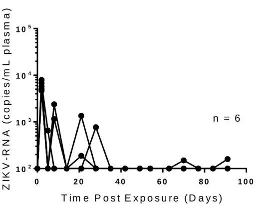
BALB/c mice do not completely clear ZIKV-RNA from the periphery when CD4+ and CD8+
T cells are depleted. Given the presence of neurological symptoms suggestive of encephalitis in the
intranasally exposed mice and the presence of detectable viremia long after exposure in intravenously exposed mice, we were very interested in the role that adaptive immune cells play in fighting ZIKV infection in BALB/c animals. To investigate the role of the adaptive immune system in BALB/c mice, we depleted CD4+ and CD8+ T cells prior to intravenous exposure (5.0 x 10⁵ FFU) with ZIKV H/PF/2013
(n=6). This depletion was maintained through the course of the experiment and regularly confirmed via flow cytometry of peripheral blood. Two days post exposure, the peripheral viral load in the six animals tested was 5.9 x 10³ ± 5.3 x 10² s.e.m. ZIKV-RNA copies/mL plasma compared to 8.1 x 10⁴ ± 1.2 x 10⁴
s.e.m. ZIKV-RNA copies/mL plasma in undepleted controls (p=0.0034 Mann-Whitney test) (Figure 1.1, Figure 1.2). Importantly, all six of the animals tested had detectable ZIKV-RNA in the periphery during the course of the experiment (91 days total) which presented as transient, low-quantity detectable viremia (Figure 1.2). This is in direct contrast to the undepleted controls (0-1 month: n=18, 1-3 months: n=12, 3-6 months: n=6), which presented only two instances of detectable viremia through 106 days post exposure (Figure 1.1).
One possible cause for this reduction in viral load with an increased frequency of detectable viremia in an immune compromised mouse is the demonstrated capacity for ZIKV to infect lymphocytes. By depleting CD4+ and CD8+ T cells, ZIKV was able to establish a somewhat more productive infection
but fewer targets of peripheral infection were provided during the initial exposure, resulting in less peripheral virus at two days post exposure compared to undepleted controls but more frequent instances of detectable viremia as the experiment progressed (Figure 1.1, Figure 1.2).
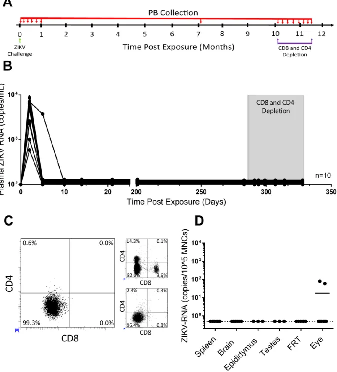
Analysis of replication and long-term persistence of ZIKV in immune competent mice.
Long-term persistence of ZIKV after peripheral clearance has not been adequately described. After considering the frequently detectable virus in our six month post exposure harvests (Table 1.1) and the impact of CD4+ and CD8+ T cell depletion on peripheral virus (Figure 1.2), we designed an experiment
7
peripheral clearance and depletion of CD4+ and CD8+ T cells. To evaluate ZIKV persistence in immune
competent mice, a group of BALB/c mice (n=10) were intravenously exposed to 5.0 x 10⁵ FFU ZIKV
H/PF/2013 (Figure 1.3A). Two days after exposure, ZIKV-RNA was detected in all animals (mean viral load of 4.9 x 10³ ± 3.2 x 10³ s.e.m. ZIKV-RNA copies/mL plasma) (Figure 1.3B). By ten days post
exposure, ZIKV-RNA was undetectable in the plasma of all animals. Plasma was monitored weekly for the first month after infection, and no viral rebound was detected. The mice were analyzed again at 201 and 283 days post exposure to confirm long-term suppression of viremia. Two male mice were found dead in their cage 130 and 268 days post exposure. Neither animal had evidence of ZIKV in plasma at the last timepoints analyzed and tissues could not be examined. To investigate the possible role of adaptive immune cell-mediated control of viral replication, starting 287 days post exposure, CD4+ and
CD8+ T cells were depleted from these animals for 42 days (Figure 1.3A). CD4+ and CD8+ T cell depletion
was confirmed in peripheral blood by flow cytometry (Figure 1.3C, left). Despite effective depletion of both CD4+ and CD8+ T cells, no viral rebound was detected in the periphery. Finally, at 329 days post ZIKV
exposure (42 days after CD4+ and CD8+ T cell depletion), necropsy was performed and flow cytometric
analysis of splenocytes was used to demonstrate efficient tissue CD4+ and CD8+ T cell depletion (Figure
1.3C bottom right). In addition, tissues where high ZIKV replication levels are observed in macaques or in humans were collected from the mice for real-time PCR analysis of localized viral persistence (brain, epididymis, testes or female reproductive tract [FRT] and eyes). No ZIKV-RNA was detected in any of the samples analyzed from spleen, brain, epididymis, testes or the FRT (Figure 1.3D). However, several hundred copies of ZIKV-RNA were detected in eyes from two mice. Our results demonstrate that despite efficient control and clearance in the periphery, in some animals ZIKV-RNA can persist in the eyes for almost a year post exposure.
Immune competent BALB/c mice lose coordination and balance following intranasal ZIKV
exposure. We performed intranasal exposures (1.0 x 10⁶ FFU ZIKV H/PF/2013) of immune competent
8
s.e.m. copies/mL plasma). At the last time point evaluated, only 2/4 BALB/c mice had detectable ZIKV-RNA (2.5 x 10⁴ ± 1.6 x 10⁴ s.e.m. copies/mL plasma), though it did increase ten-fold in the mice where it
remained detectable.
Mice were scored for neurological symptoms each day for a period of nine days after exposure: 1 – loss of balance/ataxia; 2 – hind-limb paralysis with forelimb clutching; 3 – hind-limb paralysis with forelimb weakness; 4 – limb paralysis and difficulty eating and drinking. Critically, loss of balance and coordination was detected in the ZIKV-exposed BALB/c animals starting three days post exposure (Figure 1.4B). Three- and four-days post exposure, loss of balance was recorded in 3/4 BALB/c mice and in 4/4 BALB/c mice from five days post exposure until the end of the experiment. The presence of
9
Discussion
The existence of infected reservoirs was reported in flavivirus infection before the recent ZIKV epidemic. Specifically, it has been reported in brain and nervous tissue up to 10 years after Siberian Tick Borne Encephalitis virus infection, in cerebrospinal fluid three weeks after infection and in PBMCs 8 months after infection with Japanese Encephalitis virus, and in donated tissue 40 days after infection and in urine six-and-a-half years after infection with West Nile virus (28-30). However, persistent ZIKV infection has a larger impact on public health because of the capacity for ZIKV to be sexually transmitted long after recovery from disease symptoms (31). ZIKV reservoirs have been confirmed in human eyes, testes, placenta, nervous tissue, and kidneys, with persistent shedding identified in vaginal secretions, urine, and semen (28, 31-37). These findings reinforce the need for a ZIKV animal model that can recapitulate human infection on an appropriate time scale.
In immune competent mice, the inability of NS5 to inhibit murine STAT2 results in a strong type I IFN response, suppression of virus replication and control of ZIKV infection (38, 39). This is consistent with our results in BALB/c mice intravenously exposed to ZIKV H/PF/2013. We determined that ZIKV can establish a persistent infection in immune competent BALB/c mice up to one-year post exposure despite initial clearance from the periphery after five days post exposure and no signs of illness at any point after infection. Critically, virus was detectable in critical tissues such as the brain, eyes, and male genital tract six months post exposure. Given the growing concern that persistent infection could magnify the risk of horizontal transmission in humans, long after the virus was believed to be cleared, a mouse model recapitulating this is of great importance (28). These findings highlight the lack of a ZIKV model for persistent infection on this time scale (28).
10
ZIKV-associated fetal microcephaly is a direct result of ZIKV infecting the brain and central nervous system (18, 19, 21). The capacity for ZIKV to establish infection in immune privileged tissues such as the testis, brain, and placenta, is well established, but infection of adult human nervous tissue is not thoroughly characterized (28). Commonly used mouse models of ZIKV infection demonstrate neurological symptoms such as limb paralysis and dragging (26). In our experiments, the intranasally exposed BALB/c mice developed ataxia, a symptom of encephalitis, just a few short days after exposure. All of the immune competent mice we tested showed loss of coordination and balance through the experiment, even when virus was not detectable in the plasma. Future experiments in this study have the potential to demonstrate large scale lymphocyte migration to the brain.
11
0 5 0 1 0 0 1 5 0 2 0 0
1 02 1 03 1 04 1 05 1 06
T im e P o s t E x p o s u r e ( d a y s )
Z
IK
V
-R
N
A
(
c
o
p
ie
s
/m
L
p
la
s
m
a
)
n = 1 8
Figure 1.1. ZIKV-RNA in plasma from BALB/c mice. Analysis of ZIKV-RNA in plasma of infected
BALB/c mice (n=18 mice). Mice were intravenously exposed to ZIKV H/PF/2013 (5.0 x 10⁵ FFU). Six mice
12
Table 1.1. Analysis of ZIKV-RNA in tissues from immune competent BALB/c mice up to one year
post exposure.
1 Month 3 Month 6 Month 1 Year
Lung 0/6 0/6 4/6 NA
Liver 0/6 0/6 6/6 NA
Spleen 3/6 0/6 5/6 0/8
Bone Marrow 0/6 0/6 4/6 NA
Brain 0/6 0/6 4/6 0/8
IEL 0/6 0/6 4/6 NA
LPL 0/6 0/6 4/6 NA
Eye 1/12 0/12 7/12 2/8
Prostate 0/6 0/6 4/6 NA
Epididymis 0/6 0/6 5/6 0/3
Seminal Vesicles 0/6 0/6 1/6 NA
Penis 0/6 0/6 5/6 NA
Testes 2/6 0/6 6/6 0/3
FRT NA NA NA 0/5
13
0 2 0 4 0 6 0 8 0 1 0 0
1 02 1 03 1 04 1 05
T im e P o s t E x p o s u r e ( D a y s )
Z
IK
V
-R
N
A
(
c
o
p
ie
s
/m
L
p
la
s
m
a
)
n = 6
Figure 1.2. ZIKV-RNA in periphery of CD4 and CD8 T cell depleted BALB/c mice. Analysis of
ZIKV-RNA in plasma from infected BALB/c mice (n=6 mice). Mice were intravenously exposed to ZIKV H/PF/2013 (5.0 x 10⁵ FFU). Mice were depleted of CD4+ and CD8+ T cells before exposure and through
the course of the experiment using anti-mCD4(GK1.5) and anti-mCD8 (2.43) antibody treatment. CD4+
and CD8+ T cell depletion was confirmed regularly by flow cytometry analysis of peripheral blood. Values
14
Figure 1.3. Control of ZIKV replication and persistence in BALB/c mice. (A) Experimental design.
BALB/c mice (5 males and 5 females) intravenously exposed to ZIKV H/PF/2013 (5.0 x 10⁵ FFU) were
monitored over time for the presence of ZIKV-RNA in peripheral blood (PB, small red arrows). Mice were depleted of CD4+ and CD8+ T cells at 286 days post-exposure using anti-mCD4+ (GK1.5) and anti-mCD8+
15
Shaded area represents the period of antibody treatment. (C) Flow cytometric analysis of peripheral blood confirming CD4+ and CD8+ T cell depletion in ZIKV-infected BALB/c mice after treatment with anti-T cell
depleting antibodies. Flow cytometric analysis showing the presence of T cells in untreated mice (top right) and demonstrating efficient depletion of splenocytes at the time of harvest (bottom right) are
included. Samples for flow cytometry analysis were gated as follows: singlets → live cells → mCD45+. (D)
ZIKV-RNA levels in tissues of BALB/c mice harvested 329 days post-exposure and 42 days after CD4+
and CD8+ T cell depletion (spleen, brain, eye, n = 8. Epididymis, testes, n = 3. Female reproductive tract n
16
0 2 4 6 8 1 0
1 02 1 03
1 04
1 05
1 06
T im e P o s t E x p o s u r e ( D a y s )
Z IK V -R N A ( c o p ie s /m L p la s m a )
B A L B /c v e h ic le B A L B /c Z I K V
0 2 4 6 8 1 0
0 1 2 3 4
T im e P o s t E x p o s u r e ( D a y s )
N e u ro lo g ic a l S y m p to m S c o re
B A L B /c v e h ic le
B A L B /c Z I K V
A
B
Figure 1.4. Intranasal exposure to ZIKV induces neurological symptoms in immune competent
BALB/c mice but not immune deficient NSG mice. (A) Analysis of ZIKV-RNA in plasma of ZIKV
exposed and vehicle control inoculated BALB/c (n=4 for each group). Mice were intranasally exposed to ZIKV H/PF/2013 (1.0 x 10⁶ FFU). Values were calculated using a standard curve.
17
Methods
Mice
BALB/c and immunodeficient NOD/SCID/γc-/- (NSG) mice were used for experiments at 12-20
weeks of age. Mice were maintained by the Division of Comparative Medicine at UNC-Chapel Hill according to protocol approved by the Institutional Animal Care and Use Committee.
Virus challenges
Stocks of ZIKV H/PF/2013 were prepared as previously described (50). Viral challenges were performed by diluting viral stocks in RPMI (Gibco, Gaithersburg, MD). Virus (300 FFU – 2.5 x 10⁵ FFU)
was administered intravenously via tail vein injection (200 µL volume). Virus (1.0 x 10⁶ FFU) for intranasal
challenge was delivered via micropipette directly into the nostrils (20 µL volume) while the mouse was held upside down.
Collection and processing of mouse bodily fluids
Mouse peripheral blood was collected longitudinally for ZIKV-RNA quantification. Peripheral blood was collected into tubes containing anti-coagulant (EDTA solution, Sigma-Aldrich, St. Louis, MD). Plasma was separated by centrifugation.
CD4+ and CD8+ T cell depletion and flow cytometry
Mouse CD4+ and CD8+ T cells were depleted by twice weekly intraperitoneal injections of 200 µg
anti-mouse CD4 (GK1.5) (Bio X Cell, West Lebanon, NJ) and 200µg anti-mouse CD8 (2.43) (Bio X Cell, West Lebanon, NJ) diluted in sterile PBS.
The antibodies used to analyze cells isolated from peripheral blood and spleens included
antibodies directed against mCD45 (APC-Cy 7, BD Pharmingen, Franklin Lakes, NJ, Cat. 559864), mCD3 (PE, BD Pharmingen, Franklin Lakes, NJ, Cat. 555275), mCD4 (APC, BD Pharmingen, Franklin Lakes, NJ, Cat. 560181), mCD8a (FITC, BD Pharmingen, Franklin Lakes, NJ, Cat. 553030), mCD19 (PE-Cy7, BD Pharmingen, Franklin Lakes, NJ, Cat. 552854) and mCD11b (PerCP, BD Pharmingen, Franklin Lakes, NJ, Cat. 550993). Live cells were distinguished by forward and side scatter profiles. Data was acquired with a BD FACSCanto flow cytometer and analyzed with BD FACS Diva software (v. 6.1.3).
18
Mouse tissues were collected essentially as previously described (42-45). Tissues collected for analysis (depending on mouse gender) included the spleen, bone marrow, lungs, liver, gastrointestinal tract, brain, eyes, FRT, epididymis, testes, prostate, penis, and seminal vesicles. For ZIKV-RNA analysis, tissues were processed into single cell suspensions as previously described (36-39). In brief, cells were isolated by forcing tissues through a 70 µm cell strainer (Falcon, Corning, NY) followed by red blood cell lysis if necessary. The liver, lung, female reproductive tract, and penis were digested in an enzyme digest cocktail prior to filtration. Liver, lung, and brain cells were purified with percoll gradients (GE Healthcare, Little Chalfont, UK). The mouse gastrointestinal tract was flushed with PBS and incubated with a dithiothreitol (Fisher Scientific, Hampton, NC) and EDTA solution to isolate cells from the intraepithelial layer and incubated with elastase (Worthington Biochemical, Lakewood, NJ) and hyaluronidase (Worthington Biochemical, NJ) to isolate the cells from the lamina propria layer (45).
ZIKV-RNA analysis
RNA was extracted from plasma (40 µL) using the QIAmp Viral RNA kit (Qiagen). Tissue RNA was extracted using RNeasy mini columns (Qiagen) according to the manufacture’s protocol including an optional treatment with RNase-free DNase. ZIKV-RNA levels in the peripheral blood plasma from infected mice were measured using a one-step quantitative real-time PCR (TaqMan® RNA to-CT 1-step kit, Applied Biosystems, Foster City, CA). The sequences of the forward and reverse primers and the TaqMan® probe for PCR amplification and detection of ZIKV RNA were: 5′CCGCTGCCCAACACAAG -3′, 5′-CCACTAACGTTCTTTTGCAGACAT --3′, and
5′-FAM-AGCCTACCT/ZEN/TGACAAGCAGTCAGACACACTCAA-Q-3′, respectively (46). ZIKV-RNA was
transcribed using a custom synthesized plasmid (Biomatik) to create a standard curve. Sample RNA was quantified by using a standard curve. All samples were run and analyzed on an ABI 7500 Fast Real Time PCR System (Applied Biosystems, Foster City, CA).
Statistical Analysis
19
To ensure appropriate blinding, the investigators involved in analyzing tissue samples for viral loads were given numbered samples and were thus unable to know treatment group identity (treated, controls). No other blinding procedures were used in the study. No statistical methods were used to pre-determined sample size. No randomization was used to allocate animals/samples to experimental groups.
20
REFERENCES1. Agrelli A, de Moura RR, Crovella S, Brandao LAC. 2019. ZIKA virus entry mechanisms in human cells. Infect Genet Evol 69:22-9.
2. Junjhon J, Lausumpao M, Supasa S, Noisakram S, Songjaeng A, Saraithong P, Chaichoun K, Utaipat U, Keelapang P, Kanjanahaluethai A, Puttikhunt C, Kasinrerk W, Malasit P, Sittisombut N. 2008. Differential modulation of prM cleavage, extracellular particle distribution, and virus
infectivity by conserved residues at nonfurin consensus positions of the dengue virus pr-M junction. J Virol 82:10776-91.
3. Cruz-Oliveira C, Freire JM, Conceicao TM, Higa LM, Castanho MARB, Da Poian AT. 2015. Receptors and routes of dengue virus entry into the host cells. FEMS Microbiol Rev 39:155-70. 4. Bressanelli S, Stuasny K, Allison SL, Stura EA, Duquerroy S, Lescar J, Heinz FX, Rey FA. 2004.
Structure of a flavivirus envelope glycoprotein in its low-pH-induced membrane fusion conformation. EMBO J 23(4):728-38.
5. Modis Y, Ogata S, Clements D, Harrison SC. 2004. Structure of the dengue virus envelope protein after membrane fusion. Nature 427(6972): 313-9.
6. Lindenbach BD, Rice CM. 2001. Flaviviridae: The Viruses and Their Replication. Fields Virolofy 4th edn. Knipe DM, Howley PM, editors. pp 991-1041.
7. Murray CL, Jones CT, Rice CM. 2008. Architects of assembly: rls of Flaviviridae non-structural proteins in virion morphogenesis. Nat Rev Microbiol 6(9):699-708.
8. Cortese M, Goellner S, Acosta EG, Neufeldt CJ, Oleksiuk O, Lampe M, Haselmann U, Funaya C, Schieber N, Ronchi P, Schorb M, Pruunsild P, Schwab Y, Chatel-Chaix L, Ruggieri A,
Bartenschlager R. 2017. Ultrastructural characterization of zika virus replication factories. Cell Rep 18(9):2113-23.
9. Youn S, Ambrose RL, Mackenzie JM, Diamond MS. 2013. Non-structural protein-1 is required for west nile virus replication complex formation and viral RNA synthesis. Virol J 10:339.
10. Lescar J, Soh S, Lee LT, Vasudevan SG, Kang C, Lim SP. 2018. The dengue virus replication complex: from RNA replication to preotein-protein interactions to evasion of innate immunity. Adv Exp Med Biol 1062:115-29.
11. Leung JY, Pijlman GP, Kondratieva N, Hyde J, Mackenzie JM, Khromykh AA. 2008. Role of nonstructural protein NS2A in flavivirus assembly. J Virol 82:4731-41.
12. Nambala P, Wen-Chi Su. 2018. Role of zika virus prM protein in viral pathogenicity and use in vaccine development. Front Microbiol 9:1797.
13. Yu IM, Zhang W, Holdaway HA, Li L, Kostyuchenko VA, Chipman PR, Kuhn RJ, Rossmann MG, Chen J. 2014. Structure of the immature dengue virus at low pH primes proteolytic maturation. Science 80(319):1834-7.
14. Rey A, Stiasny K, Heinz FX. 2017. Flavivirus structural heterogeneity: implications for cell entry. Curr Opin Virol 24:132-9.
21
16. Mercer J, Helenius A. 2010. Apoptotic mimicry: phosphatidylserine-mediated micropinocytosis of vaccinia virus. Ann NY Acad Sci 1209:49-55.
17. Kaksonen M, Roux A. 2018. Mechanisms of clathrin-mediated endocytosis. Nat Rev Mol Cell Biol 19(5):313-26.
18. Yoon KJ, Song G, Qian X, Pan J, Xu D, Rho HS, Kim NS, Habela C, Zheng L, Jacob F, Zhang F, Lee EM, Huang WK, Ringeling FR, Vissers C, Li C, Yuan L, Kang K, Kim S, Yeo J, Cheng Y, Liu S, Wen Z, Qin CF, Wu Q, Christian KM, Tang H, Jin P, Xu Z, Qian J, Zhu H, Song H, Ming GL. 2017. Zika-virus-encoded NS2A disrupts mammalian cortical neurogenesis by degrading adherens junction proteins. Cell Stem Cell 21:349-58.
19. Cao B, Diamond MS, Mysorekar IU. 2017. Maternal-Fetal Transmission of Zika Virus: Routes and Signals for Infection. J Interferon Cytokine Res 37(7):287-94.
20. Coelho AVC, Crovella S. 2017. Microcephaly Prevalence in Infants Born to Zika Virus-Infected Women: A Systematic Review and Meta-Analysis. Int J Mol Sci 18(8):1714.
21. De Araújo TVB, Rodrigues LC, de Alencar Ximenes RA, de Barros Miranda-Filho D, Montarroyos UR, de Melo APL, Valongueiro S, de Albuquerque FPM, Souza WV, Braga C, et al. 2016. Association between zika virus infection and microcephaly in Brazil, January to May, 2016: preliminary report of a case-control study. Lancet Infect Dis 16:1356–63.
22. Dirlikov E, Major CG, Medina NA, Lugo-Robles R, Matos D, Muñoz-Jordan JL, Colon-Sanchez C, Garcia M, Olivero-Segarra M, Malave G, Rodríguez-Vega GM, Thomas DL, Waterman SH, Sejvar JJ, Luciano CA, Sharp TM, Rivera-García B. 2018. Clinical Features of Guillain-Barré Syndrome With vs Without Zika Virus Infection, Puerto Rico, 2016. JAMA Neurol 75(9):1089–97.
23. Rivera-Concepcion JR, Betancourt JP, Cerra J, Reyes E. 2018. the zika virus: an association to guillain-Barré syndrome in the united states – a case report. P R Health Sci J. 37(Special Issue): S93-5.
24. Hermanns K, Gohner C, Kopp A, Schmidt A, Merz WM, Markert UR, Junglen S, Drosten C. 2018. Zika virus infection in human placental tissue explants is enhanced in the presence of dengue virus antibodies in-vitro. Emerg Microbes Infect 7(1):198.
25. Dejnirattisai W, Supasa P, Wongwiwat W, Rouvinski A, Barba-Spaeth G, Duangchinda T,
Sakuntabhai A, Cao-Lormeau V, Malasit P, Rey FA, Mongkolspaya J, Screaton G. 2016. Dengue virus sero-cross reactivity drives antibody-dependent enhancement of infection with zika virus. Nat Immunol 17(9):1102-8.
26. Lazear HM, Govero J, Smith AM, Platt DJ, Fernandez E, Miner JJ, Diamond MS.2016. A Mouse Model of Zika Virus Pathogenesis. Cell Host Microbe 19:720-30.
27. Rossi SL, Tesh RB, Azar SR, Muruato AE, Hanley KA, Auguste AJ, Langsjoen RM, Paessler S, Vasilakis N, Weaver SC.2016. Characterization of a Novel Murine Model to Study Zika Virus. Am J Trop Med Hyg 94:1362-9.
28. Kalkeri R, Krishna KM. 2017. Zika virus reservoirs: implications for transmission, future outbreaks, drug and vaccine development [version 1; referees: 2 approved]. F1000Research 6:1850.
22
30. Ravi V, Desai AS, Shenoy PK, Satishchandra P, Chandramuki A, Gourie-Devi M. 1993.
Persistence of Japanese encephalitis virus in the human nervous system. J Med Virol 40(4):326-9.
31. Atkinson B, Thorburn F, Petridou C, Bailey D, Hewson R, Simpson AJ, Brooks TJ, Aarons EJ. 2017. Presence and persistence of zika virus RNA in semen, United Kingdom, 2016. Emerg Inf Dis 23(4):611-5.
32. Ma W, Li S, Jia L, Zhang F, Zhang Y, Zhang J, Wong G, Zhang S, Lu X, Liu M, Yan J, Li W, Qin C, Han D, Qin C, Wang N, Li X, Gao GF. 2017. Zika virus causes testis damage and leads to male infertility in mice. Cell 167(6):1511-24.
33. Bhatnagar J, Rabenack DB, Martines RB, Reagan-Steiner S, Ermias Y, Estetter LB, Suzuki T, Ritter J, Keating MK, Hale G, Gary J, Muehlenbachs A, Lambert A, Lanciotti R, Oduyebo T, Meaney-Delman D, Bolaños F, Saad EA, Shieh WJ, Zaki SR. 2017. Zika virus RNA replication and persistence in brain and placental tissue. Emerg Infect Dis 23(3):405-11.
34. Kodati S, Palmore TN, Spellman FA, Cunningjam D, Weistrop B, Sen HN. 2017. Bilateral posterior uveitis associated with zika virus infection. Lancet 389(10064):125-6.
35. Nicastri E, Castilletti C, Liuzzi G, Iannetta M, Capobianchi MR, Ippolito G. 2016. Persistent detection of zika virus RNA in semen for six months after symptom onset in a traveler returning from Haiti to Italy, February 2016. Euro Surveill 21(32):30314.
36. Rossini G, Gaibani P, Vocale C, Cagarelli R, Landini MP. 2017. Comparison of zika virus (ZIKV) RNA detection in plasma, whole blood and urine – Case series of travel-associated ZIKV infection imported to Italy, 2016. J Infect 75(3):242-5.
37. Sun JW, Zhong H, Guan D, Zhang H, Tan Q, Ke C. 2016. Presence of zika virus in conjunctivital fluid. Opthamol 134(11):1330-2.
38. Grant A, Ponia SS, Tripathi S, Balasubramaniam V, Miorin L, Sourisseau M, Schwarz MC, Sáchez-Seco MP, Evans MJ, Best SM, García-Sastre A. 2016. Zika virus targets human STAT2 to inhibit type I interferon signaling. Cell Host Microbe 19(6):882-90.
39. Kumar A, Hou S, Airo AM, Limonta D, Mancinelli V, Branton W, Power C, Hobman TC. 2016. Zika virus inhibits type-I interferon production and downstream signaling. EMBO Rep
17(12):1766-75.
40. Miner JJ, Sene A, Richner JM, Smith AM, Santeford A, Ban N, Weger-Lucarelli J, Manzella F, Rückert C, Govero J, Noguchi KK, Ebel GD, Diamond MS, Apte RS. 2016. Zika virus infection in mice causes panuveitis with shedding of virus in tears. Cell Rep 16(12):3208-18
41. Zhao Z, Yang M, Azar SR, Soong L, Weaver SC, Sun J, Chen Y, Rossi SL, Cai J. 2017. Viral retinopathy in experimental models of Zika infection. Invest Ophthalmol Vis Sci 58(10):4355-65. 42. Denton PW, Estes JD, Sun Z, Othieno FA, Wei BL, Wege AK, Powell DA, Payne D, Haase AT,
Garcia JV.2008. Antiretroviral pre-exposure prophylaxis prevents vaginal transmission of HIV-1 in humanized BLT mice. PLoS Med 5:e16.
43. Olesen R, Wahl A, Denton PW, Garcia JV.2011. Immune reconstitution of the female
23
44. Denton PW, Nochi T, Lim A, Krisko JF, Martinez-Torres F, Choudhary SK, Wahl A, Olesen R, Zou W, Di Santo JP, Margolis DM, Garcia JV.2012. IL-2 receptor gamma-chain molecule is critical for intestinal T-cell reconstitution in humanized mice. Mucosal Immunol 5:555-66.
45. Shanmugasundaram U, Kovarova M, Ho PT, Schramm N, Wahl A, Parniak MA, Garcia JV.2016. Efficient Inhibition of HIV Replication in the Gastrointestinal and Female Reproductive Tracts of Humanized BLT Mice by EFdA. PLoS One 11:e0159517.
24
CHAPTER 2: PHARMACOLOGICAL AND IMMUNOLOGICAL CONTROL OF ZIKA VIRUS REPLICATION IN MICE DEFICIENT IN ADAPTIVE IMMUNE RESPONSES – REFORMATTED
MANUSCRIPT DRAFT
Introduction
Zika virus (ZIKV) is a mosquito-transmitted small-enveloped positive-stranded RNA virus from the Flavivirus genus in the Flaviviridae family that has emerged as a human pathogen with epidemic potential (1). Until 2007, only sporadic outbreaks of ZIKV infection involving no more than a few persons had occurred, usually resulting in a mild infection causing a self-limiting fever, headache, myalgia, rash, and conjunctivitis (1). However, recent outbreaks in Micronesia in 2007 (2, 3), French Polynesia in 2013-2014 (4, 5), and the Americas in 2015-2016 (6-8) revealed that ZIKV infections, can be prolonged and cause more severe clinical consequences including Guillain-Barré syndrome in adults and microcephaly and congenital malformations in fetuses and newborn infants (9). Unlike other flaviviruses, ZIKV has the potential for significant horizontal transmission due to shedding in bodily fluids long after symptom onset (10-14). Given the severe clinical consequences and potential for ZIKV spread, vaccine development is important. However, development of clinical strategies needed for treatment during epidemics when prevention of infection is no longer an option is paramount. Animal models capable of recapitulating chronic and persistent ZIKV infection will be critical to understand pathogenic mechanisms of ZIKV infection and to evaluate novel treatment strategies.
25
Mice lacking the interferon alpha/beta receptor 1 (Ifnar1), like A129 mice, Ifnar1-/- C57BL/6 mice, mice deficient in Irf3, Irf5, and Irf7 or STAT2 deficient mice support ZIKV infection (16, 17, 20). When inoculated with African (MR 766 or Dakar 1984), Asian (H/PF/2013), or American (Brazil Paraiba_2015) ZIKV strains (16, 17, 20-22) these mice develop disease symptoms including hind limb weakness, paralysis, and death. Mice deficient in both type I and type II IFN receptors (AG129) show greater susceptibility and more severe disease following ZIKV infection (17, 23-26). Although severity of disease and lethality is age-dependent in these models, most mice die within 1 month of ZIKV infection (16, 17, 27).
In an alternative approach, type I IFN signaling in wild type (WT) C57BL/6 mice is suppressed with anti-IFNAR1 monoclonal antibody (mAb) prior to and after virus inoculation. Suppression, rather than complete abrogation, of the type I IFN response in this manner may more accurately reflect ZIKV infection and transmission in humans. Thisalso allows the analysis of other components of the immune response during ZIKV transmission (26). Anti-IFNAR1 mAb treated mice inoculated with ZIKV Dakar strains exhibited high levels of ZIKV-RNA in serum, weight loss and mortality compared to control mice (28). However, a less severe phenotype without weight loss or death after infection was observed when anti-IFNAR1 mAb treated mice were inoculated with an Asian ZIKV strain (H/PF/2013) (16). Similarly, WT mice treated with the anti-inflammatory steroid dexamethasone prior to and after intraperitoneal inoculation with a Puerto Rican ZIKV strain (PRVABC59) experienced weight loss, viremia, and a disseminated infection. Dexamethasone withdrawal after infection led to rapid deterioration of the mice that was associated with inflammation and injury in the brain, kidneys, and testes (29). An additional mouse model of ZIKV infection is the highly immune deficient anti-IFNAR1 mAb treated Rag1−/− (AIR) mouse. Rag1−/− mice lack adaptive immune responses but are not susceptible to ZIKV infection. The AIR variant requires anti-IFNAR1 antibody administration every 2-4 days and is permissive to ZIKV infection, with virus in the testes, spleen, and brain (30). Disease progression is slower in AIR mice compared to other mouse models permissive to ZIKV infection, severe weight loss is not observed until 14-17 dpi (22, 30). While the AIR model might be somewhat more relevant to the study of acute infection than the other models described above, none of those mouse strains seem to model chronic and
26
27
Results
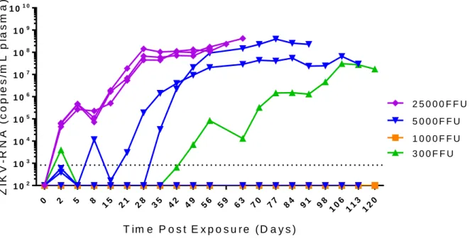
Immune deficient mice lacking T cells, B cells, and NK cells maintain high levels of
ZIKV-RNA in the periphery. W intravenously exposed immune deficient NSG mice that lack T cells, B cells,
and NK cells using decreasing inoculum doses; 2.5 x 10⁴ FFU, 5.0 x 10³ FFU, 1.0 x 10³ FFU, and 0.3 x
103 FFU ZIKV H/PF/2013. As NSG mice maintained a robust peripheral infection even at low inoculum
doses. At 0.3 x 10³ FFU, 2/3 mice had detectable ZIKV-RNA. One mouse had only a low quantity of
detectable virus two days post exposure which did not become detectable again. ZIKV-RNA was undetectable in the second mouse at that inoculum dose until 42 days post exposure, at which point it established a robust infection which was maintained through the course of the experiment (Figure 2.1). At 1.0 x 10³ FFU, 0/3 mice had detectable virus through 120 days post exposure. At 5.0 x 10³ FFU, 2/3 mice
had detectable virus by 35 days post exposure that replicated efficiently reaching as high as 108
ZIKV-RNA copies/mL plasma. The 2.5 x 10⁴ FFU group was the only one to have a 100% rate of infection by
two days post exposure. This was maintained for the course of the experiment and the mice achieved a similar peak viral load to the infected mice in the 5.0 x 10³ FFU group. Most striking, however, was the
protracted survival of the NSG mice despite highly productive infections compared to other commonly used mouse models of ZIKV infection.
The minimum inoculum dose that had 100% rate of infection in our samples sizes of n=3 was 2.5 x 10⁴ FFU ZIKV H/PF/2013. To ensure consistency of infection and pathology, we used a minimum of
10X that amount, 2.5 x 10⁵ FFU ZIKV H/PF/2013 in future experiments.
Sustained high-level systemic replication of ZIKV in immune deficient mice. To further
evaluate the importance of the adaptive immune system in ZIKV infection, we intravenously infected one mouse strain that is deficient in T cells and B cells (NOD/SCID) and two mouse strains that are deficient in T cells, B cells, and NK cells (NOG and NSG) with 0.5-1.0 x 10⁶ FFU of ZIKV H/PF/2013 (n = 4, 4, and
3, mice respectively, all females). Approximately 1000-fold higher levels of ZIKV-RNA in plasma
28
conditions, we observed similar mean levels of plasma ZIKV-RNA and survival in male and female mice. The logrank test for a sex effect was not statistically significant (p > 0.05) (Supplementary Figure 2.1A). The replication competence of the ZIKV found in plasma from infected NSG mice was verified both in vitro and in vivo. For in vitro analysis, serum from infected NSG mice (3uL) was added to Vero cells, and ZIKV-RNA was quantified in culture medium 24h, 48h, and 96h later. Levels of ZIKV-ZIKV-RNA in culture medium increased exponentially over time (Supplementary Figure 2.2A), indicating efficient replication of ZIKV from infected mice in VERO cell culture. For in vivo testing, serum was collected from 3 ZIKV infected NSG mice on days 21 and 28 post-inoculation. Serum was pooled, and 60 µl used for inoculation of two naïve NSG mice (Supplementary Figure 2.2B). Levels of ZIKV-RNA in plasma of serum-exposed mice increased rapidly and were maintained throughout the experiment (56 and 65 days, respectively) (Supplementary Figure 2.2C) providing further evidence that ZIKV in infected NSG mice was replication competent.
In humans, an important characteristic of ZIKV infection is the presence of virus in bodily
secretions (saliva, vaginal secretions, breast milk, urine, and semen)(31-34). We evaluated the presence of virus in the saliva of infected mice. High levels of ZIKV-RNA were found in saliva (Figure 2E).
Similarly, ZIKV-RNA was consistently present in the urine and cervicovaginal secretions of infected mice (Supplementary Table 2.1).
In contrast to BALB/c mice that survived for almost a year post infection, over time, immune deficient mice succumbed to infection (Figure 2.2F). All NOG mice died within the first 47 days post-infection (half-life 26 days post-exposure). The half-life of NOD/SCID and NSG mice was four weeks longer (54 and 56 days, respectively). ZIKV-infected male and female mice were similar in survival (Supplementary Figure 2.1B). The logrank test for a sex difference was not statistically significant (p = 0.83).
29
Together, these results demonstrate the susceptibility of immune deficient mouse strains to ZIKV infection without the need for preconditioning or any other type of intervention like anti-interferon treatment,
characterized by robust and sustained replication with a protracted half-life.
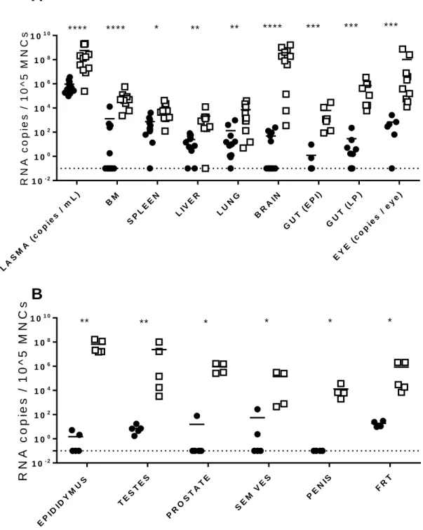
Systemic replication of ZIKV in tissues from immune deficient mice. To establish the
systemic replication of ZIKVduring acute and chronic infection, cell-associated ZIKV-RNA in infected NSG mice (n = 11 acute, n = 12 chronic) was evaluated using quantitative real-time PCR analysis from bone marrow, spleen, liver, lung, brain, gut epithelium, gut lamina propria, and the eye (Figure 2.3A). During acute infection (two days post exposure), high levels of ZIKV-RNA were detected in the plasma of all animals. However, levels of cell-associated ZIKV-RNA were variable in the different tissues analyzed. For example, whereas ZIKV-RNA was readily detected in the spleen, liver, lung, gut lamina propria and eyes from the majority of mice, ZIKV-RNA was only detected in the bone marrow and brain of
approximately half of the animals. In contrast, during chronic ZIKV infection (27-73 days post exposure) statistically significantly higher levels of virus were found in all tissues analyzed. Of these tissues, the brain (2.50 x 10⁸ ± 1.05 x 10⁸ s.e.m. ZIKV-RNA copies per 105 cells) and the eye (1.14 x 10⁸ ± 0.84 x 10⁸
s.e.m. ZIKV-RNA copies per eye) had the highest levels of ZIKV-RNA. These results demonstrate that ZIKV rapidly establishes a systemic infection in immune deficient mice that is maintained at very high levels in all tissues analyzed.
ZIKV replication in the male and female reproductive tracts. Given the importance of sexual
transmission of ZIKV, we also determined the presence of ZIKV in the male genital tract (testes,
30
the levels of ZIKV-RNA in the FRT were statistically significantly higher during chronic infection
(p=0.0159, Mann Whitney test). These results demonstrate that ZIKV is consistently present in the male and female reproductive tracts during acute ZIKV-infection and that ZIKV replication in sustained at high levels in these compartments during chronic infection.
7-Deaza-7-fluoro-2’-C-methyl-adenoside (DFMA) reduces viral burden and improves
survival after ZIKV infection. Currently there are no approved treatments for ZIKV infection. Recently,
the nucleoside DFMA (Supplementary Figure 2.3) was reported to have anti-ZIKV activity (35). The ability of ZIKV to replicate efficiently and for prolong periods of time in immune deficient mice allows for the in vivo evaluation of novel viral inhibitors like DFMA. Beginning two days prior to infection with ZIKV
H/PF/2013 (2.5 x 10⁵ FFU) (Figure 2.4), NSG mice were administered DFMA (n = 6, 10 mg/kg per day,
i.p.) or vehicle (n = 4) daily for 21 days (until 19 days post-exposure with ZIKV). Plasma ZIKV-RNA levels were monitored over time. At two days post exposure, the levels of viremia in DFMA treated and control groups were similar. However, as early as four days post-infection, ZIKV-RNA levels in the plasma of DFMA treated mice (9.41 x 10⁵ ± 1.40 x 10⁵ s.e.m. ZIKV-RNA) were statistically significantly lower
compared to control animals (2.00 x 10⁶ ± 2.90 x 10⁵ s.e.m. ZIKV-RNA) (Figure 2.4, panels A and B).
Statistically significantly lower levels of ZIKV-RNA were consistently observed in the plasma of DFMA treated mice compared to controls during the duration of treatment. Surprisingly, although DFMA
treatment was discontinued after 21 days, statistically significantly lower levels of viremia were observed in DFMA treated mice up to three weeks post-treatment discontinuation (Figure 2.4, A and B). All 4 animals in the control group succumbed to infection by 57 days post-exposure (Figure 2.4, C and D). Only 2/6 DFMA treated animals succumbed to infection (p < 0.05) (Figure 2.4, C and D). These results demonstrate that DFMA treatment reduces viremia and reduces mortality of ZIKV infected mice.
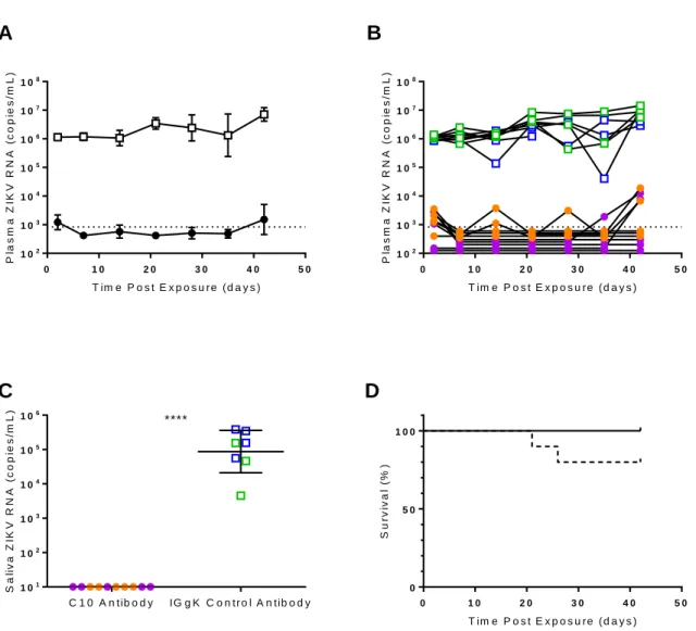
Pretreatment with C10, a neutralizing anti-ZIKV antibody, markedly reduces virus
replication, shedding and overall plasma viral burden. C10 is a dengue virus serotype
cross-neutralizing monoclonal antibody isolated from a dengue patient. It was previously shown to neutralize ZIKV in cell culture and reduce ZIKV-induced morbidity and mortality in a type I/II interferon receptor-knockout murine model (36). To investigate the effect of C10 pre-exposure prophylaxis on the
31
of C10 intraperitoneally (62.5 µg, n = 10) or control antibody (62.5 µg IgG, n = 9). Mice were exposed to ZIKV H/PF/2013 intravenously (2.5 x 10 5 FFU) 18 h after treatment and ZIKV infection was monitored in
peripheral blood for six weeks.
All mice treated with control antibody became infected. High levels of ZIKV-RNA were detected in the plasma of all control animals by two days post-exposure (1.15 x 10⁶ ± 5.23 x 10⁴ s.e.m. ZIKV-RNA
copies/mL) and viremia was maintained for six weeks (last time point analyzed) (Figure 2.5, panels A and B). In stark contrast, there was no evidence of peripheral blood infection in 7/10 C10-treated mice. Low levels of ZIKV-RNA were only transiently observed immediately after infection in three animals.
Specifically, ZIKV-RNA was not detected in the plasma from 7/10 C10-treated mice two days post-exposure and low levels (1,722 ± 307 copies/mL) were detected in three ZIKV-positive mice (Figure 2.5B). Seven days post exposure, plasma ZIKV-RNA levels were undetectable in all treated mice (Figure 2.5B). ZIKV-RNA was undetectable through 35 days post exposure in most of the animals, with only three transient instances of detectable viral-RNA in 2/10 animals. By six weeks post exposure, only 4/10 mice had low but detectable levels of ZIKV-RNA in plasma (5,125 ± 2,023 copies per mL).
C10 pre-exposure prophylaxis also efficiently inhibited ZIKV-RNA shedding. ZIKV-RNA levels were undetectable in saliva collected from all (10/10) C10-treated mice 30 days post ZIKV exposure. In contrast, high levels of ZIKV-RNA were present in the saliva of control mice (1.64 x 10⁵ ± 5.62 x 10⁴ s.e.m
copies/mL) (Figure 2.5C). During the course of the experiment (42 days), no C10-treated mice
succumbed to infection or showed any signs of illness. However, 2/9 control mice succumbed to infection by 26 days post-exposure (Figure 2.5D). These data demonstrate that a single dose of C10 administered prior to ZIKV exposure effectively inhibits ZIKV replication in vivo over an extended period of time and markedly reduces (>2,000-fold) ZIKV-RNA levels in plasma and saliva (p < 0.0001).
A single dose of C10 greatly reduces ZIKV replication in tissues. Successful ZIKV therapy
32
the gut lamina propria, brain, and eyes. In sharp contrast, almost all tissues analyzed from the treated mice had low levels of viral RNA or levels that were below the LOD. In most tissues of C10-treated mice, cell-associated ZIKV-RNA levels were 2-5 logs lower compared to control animals (Figure 2.6A). These results show that C10 effectively reduces the systemic levels of cell-associated ZIKV-RNA (Figure 2.6A).
C10 efficiently suppresses ZIKV replication in the male and female reproductive tracts.
Because of their relevance to sexual ZIKV transmission, we evaluated the effect of C10 pre-treatment on the levels of virus in the different organs of the male genital tract (testes, epididymis, prostate, penis, and seminal vesicles) and FRT. ZIKV-RNA was readily detected in the epididymis, penis, prostate, seminal vesicles and testes of all control male animals (Figure 2.6B). In contrast, ZIKV-RNA levels were
33
Discussion
During human ZIKV infection, the ZIKV NS5 protein inhibits STAT2, thereby suppressing the type I IFN response to ZIKV allowing for viral replication and dissemination. As a result, replication-competent infection has been verified in the female reproductive tract, the placenta and fetal tissue, in neural
progenitor and adult neural cells, and in immune privileged tissues such as the testes and the eyes. ZIKV shedding has also been observed in humans in breast milk, saliva, urine, semen, and cervical mucus, which may contribute to vectorless transmission (37). In newborns, ZIKV infection results in severe eye disease characterized by optic neuritis, chorioretinal atrophy and blindness (9, 38). In adults, ZIKV infection can result in conjunctivitis and uveitis (39, 40).
Growing evidence suggests that the immune response to ZIKV in mice is more complex than just type I interferon and that other components of the immune system may be able to control ZIKV infection. For example, WT C57BL/6 mice treated with anti-IFNAR antibodies have a suppressed, but not
completely deficient IFN response, and develop viremia when inoculated with Asian ZIKV strains. However, they do not lose weight or develop neurologic disease (16). Mice deficient in MAVS, an adaptor of cytosolic RIG-I–like receptors signaling, develop an acute infection after ZIKV exposure, but only experience significant weight loss when their CD4+ and CD8+ T cells are depleted (22). ZIKV-infected
AIR mice, Rag1-/- mice deficient in functional T and B cells and IFN responses suppressed with anti-IFNAR Abs, have high levels of viral RNA in the spleen, lymph nodes, and brain and exhibit significant weight loss. Thus, both adaptive and innate immune responses appear to be required to control ZIKV replication, its spread and the severity of disease symptoms.
As deficiency in the type I interferon pathway in mice results in severe ZIKV disease and death, mouse models with a deficient adaptive immune response and impaired innate immune response could prove valuable for the study of ZIKV infection with a delayed onset of disease. NSG mice have a scid mutation on the NOD/ShiLtJ genetic background and a complete null allele of the IL2 receptor common gamma chain (IL2rgnull). This renders NSG mice B and T cell deficient, prevents cytokine signaling
34
days post-infection that was sustained for up to 91 days (last time point analyzed). ZIKV-RNA was detected in multiple tissues as early as 2 days post-infection, suggesting rapid systemic dissemination. Although the majority of ZIKV-infected NSG mice (75%) eventually succumbed to infection, the half-life of ZIKV-infected NSG mice was 56 days, much longer than other murine models, where the majority of the animals died within 30 days post ZIKV infection (16, 17, 27). Immune deficient NOG mice are
phenotypically similar to NSG mice but they express a truncated form of the protein instead of a deletion of the common gamma chain receptor. NOG mice intravenously inoculated with ZIKV H/PF/2013 developed very similar viremia compared to NSG mice, except that all of the animals succumbed to infection by 26 days post-infection. NOD/SCID mice are the parental strain of both NSG and NOG mice and have an intact common gamma chain. When infected with ZIKV, NOD/SCID mice also developed sustained plasma viremia but had a lower initial plasma viral load compared to NSG mice. However, during chronic infection, their plasma viral load, half-life and survival rate was similar to NSG mice. These results suggest a minimal role for the common gamma chain in the progression of ZIKV infection in NSG or NOG mice.
ZIKV infection of immune deficient mice lacking T cells, B cells and NK cells is therefore
characterized by: 1) a rapid increase in plasma viremia and subsequent maintenance of a high viral load that allows for the evaluation of therapeutic strategies targeting ZIKV infection during acute and chronic stage of infection; 2) early compartmentalization resulting in very high ZIKV loads in the brain, eye, testes, epididymis, and FRT during chronic stage of infection which is useful for the evaluation of tissue
penetration and efficacy of ZIKV therapeutics; 3) ZIKV shedding in saliva, cervicovaginal secretions, and urine, which allows assessment of the role of therapeutics in prevention of sexual and non-sexual ZIKV transmission as well as persistence; and 4) Immune deficient mice do not need anti-interferon treatment before or during ZIKV infection, greatly simplifying the use of this model and lowering its cost.
35
monoclonal antibody (36, 42), prior to ZIKV H/PF/2013 infection had dramatically reduced levels of plasma viremia immediately after exposure. ZIKV-RNA levels in plasma were below or near the LOD for five weeks after infection. Importantly, C10 treatment also prevented ZIKV-RNA shedding in saliva and was very effective in reducing ZIKV-RNA levels in immune privileged tissues including the brain, eye and genital tract. Immune privileged organs are critical to the study of ZIKV drug effectiveness because they have been noted for having some of the highest levels of ZIKV during infection. These results show that C10 treatment could be used as an approach to control life-threatening ZIKV infection (43). It also points to the possibility of repurposing existing treatment for ZIKV (44, 45). This is supported by recent studies showing that sofosbuvir, an FDA-approved nucleotide analog inhibitor of the hepatitis C RNA-dependent RNA polymerase has protective effect against ZIKV in vitro and in vivo (46-49).
36
Author contributions: Conceptualization, N.J.S, A.S.C., R.F.S., A.W., R.S.B., M.K., and J.V.G.;
Investigation, N.J.S, A.S.C, W.O.T., R.A.S., P.T.H, and J.B.H., Writing – Original Draft, N.J.S., M.K., and J.V.G.; Writing – Review & Editing, A.S.C., R.F.S., A.W., R.S.B. and J.V.G; Funding Acquisition,
R.S.B.and R.F.S.; Resources, A.S.C., F.A., L.B, R.S.B., and R.F.S., Supervision, R.S.B., R.F.S., and J.V.G.
37
0 2 5 8 15 21 28 35 42 49 56 59 63 70 77 84 91 98 10
6 11
3 12
0 1 02
1 03
1 04
1 05 1 06 1 07
1 08
1 09 1 01 0
T im e P o s t E x p o s u r e ( D a y s )
Z
IK
V
-R
N
A
(
c
o
p
ie
s
/m
L
p
la
s
m
a
)
3 0 0 F F U 2 5 0 0 0 F F U
5 0 0 0 F F U
1 0 0 0 F F U
Figure 2.1. Immune deficient NSG mice are permissive to ZIKV infection. Analysis of ZIKV-RNA in
plasma NSG mice exposed to ZIKV H/PF/2013 (n=3 mice in each group). Mice were intravenously exposed to ZIKV H/PF/2013 using 2.5 x 10⁴ FFU (Purple diamonds), 5.0 x 10³ FFU (Blue inverted
triangles), 1.0 x 10³ FFU (Orange squares), and 0.3 x 10³ FFU (Green triangles). The dashed line
38
0 2 0 4 0 6 0 8 0
1 02
1 03
1 04
1 05
1 06
1 07
1 08
1 09
T im e P o s t E x p o s u r e ( D a y s )
P la s m a Z IK V -R N A ( c o p ie s /m L )
0 2 0 4 0 6 0 8 0
1 02
1 03
1 04
1 05
1 06
1 07
1 08
1 09
T im e P o s t E x p o s u r e ( D a y s )
P L a s m a Z IK V -R N A ( c o p ie s /m L )
0 2 0 4 0 6 0 8 0
1 02
1 03
1 04
1 05
1 06
1 07
1 08
1 09
T im e P o s t E x p o s u r e ( D a y s )
P la s m a Z IK V -R N A ( c o p ie s /m L )
0 2 0 4 0 6 0 8 0
1 02
1 03
1 04
1 05
1 06
1 07
1 08
1 09
T im e P o s t E x p o s u r e ( D a y s )
P la s m a Z IK V -R N A ( c o p ie s /m L )
0 1 0 2 0 3 0 4 0 5 0 6 0 7 0
1 02
1 03
1 04
1 05
1 06
1 07
T im e P o s t E x p o s u r e ( D a y s )
S a li v a Z IK V -R N A ( c o p ie s /m L )
0 5 0 1 0 0
0 5 0 1 0 0
T im e P o s t E x p o s u r e ( D a y s )
S u rv iv a l (% )
0 5 1 0 1 5 2 0 2 5
1 02
1 03
1 04
1 05
1 06
1 07
T im e P o s t E x p o s u r e ( D a y s )
P la s m a Z IK V -R N A ( c o p ie s /m L )
0 5 1 0 1 5 2 0 2 5
1 02
1 03
1 04
1 05
1 06
1 07
T im e P o s t E x p o s u r e ( D a y s )
P la s m a Z IK V -R N A ( c o p ie s /m L )
0 5 1 0 1 5 2 0 2 5
1 02
1 03
1 04
1 05
1 06
1 07
T im e P o s t E x p o s u r e ( D a y s )
P la s m a Z IK V -R N A ( c o p ie s /m L )
C
B
A
D
E
F
G
H
I
Figure 2.2. Sustained plasma viremia and viral shedding in the saliva of ZIKV-infected immune
deficient mice. ZIKV-RNA levels in the plasma of (A) NOD/SCID (n = 4 females), (B) NOG (n = 4
females) and (C-D) NSG (n = 3 females in panel C and n = 3 males in panel D) immune deficient mice intravenously exposed to ZIKV H/PF/2013 (0.5 – 1.0 x 10⁶ FFU). (E) ZIKV-RNA levels in saliva of
NOD/SCID (n = 4 females, green triangles), NOG (n = 4 females, blue squares) and NSG (n = 3 females, purple open circles and n = 3 males, purple closed circles) mice. (F) Kaplan-Meier plot comparing survival of ZIKV-infected BALB/c (n = 10 mice, black line) and immune deficient NOD/SCID (n = 4 mice, green line) NOG (n = 4 mice, blue line) and NSG (n = 6 mice, purple line) mice. ZIKV-RNA concentration in plasma from NSG mice intravenously exposed to ZIKV strain (G) PRVABC59 (n = 4 mice), (H) SPH2015 (n = 4 mice), and (I) BeH 819015 (n = 4 mice) (5.0 x 10⁵ FFU). ZIKV-RNA was quantified by RT-PCR and
39
EP IDID YM US TE ST ES PR OS TA TE SE M VES
PE
NIS FR
T
1 0- 2
1 00 1 02
1 04
1 06 1 08
1 01 0
R
N
A
c
o
p
ie
s
/
1
0
^
5
M
N
C
s
** ** * * * *A
B
PL AS MA (c op ies / mL) BM SP LE EN LIV ER LU NG BR AIN GU
T ( EP
I)
GU T (
LP )
EY E (
co pie
s / ey
e)
1 0- 2
1 00 1 02
1 04
1 06 1 08
1 01 0
R N A c o p ie s / 1 0 ^ 5 M N C
s **** **** * ** ** **** *** *** ***
Figure 2.3. Analysis of systemic infection in immune deficient mice exposed to ZIKV. NSG mice
were intravenously exposed to ZIKV H/PF/2013 (0.25 – 1 x 10⁶ FFU) and tissues collected during acute