Associations
Among
25-Hydroxyvitamin
D
Levels,
Lung
Function,
and
Exacerbation
Outcomes
in
COPD
An
Analysis
of
the
SPIROMICS
Cohort
RobertM.Burkes,MD;AgatheS.Ceppe,MS;ClaireM.Doerschuk,MD;DavidCouper,PhD;EricA.Hoffman,PhD; AlejandroP.Comellas,MD;R.GrahamBarr,MD,PhD;JerryA.Krishnan,MD,PhD;ChristopherCooper,MD; WassimW.Labaki,MD;VictorE.Ortega,MD,PhD;J.MichaelWells,MD;GerardJ.Criner,MD;
PrescottG.Woodruff,MD,MPH;RussellP.Bowler,MD,PhD;CherylS.Pirozzi,MD;NadiaN.Hansel,MD;
RobertA.Wise,MD;ToddT.Brown,MD,PhD;andM.BradleyDrummond,MD,MHS;fortheSPIROMICSInvestigators*
BACKGROUND: The relationship between 25-hydroxyvitamin D (25-OH-vitamin D) and COPD outcomes remains unclear. Using the Subpopulations and Intermediate Outcome Measures in COPD Study (SPIROMICS), we determined associations among baseline 25-OH-vitamin D and cross-sectional and longitudinal lung function and COPD exacerbations. METHODS: Serum 25-OH-vitamin D level was measured in stored samples from 1,609 SPI-ROMICS participants with COPD. 25-OH-vitamin D levels were modeled continuously and dichotomized as deficient (<20 ng/mL) vs not deficient ($20 ng/mL). Outcomes of interest included % predicted FEV1 (current and 1-year longitudinal decline) and COPD exacerba-tions (separately any and severe, occurring in prior year and first year of follow-up). RESULTS:Vitamin D deficiency was present in 21% of the cohort and was more prevalent in the younger, active smokers, and blacks. Vitamin D deficiency was independently associated with lower % predicted FEV1(by 4.11%) at enrollment (95% CI,–6.90% to–1.34% predicted FEV1;P¼.004), 1.27% predicted greater rate of FEV1decline after 1 year (95% CI,–2.32% to –0.22% predicted/y;P¼.02), and higher odds of any COPD exacerbation in the prior year (OR, 1.32; 95% CI, 1.00-1.74;P¼.049). Each 10-ng/mL decrease in 25-OH-vitamin D was associated with lower baseline lung function (–1.04% predicted; 95% CI, –1.96% to –0.12% predicted; P ¼ .03) and increased odds of any exacerbation in the year before enrollment (OR, 1.11; 95% CI, 1.01-1.22;P ¼.04).
CONCLUSIONS:Vitamin D deficiency is associated with worse cross-sectional and longitudinal lung function and increased odds of prior COPD exacerbations. Thesefindings identify 25-OH-vitamin D levels as a potentially useful marker of adverse COPD-related outcomes.
CHEST 2020; 157(4):856-865
KEY WORDS:COPD; COPD epidemiology; COPD exacerbations; lung function; vitamin D
FOR EDITORIAL COMMENT, SEE PAGE 755
AFFILIATIONS: From the Division of Pulmonary Diseases and Critical Care Medicine (Drs Burkes, Doerschuk, and Drummond), University of North Carolina at Chapel Hill, Chapel Hill, NC; the Marsico Lung Institute (Ms Ceppe and Drs Doerschuk and Drummond), University of North Carolina at Chapel Hill, Chapel Hill, NC; the University of North Carolina Gillings School of Global Public Health (Dr Couper),
ABBREVIATIONS: 25-OH-vitamin D = 25-hydroxyvitamin D;
Nutritional and vitamin deficiencies are prevalent in COPD.1-3Relationships between 25-hydroxyvitamin D (25-OH-vitamin D) and COPD outcomes are of interest because of the effects of 25-OH-vitamin D on the immunologic and musculoskeletal systems, which may contribute to respiratory outcomes.4 Vitamin D deficiency (VDD) (25-OH-vitamin D level<20 ng/mL5) has been implicated as a risk factor for the development and severity of airflow limitation6-8and physical function in those with COPD.9-14Analyses of the general US population in the National Health and Nutrition Examination Study have reported lower lung function and increased chronic bronchitis in vitamin D-deficient participants.8,15 However, longitudinal declines in FEV1 among individuals with COPD are heterogeneous.16Prior studies limited to those without COPD17,18have not described an association between vitamin D levels and lung function decline. To date, the impact of 25-OH-vitamin D levels on individuals with established COPD is not established.
Randomized controlled trials have failed to show lung function improvement,19decreased risk of
exacerbation,19,20or improved COPD symptoms with vitamin D supplementation.21These trialfindings are limited by including participants at or near vitamin D sufficiency.19,20Despite these trialfindings, participants with lower vitamin D levels may benefit from
supplementation.4,22,23As equipoise remains regarding the role VDD may play in prospective COPD outcomes, an analysis of large, well-characterized COPD-specific cohorts with detailed and standardized clinical phenotyping and longitudinal follow-up may be of particular interest to clinicians who treat patients with COPD and VDD.
We use the detailed demographic, clinical, and spirometric data from the multicenter, prospective Subpopulations and Intermediate Outcome Measures in COPD Study (SPIROMICS)24cohort to characterize cross-sectional and longitudinal independent
associations among 25-OH-vitamin D, lung function, and acute exacerbations of COPD (AECOPD). We hypothesize that VDD in the SPIROMICS cohort will be associated with poorer baseline and longitudinal lung function and greater risk of AECOPD at enrollment and over the first year of the SPIROMICS study.
Methods
Study Participants
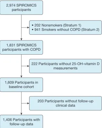
SPIROMICS is a multicenter, observational, prospective, cohort study that includes current or former smokers ($20 pack-years), with or without chronic airflow obstruction, between the ages of 40 and 80 years, and nonsmoking control subjects recruited from 12 clinical centers (n ¼ 2,974).24,25Participants included in this analysis had spirometry-confirmed COPD (FEV1/FVC < 0.70) and available baseline clinical data (n¼1,609) (Fig 1). Institutional review boards at each center approved SPIROMICS and participants provided informed, written consent (e-Table 1).
Data Collection
The SPIROMICS cohort provided participant-reported demographic data, medical history, smoking history, and serum samples at enrollment. Postbronchodilator FEV1 and FVC were determined at enrollment and at the 1-year follow-up visit. Use of vitamin D supplementation was determined via patient report without specific ascertainment of formulation. An AECOPD was defined as the report of any worsening of COPD symptoms requiring antibiotics or steroids. Severe AECOPD was defined as the report of worsening COPD symptoms requiring an ED visit or hospitalization.26Baseline AECOPD data were obtained through participant self-report at the time of enrollment. Longitudinal AECOPD data were collected by self-report during quarterly telephone calls and at a yearly follow-up visit. CT scan metrics (percent emphysema, functional small-airway disease [FSAD], airway wall thickness at an internal perimeter of 10 mm [Pi10] for airways# 20 mm) were assessed as previously Chapel Hill, NC; the Department of Radiology (Dr Hoffman),
Uni-versity of Iowa Carver College of Medicine, Iowa City, IA; the Division of Pulmonary, Critical Care, and Occupational Medicine (Dr Comel-las), University of Iowa Carver College of Medicine, Iowa City, IA; the Department of Epidemiology (Dr Barr), Columbia University, New York, NY; the Division of Pulmonary, Critical Care, Sleep, and Allergy Medicine (Dr Krishnan), University of Illinois at Chicago, Chicago, IL; the Division of Pulmonary and Critical Care Medicine (Dr Cooper), David Geffen School of Medicine at the University of California, Los Angeles, Los Angeles, CA; the Division of Pulmonary and Critical Care Medicine (Dr Labaki), University of Michigan, Ann Arbor, MI; the Section of Pulmonary, Critical Care, Allergy, and Immunologic Disease (Dr Ortega), Wake Forest University, Winston-Salem, NC; the Division of Pulmonary, Allergy, and Critical Care Medicine (Dr Wells), Uni-versity of Alabama at Birmingham, Birmingham, AL; the Division of Thoracic Medicine and Surgery (Dr Criner), Temple University, Phil-adelphia, PA; the Division of Pulmonary Critical Care, Allergy, and Sleep Medicine (Dr Woodruff), University of California, San Francisco, San Francisco, CA; the Division of Pulmonary, Critical Care, and Sleep Medicine (Dr Bowler), National Jewish Health, Denver, CO; the Divi-sion of Pulmonary and Critical Care Medicine (Dr Pirozzi), University of Utah, Salt Lake City, UT; the Division of Pulmonary and Critical Care Medicine (Drs Hansel and Wise), Johns Hopkins University, Baltimore, MD; and the Division of Endocrinology, Diabetes, and Metabolism (Dr Brown), Johns Hopkins University, Baltimore, MD.
*Collaborators from the SPIROMICS study are listed in the Acknowledgments.
FUNDING/SUPPORT:R. M. B. has received support from the National
Institutes of Health, National Heart, Lung, and Blood Institute (NIH-NHLBI) [Grant F32HL143867-01] related to this work. M. B. D. has received support from the NIH-NHLBI [Grant R01HL125432-01A1] related to this work. For SPIROMICS funding, see Acknowledgments.
CORRESPONDENCETO: Robert M. Burkes, MD, 130 Mason Farm Rd,
CB #7020, BioInformatics, Chapel Hill, NC 27599; e-mail: robert. [email protected]
defined.27-29Serum 25-OH-vitamin D levels were measured post hoc
from stored baseline serum samples, using radioimmunoassay (enzyme immunoassay [IDS]; intraassay coefficient of variance, 8.14%; limit of detection, 3.7 ng/mL).
Statistical Methods
Two-sample Studentt-test or Kruskal-Wallis testing andc2 testing were performed to identify relationships between VDD and continuous or categorical variables, respectively. 25-OH-vitamin D levels were modeled continuously and as VDD (< 20 ng/mL) vs not deficient ($ 20 ng/mL).5 Multilevel linear regression modeling was used to determine relationships between 25-OH-vitamin D levels and % predicted FEV1 at baseline as well as the rate of change over 1 year. Exacerbations were modeled in binary fashion (0 vs$1 episode). Logistic regression analysis was used to determine the association between 25-OH vitamin D and the odds of experiencing an AECOPD in the year before and after enrollment. Severe AECOPD in the year before and after enrollment was also separately explored as outcomes. Covariates in the multivariable model were selected on the basis of clinical relevance.30 Covariates in linear regression include pack-years of smoking (per 10 pack-years), smoking status, and season of 25-OH-vitamin D blood draw (spring, summer, and fall with winter as referent). As age, sex, race, and height are incorporated into the % predicted FEV1 calculation, these covariates were not separately included in % predicted FEV1 models. Covariates in logistic regression models include age (per 10 years), race, sex, pack-years of smoking (per 10 pack-years), current smoking status, and season of blood draw. The association between 25-OH vitamin D and CT scan metrics was assessed in bivariate and multivariable linear regression models. Adjusted CT models included age, race, sex, BMI, current smoking, pack-years of smoking, and site of study. Total lung capacity was also included for the models incorporating Pi10. Sensitivity analyses were performed by incorporating reported yearly income (< $50,000/y vs $ $50,000/y) into statistically significant models. Baseline postbronchodilator % predicted FEV1 was added to statistically significant AECOPD models (with removal of age, race, and sex to avoid collinearity). Main modeling approaches were performed restricting to participants not reporting vitamin D supplementation. For all comparisons, P < .05 was considered significant; CT scan associations were corrected for multiple comparisons. Statistical analysis was performed with SAS version 9.4 (SAS Institute).
Results
Cohort Characteristics
There were 1,609 participants in the analytical cohort (Table 1). Participants had a mean age of 65 years, 42% were female, and 14% were black. Severe COPD (Global Initiative for Chronic Obstructive Lung Disease [GOLD] stage III-IV) was present in 34% of participants, and 31% reported one or more AECOPD in the year before enrollment. All participants had 25-OH-vitamin D levels above the limit of detection (>3.7 ng/mL). The median vitamin D level in the cohort was 28.9 ng/mL (quartile 1-quartile 3, 21.3-36.5 ng/mL), with 338 (21%) having VDD. By vitamin D category, 1% had vitamin D< 10 ng/mL, 20% between$10 and<20 ng/mL,
33% between$20 and<30 ng/mL, and 46%$30 ng/ mL.Figure 2shows the distribution of vitamin D levels stratified by GOLD FEV1severity. A total of 24% of the cohort reported some form of oral vitamin D
supplementation (6% in the VDD group and 29% in the
vitamin D-nondeficient group). VDD was associated with younger age (62 vs 66 years;P<.0001), black race (29% vs 10%;P<.0001), and current smoking (48% vs 30%;P<.0001). The VDD group had lower postbronchodilator % predicted FEV1at baseline (57.5% vs 62.0% predicted;P¼.001) and at 1 year (56.7% vs 63.4% predicted;P<.001). There was a greater proportion of baseline visits occurring during winter among VDD participants compared with non-VDD participants (27% vs 18%;P¼.003). A greater proportion of participants with VDD experienced one or more AECOPD in the year before enrollment (39% vs 30%;P¼
.005), as well as one or more severe AECOPD in the year before enrollment (22% vs 14%;P<.001).
25-OH-Vitamin D and FEV1Associations
In bivariate linear regression, a 10-ng/mL decrease in 25-OH-vitamin D was associated with lower % predicted FEV1 (–1.14% predicted; 95% CI,–2.26% to
2,974 SPIROMICS participants
• 202 Nonsmokers (Stratum 1)
• 941 Smokers without COPD (Stratum 2)
1,831 SPIROMICS participants with COPD
222 Participants without 25-OH-vitamin D measurements
203 Participants without follow-up clinical data
1,609 Participants in baseline cohort
1,406 Participants with follow-up data
TABLE 1] Cohort Characteristics
Total Vitamin D<20 Vitamin D$20 P Value
Patients 1,609 338 1,271 .
Age, y 65.3 (7.9) 62.0 (7.9) 66.1 (7.7) <.001
Sex, female 673 (42) 154 (46) 519 (41) .12
Black race 226 (14) 97 (29) 139 (10) <.001
BMI, kg/m2 27.3 (5.3) 27.4 (5.7) 27.3 (5.1) .71
Current smoker 529 (33) 161 (48) 368 (30) <.001
Pack-years smoking, median (Q1-Q3) 46 (35-62) 45 (35-60) 48 (36-63) .20
History of asthma 363 (24) 88 (28) 275 (23) .055
Yearly income<$50,000 819 (51.0) 211 (62.6) 608 (47.8) <.001
FEV1/FVC ratio, baseline 0.51 (0.13) 0.50 (0.13) 0.51 (0.13) .13
FEV1% predicted, post-BD, baseline 61.1 (23.1) 57.5 (22.7) 62.0 (23.1) .001
FVC % predicted, post-BD, baseline 88.8 (19.9) 86.1 (19.4) 89.5 (20) .005
GOLD FEV1severity
Stage I 346 (22) 59 (17) 287 (23) .115
Stage II 712 (44) 145 (43) 567 (45)
Stage III 383 (24) 92 (27) 291 (23)
Stage IV 159 (10) 40 (12) 119 (9)
Vitamin D supplementation 383 (24) 20 (6) 363 (29) <.001
Vitamin D category
<10 ng/mL 24 (1.05) 24 (7) 0 (0) N/A
10 to<20 ng/mL 314 (19.5) 314 (93) 0 (0)
20 to<30 ng/mL 535 (33.2) 0 (0) 535 (42)
$30 ng/mL 736 (45.7) 0 (0) 736 (58)
Season of baseline visit
Spring 441 (27) 94 (28) 347 (27) .003
Summer 423 (26) 72 (21) 351 (28)
Fall 414 (26) 80 (24) 334 (26)
Winter 330 (21) 92 (27) 238 (18)
FEV1% predicted, post-BD, 1 y 61.1 (23.1) 56.7 (22.4) 63.4 (22.6) <.001
AECOPD in previous year, any
0 1,092 (69) 207 (61) 885 (70) .005
1 273 (17) 75 (23) 198 (16)
2þ 224 (14) 53 (16) 171 (14)
AECOPD in previous year, severe
0 1,336 (84) 258 (78) 1,078 (86) <.001
1 173 (11) 50 (15) 123 (10)
2þ 76 (5) 24 (7) 52 (4)
AECOPD infirst year, any
0 1,125 (74) 226 (72) 899 (74) .35
1 226 (15) 48 (15) 178 (15)
2þ 172 (11) 40 (13) 132 (11)
AECOPD infirst year, severe
0 1,324 (87) 273 (86) 1,051 (88) .59
1 123 (8) 24 (8) 99 (8)
2þ 71 (5) 22 (6) 50 (4)
All values represent No. (%) or mean (SD) unless otherwise indicated. P value, comparing vitamin D strata. AECOPD¼acute exacerbation of COPD; BD¼
–0.28% predicted;P¼.01) at enrollment. Similarly, VDD was associated with lower % predicted FEV1 (–4.98% predicted; 95% CI,–7.66% to–2.30% predicted;
P ¼.003) at enrollment. Multivariable multilevel linear regression modeling, adjusting for covariates described in Methods, was performed (Table 2). In this model, a 10-ng/mL lower vitamin D level at baseline was associated with lower % predicted FEV1
(–1.04% predicted; 95% CI,–1.96% to–0.12% predicted;
P¼.03) at enrollment. Continuous 25-OH-vitamin D was not associated with a more rapid rate of lung function decline in thefirst year of follow-up. In multivariate modeling, VDD was associated with lower % predicted FEV1(–4.11% predicted; 95% CI,–6.90% to –1.34% predicted;P ¼.004) at baseline. VDD was associated with a significantly greater rate of % predicted FEV1 decline over thefirst year of follow-up
(–1.27% predicted; 95% CI,–2.32% to–0.22% predicted;
P¼.02).
25-OH-Vitamin D and AECOPD Associations
In bivariate analysis, a 10-ng/mL decrease in 25-OH-vitamin D was associated with 14% higher odds of an AECOPD in the year before enrollment (95% CI, 1.04-1.24; P ¼ .005). Similarly, VDD was associated with 48% increased odds of an AECOPD in the year before enrollment (OR, 1.48; 95% CI, 1.15-1.91;P ¼
.002). In addition, a 10-ng/mL decrease in vitamin D was associated with a 17% increase in the odds of a severe AECOPD in the year before enrollment (OR, 1.17; 95% CI, 1.04-1.32; P ¼ .01). VDD was associated with a 77% increase in the odds of a severe AECOPD in the year before enrollment (OR, 1.77; 95% CI, 1.30-2.39; P < .0001) in bivariate analysis.
0
Total Stage I Stage II
<10 ng/mL 10 to <20 ng/mL 20 to <30 ng/mL ≥30 ng/mL
Stage III Stage IV
% Participants
20 40 60 80 100
Figure 2–Distribution of vitamin D levels in the SPIROMICS cohort, stratified by GOLD FEV1stage. GOLD¼Global Initiative for Chronic Obstructive Lung Disease. SeeFigure 1caption for expansion of other abbreviation.
TABLE 2] Multilevel Linear Regression Modelingaof Associations Between 25-Hydroxyvitamin D and FEV 1 % Predicted at Baseline and First Year of Follow-Up
FEV1% Predicted (Continuous Vitamin D) FEV1% Predicted (Vitamin D Deficiency Status)
FEV1
(% Predicted) 95% CI P Value
FEV1
(% Predicted) 95% CI P Value
25-OH-vitamin D
(per 10-ng/mL decrease)
–1.04 –1.96 to–0.12 .03 . . .
Annual FEV1rate of change, % predicted
(per 10-ng/mL decrease)
–0.19 –0.53 to 0.15 .28 . . .
25-OH-vitamin D (<20 vs$20 ng/mL) . . . –4.11 –6.90 to–1.34 .004
Annual FEV1rate of change, % predicted
(<20 vs$20 ng/mL)
. . . –1.27 –2.32 to–0.22 .02
Visit 1 vs baseline –1.19 –0.15 to 0.53 .04 –0.36 –0.83 to 0.12 .14
Current smoking –0.37 –1.69 to 0.96 .58 –0.27 –1.59 to 1.06 .69
Pack-years smoking (per 10 pack-years)
–0.16 –0.56 to 0.25 .45 –0.16 –0.57 to 0.24 .43
Season of blood draw
Spring 3.04 –0.23 to 6.31 .07 3.07 –0.20 to 6.33 .07
Summer 5.56 2.26 to 8.85 .001 5.38 2.09 to 8.67 .001
Fall –1.88 –5.19 to 1.42 .26 –2.02 –5.32 to 1.28 .23
Winter (Ref) . . (Ref) . .
In multivariable analysis adjusted for covariates described in Methods, every 10-ng/mL decrease in 25-OH-vitamin D was associated with an 11% increase in the odds of an AECOPD in the year before enrollment (OR, 1.11; 95% CI, 1.01-1.22;P¼.04) (Table 3). Similarly, VDD was associated with 32% higher odds of an AECOPD in the year before enrollment (OR, 1.32; 95% CI, 1.00-1.74;P¼.049). Neither continuous nor dichotomized vitamin D levels were associated with prior severe AECOPD in adjusted analyses (e-Table 2).
There were no associations between continuous 25-OH-vitamin D or VDD and the odds of an AECOPD at 1 year in bivariate or multivariable analysis (e-Table 3). Moreover, there was no association between vitamin D levels (bivariate or multivariable) and a severe AECOPD at 1 year (e-Table 4).
CT Analysis
In comparing VDD with nondeficient participants, there were no differences in percent emphysema
(11.6% vs 11.3%; adjustedP>.99), FSAD
(26.5% vs 27.4%; adjustedP>.99), and Pi10 (3.73 vs 3.72 mm; adjustedP¼.22). Modeling 25-OH-vitamin D continuously (per 10-ng/mL increase), there was no association with percent emphysema (0.065%; adjustedP >.99) or FSAD (0.64%;P¼.18). For every 10-ng/mL increase in 25-OH-vitamin D, Pi10 increased 0.004 mm (95% CI, 0.007-0.002;P¼.03). However, this association was attenuated in multivariable modeling (P¼.11).
Sensitivity Analyses
Yearly self-reported income category (reported by 82% of the analytical cohort) was added to the statistically significant FEV1and AECOPD models. Inclusion of income in FEV1models did not attenuate the relationship between VDD and baseline or
longitudinal % predicted FEV1. Inclusion of income did attenuate the relationship between continuous vitamin D level and baseline % predicted FEV1(per 10-ng/mL decrease in 25-OH-vitamin D:–0.43% predicted FEV1; 95% CI,–0.57% to 0.37% predicted;P¼.43). Income also attenuated the relationship between VDD and prior AECOPD (OR, 1.20; 95% CI, 0.87-1.63;P¼.27) and the relationship between continuous vitamin D and prior AECOPD (OR, 0.95; 95% CI, 0.85-1.07;P¼.41). Inclusion of baseline lung function into AECOPD models did not attenuate the association between continuous 25-OH-vitamin D or VDD with prior AECOPD.
Sensitivity analysis restricting the analytical cohort to the 76% of participants not reporting vitamin D supplementation (n¼1,226) was performed (e-Table 5). The associations between continuous vitamin D and baseline lung function and any prior exacerbation were not attenuated. Similarly, the associations between VDD and baseline lung function and prior COPD
exacerbations were not attenuated. However, restricting the analysis to participants not reporting
supplementation attenuated the association between VDD and rate of lung function decline
TABLE 3] Logistic Regression Modelingaof Associations Between Vitamin D and Odds of COPD Exacerbation in the Year Before Enrollment
Odds of COPD Exacerbation in Prior Year (Continuous Vitamin D)
Odds of COPD Exacerbation in Prior Year (Vitamin D Deficiency Status)
OR 95% CI P Value OR 95% CI P Value
25-OH-vitamin D (per 10-ng/mL decrease) 1.11 1.01-1.22 .04 . . .
25-OH-vitamin D (<20 vs$20 ng/mL) . . . 1.32 1.00-1.74 .049
Age (per 10 y) 0.60 0.52-0.71 <.001 0.60 0.54-0.74 <.001
Sex, female 1.73 1.38-2.16 <.001 1.67 1.34-2.10 <.001
Race, black 1.28 0.93-1.77 .09 1.28 0.93-1.77 .09
Current smoking 0.49 0.37-0.63 <.001 0.48 0.37-0.63 <.001
Pack-years smoked (per 10 pack-years) 0.97 0.93-1.02 .19 0.97 0.93-1.02 .19
Season of blood draw
Spring 0.89 0.65-1.23 .96 0.88 0.64-1.21 .63
Summer 0.91 0.66-1.26 .99 0.91 0.66-1.26 .91
Fall 0.90 0.65-1.25 .97 0.90 0.65-1.25 .81
Winter (ref) . . (ref) . .
(–1.05% predicted; 95% CI,–2.21 to 0.12% predicted;
P ¼.09).
Discussion
This study presents the relationship between 25-OH-vitamin D levels and COPD outcomes among 1,609 participants in the SPIROMICS cohort. VDD was seen in 21% of the study cohort and was associated with black race, younger age, and current smoking. VDD was independently associated with lower FEV1at baseline, a greater rate of lung function decrement after 1 year, and experiencing AECOPD in the year before study enrollment. These findings demonstrate the association between VDD and COPD outcomes, and indicate the need for a future randomized study analyzing whether attaining vitamin D sufficiency prevents adverse COPD outcomes.
Patients with COPD are more likely to have VDD, including when compared with age- and sex-matched control subjects.12,31This relationship may be due to heavier smoking history, current smoking, more advanced pulmonary disease, or a lower BMI.12Our study reports lower baseline lung function, increased rate of lung function decline, and higher odds of reported prior AECOPD independent of these factors. A randomized study has shown no effect on lung function decline with supplementation in individuals with COPD and baseline 25-OH-vitamin D near 20 ng/mL.19 However, understanding relationships between VDD and clinical outcomes may inform the importance of remedying VDD in people with COPD.
Our analysis demonstrates that VDD is independently associated with lower % predicted FEV1(by 4.11%) at study enrollment. These findings are consistent with previous observational studies associating VDD with lower FEV1and higher incident COPD.8,12,13,32Our study extends these observations by showing that VDD was associated with a 1.27/y greater decrease in FEV1 % predicted over 1 year in our study. This differs from other cohort studies of those with and without COPD, where VDD has not been shown to be associated with greater lung function decline.17,18Our cohort,
exclusively those with spirometry-confirmed COPD, may contain participants with differential inflammatory profiles. Although the mechanism cannot be determined from this analysis, altered inflammatory responses to noxious stimulants and decreased innate immune system effectiveness may be a potential explaination.33-35 Vitamin D sufficiency has been associated with a
protective effect against lung function decline in smokers,18suggesting an antiinflammatory role for vitamin D in the airways.36Smoking status or history did not independently contribute to FEV1decline in our models. Future investigation in smokers with preserved lung function may further inform the interaction of smoking, vitamin D status, and lung function.
Beyond the immunologic role of vitamin D, VDD is associated with demographic factors, poorer health status, and poor diet37and may be an indicator of poor general health in those with lower lung function or rapidly progressing COPD. These potential
explanations are supported by the attenuation of some of our results by inclusion of income as a surrogate for socioeconomic status into models. These findings illustrate the potential for reverse causality in the relationship between 25-OH-vitamin D levels and COPD outcomes.
This study demonstrates an increase in the odds of experiencing an AECOPD in the year before enrollment in those with VDD. An association between VDD and any or severe AECOPD in the year after enrollment was not observed. However, the numerically higher odds of experiencing an AECOPD in the year after study enrollment in those with low 25-OH-vitamin D is similar to another study,23and the potential to observe statistically significant associations may be hindered by the low number of exacerbations and severely vitamin D-deficient participants in the SPIROMICS cohort.26 Studies regarding AECOPD and VDD have differential results, with meta-analyses suggesting that VDD is not significantly associated with an increased risk of AECOPD.4,38In our models, the association between VDD and prior AECOPD is not attenuated by the addition of % predicted FEV1, suggesting VDD was not mediating exacerbation risk through an association with lung function. Implementing vitamin D
supplementation as an intervention to prevent AECOPD has been investigated,20,39,40with those most likely to benefit being patients with 25-OH-vitamin D levels< 10 ng/mL.19,20,23Our analysis highlights the need for further studies of COPD populations to identify who may benefit from 25-OH-vitamin D supplementation.
rather than chart-adjudicated events. These events were collected with greater frequency in the year after enrollment, potentially introducing differential recall bias contributing to the differential associations of vitamin D on prior vs 1-year AECOPD assessments. Moreover, reliable outdoor time, frailty, and diet metrics were not assessed. As with any cohort study, those too unwell to present for study visits would not be represented, reducing the inclusion of those with decreased outdoor time and higher risk for VDD. Vitamin D supplementation was collected by participant report without ascertainment of dosage. Longitudinal lung function is attenuated in restricted analysis of those not receiving vitamin D supplementation. As vitamin D supplementation data were collected by patient report in SPIROMICS and the dosage was not adjudicated, this effect may be due to reducing the analytical cohort size. Radioimmunoassay analysis used in this study is not the “gold standard”for assessing 25-OH-vitamin D levels. Although the kit manufacturer does participate in the Centers for Disease Control and Prevention Vitamin D Standardization Certification Program, the performing laboratory did not participate in vitamin D external quality assessment. However, all testing included
standard curves, kit controls, and internal laboratory controls. This method is used to assess 25-OH-vitamin D levels in the clinical setting, making our results relevant to clinical practice. We do not have longitudinal 25-OH-vitamin D levels to assess the impact of varying vitamin D levels on lung function decline estimates over multiple years. Effect size of ourfindings cannot be reliably reported. The predominance of white participants may limit generalizability to other populations with COPD and greater racial diversity.
Conclusion
We have observed VDD in approximately one of five participants with COPD in a multicenter cohort. Active smokers, blacks, and younger participants were more likely to have VDD. Lower 25-OH-vitamin D levels are independently associated with lower baseline lung function as well as greater odds of an AECOPD in the year before enrollment. VDD was associated with a greater rate of lung function decline over 1 year. These findings describe potential adverse effects of VDD on lung function decline in those with COPD.
Acknowledgments
Author contributions:R. M. B. had access to
the data and is the guarantor for the content of the manuscript including data and analysis.R. M. B. and M. B. D. had full access to the data and take responsibility for the integrity of the data and accuracy of analysis presented herein. M. B. D. and R. M. B. contributed substantially to the design of the study, data interpretation, and drafting of the manuscript. A. S. C., M. B. D., and R. M. B. contributed to data analysis. D. C., E. A. H., A. P. C., R. G. B., J. A. K., C. C., W. W. L., V. E. O., J. M. W., G. J. C., P. G. W., R. P. B., C. S. P., N. N. H., and M. B. D. contributed to data collection in the SPIROMICS cohort. C. M. D., D. C., E. A. H., A. P. C., R. G. B., J. A. K., C. C., W. W. L., V. E. O., J. M. W., G. J. C., P. G. W., R. P. B., C. S. P., N. N. H., R. A. W., and T. T. B. critically appraised drafts of the manuscript for critically important intellectual content and contributed substantially to revisions of the manuscript. All authors agreed on the manuscript in its
final, submitted form.
Financial/nonfinancial disclosures:The
authors have reported toCHESTthe following: R. M. B. has received a grant from the NIH-NHLBI related to this work. C. M. D. has received grants from the NIH and the Department of Defense unrelated to this work. D. C. reports funding from the NIH and the COPD Foundation unrelated to this work. E. A. H. is a founder of and
shareholder in VIDA Diagnostics, a company commercializing lung image analysis software developed, in part, at the University of Iowa; activities that are unrelated to this study. A. P. C. reports funds from the NIH-NHLBI, compensated and noncompensated work for VIDA, and personal fees from
GlaxoSmithKline (GSK), all unrelated to this work. J. A. K. reports grant funding from the NIH-NHLBI unrelated to this work. C. C. reports working part-time on external scientific engagement and internal medical education for the GSK Global Respiratory Franchise, all unrelated to this work. J. M. W. reports grants from the NHLBI, NIH-NCATS (National Center for Advancing Translational Sciences), Beyer, Boehringer-Ingelheim, Mereo BioPharma, andfinancial relationships with Boehringer-Ingelheim, Mereo BioPharma, and PRA, all unrelated to this work. G. J. C. receives grants from Boehringer-Ingelheim, Novartis, Astra Zeneca, Respironics, MedImmune, Actelion, Forest, Pearl, Ikaria, Aeris, PneumRx, Pulmonx, and other fees from HGE Health Care Solutions Inc, Almirall, Boehringer-Ingelheim, and Nuvaira, all unrelated to this work. P. G. W. reports personal fees from Theravance, GSK, NGM Pharmaceuticals, Amgen, Glenmark Pharmaceuticals, Regeneron, Sanofi, Clarus Ventures, 23andMe, Astra Zeneca, all unrelated to this work. R. P. B. served on the advisory boards (GlaxoSmithKline, Boehringer Ingelheim, and Mylan Pharmaceuticals) and received
research grants from GSK and Boehringer-Ingelheim, all activities unrelated to this work. N. N. H. reports grants and personal fees from AstraZeneca; grants from Boehringer-Ingelheim, the NIH, and the COPD Foundation; and personal fees from Mylan; all unrelated to this work. R. A. W. reports grants and personal fees from AstraZeneca/Medimmune, Boehringer-Ingelheim, and GSK outside the submitted work. He reports grants from Pearl Therapeutics and Sanofi-Aventis outside the submitted work. He reports personal fees from ContraFect, Pulmonx, Roche, Spiration, Sunovion, Merck, Circassia, Pneuma, Verona, Mylan/Theravance, Propeller Health, AbbVie, and GSK, all unrelated to this work. T. T. B. reports personal fees from Gilead, ViiV, Merck, EMS-Serono, and Theratechnologies, all unrelated to this work. M. B. D. reports grants from the NIH-NHLBI during the conduct of the study related to this work as well as personal fees from Boehringer-Ingelheim, GSK, AstraZeneca, Mylan-Theravance, Novavax, Parion, Midmark, and Philips and grants from the Department of Defense and Boehringer-Ingelheim, unrelated to this work. None declared: A. S. C., R. G. B., W. W. L., V. E. O., and C. S. P.
Role of sponsors:Industry sponsors had no
*SPIROMICS collaborators:The authors acknowledge the following current and former investigators of the SPIROMICS sites and reading centers: Neil E. Alexis, MD; Wayne H. Anderson, PhD; Mehrdad Arjomandi, MD; Igor Barjaktarevic, MD, PhD; R. Graham Barr, MD, DrPH; Lori A. Bateman, MSc; Surya P. Bhatt, MD; Eugene R. Bleecker, MD; Richard C. Boucher, MD; Russell P. Bowler, MD, PhD; Stephanie A. Christenson, MD; Alejandro P. Comellas, MD; Christopher B. Cooper, MD, PhD; David J. Couper, PhD; Gerard J. Criner, MD; Ronald G. Crystal, MD; Jeffrey L. Curtis, MD; Claire M. Doerschuk, MD; Mark T. Dransfield, MD; Brad Drummond, MD; Christine M. Freeman, PhD; Craig Galban, PhD; MeiLan K. Han, MD, MS; Nadia N. Hansel, MD, MPH; Annette T. Hastie, PhD; Eric A. Hoffman, PhD; Yvonne Huang, MD; Robert J. Kaner, MD; Richard E. Kanner, MD; Eric C. Kleerup, MD; Jerry A. Krishnan, MD, PhD; Lisa M. LaVange, PhD; Stephen C. Lazarus, MD; Fernando J. Martinez, MD, MS; Deborah A. Meyers, PhD; Wendy C. Moore, MD; John D. Newell Jr, MD; Robert Paine III, MD; Laura Paulin, MD, MHS; Stephen P. Peters, MD, PhD; Cheryl Pirozzi, MD; Nirupama Putcha, MD, MHS; Elizabeth C. Oelsner, MD, MPH; Wanda K. O’Neal, PhD; Victor E. Ortega, MD, PhD; Sanjeev Raman, MBBS, MD; Stephen I. Rennard, MD; Donald P. Tashkin, MD; J. Michael Wells, MD; Robert A. Wise, MD; and Prescott G. Woodruff, MD, MPH. The project officers from the Lung Division of the National Heart, Lung, and Blood Institute were Lisa Postow, PhD, and Lisa Viviano, BSN.
Other contributions:The authors thank the
SPIROMICS participants and participating physicians, investigators, and staff for making this research possible. More information about the study and how to access SPIROMICS data may be found atwww. spiromics.org. SPIROMICS was supported by contracts from the NIH/NHLBI
[HHSN268200900013C, HHSN268200900014C, HHSN268200900015C, HHSN268200900016C, HHSN268200900017C, HHSN268200900018C, HHSN268200900019C,
HHSN268200900020C], by a grant from the NIH/NHLBI [U01 HL137880], and supplemented by contributions made through the Foundation for the NIH and the COPD Foundation from AstraZeneca/ MedImmune; Bayer; Bellerophon Therapeutics; Boehringer-Ingelheim Pharmaceuticals, Inc.; Chiesi Farmaceutici S.p.A.; Forest Research Institute, Inc.; GlaxoSmithKline; Grifols Therapeutics, Inc.; Ikaria, Inc.; Novartis Pharmaceuticals Corporation; Nycomed GmbH; ProterixBio; Regeneron Pharmaceuticals, Inc.; Sanofi; Sunovion; Takeda Pharmaceutical Company; and Theravance Biopharma and Mylan.
Additional information:Thee-Tablescan
be found in theSupplemental Materials
section of the online article.
References
1. Horadagoda C, Dinihan T, Roberts M, Kairaitis K. Body composition and micronutrient deficiencies in patients with an acute exacerbation of chronic obstructive pulmonary disease.Intern Med J. 2017;47(9):1057-1063. 2. Rawal G, Yadav S. Nutrition in chronic
obstructive pulmonary disease: a review.
J Transl Intern Med. 2015;3(4):151-154. 3. Persson LJ, Aanerud M, Hiemstra PS,
Hardie JA, Bakke PS, Eagan TM. Chronic obstructive pulmonary disease is associated with low levels of vitamin D.
PLoS One. 2012;7(6):e38934. 4. Zhu M, Wang T, Wang C, Ji Y. The
association between vitamin D and COPD risk, severity, and exacerbation: an updated systematic review and meta-analysis.Int J Chron Obstruct Pulmon Dis. 2016;11:2597-2607.
5. Holick MF, Binkley NC, Bischoff-Ferrari HA, et al. Evaluation, treatment, and prevention of vitamin D deficiency: an Endocrine Society clinical practice guideline.J Clin Endocrinol Metab. 2011;96(7):1911-1930.
6. Herr C, Greulich T, Koczulla RA, et al. The role of vitamin D in pulmonary disease: COPD, asthma, infection, and cancer.Respir Res. 2011;12:31. 7. Zosky GR, Berry LJ, Elliot JG, James AL,
Gorman S, Hart PH. Vitamin D deficiency causes deficits in lung function and alters lung structure.Am J Respir Crit Care Med. 2011;183(10):1336-1343.
8. Black PN, Scragg R. Relationship between serum 25-hydroxyvitamin D and pulmonary function in the third national health and nutrition examination survey.
Chest. 2005;128(6):3792-3798. 9. Tsiligianni IG, van der Molen T.
A systematic review of the role of vitamin insufficiencies and supplementation in COPD.Respir Res. 2010;11(1). 171-171. 10. Janssens W, Lehouck A, Carremans C,
Bouillon R, Mathieu C, Decramer M. Vitamin D beyond bones in chronic obstructive pulmonary disease: time to act.
Am J Respir Crit Care Med. 2009;179(8): 630-636.
11. Skaaby T, Husemoen LL, Thuesen BH, et al. Vitamin D status and chronic obstructive pulmonary disease: a prospective general population study.
PLoS One. 2014;9(3):e90654.
12. Sanket S, Madireddi J, Stanley W, Sura P, Prabhu M. Relation between vitamin D deficiency and severity of chronic obstructive pulmonary disease: a case control study.J Clin Diagn Res. 2016;10(1):OC16-OC19. 13. Mendy A, Forno E, Niyonsenga T,
Gasana J. Blood biomarkers as predictors of long-term mortality in COPD.Clin Respir J. 2018;12(5):1891-1899. 14. Persson LJ, Aanerud M, Hiemstra PS,
et al. Vitamin D, vitamin D binding protein, and longitudinal outcomes in COPD.PLoS One. 2015;10(3):e0121622.
15. Zhao G, Ford ES, Tsai J, Li C, Croft JB. Low concentrations of serum 25-hydroxyvitamin D associated with increased risk for chronic bronchitis among US adults.Br J Nutr. 2012;107(9): 1386-1392.
16. Vestbo J, Edwards LD, Scanlon PD, et al. Changes in forced expiratory volume in 1 second over time in COPD.N Engl J Med. 2011;365(13):1184-1192.
17. Kunisaki KM, Niewoehner DE, Singh RJ, Connett JE. Vitamin D status and longitudinal lung function decline in the Lung Health Study.Eur Respir J. 2011;37(2):238-243.
18. Lange NE, Sparrow D, Vokonas P, Litonjua AA. Vitamin D deficiency, smoking, and lung function in the Normative Aging Study.Am J Respir Crit Care Med. 2012;186(7):616-621. 19. Lehouck A, Mathieu C, Carremans C,
et al. High doses of vitamin D to reduce exacerbations in chronic obstructive pulmonary disease: a randomized trial.
Ann Intern Med. 2012;156(2):105-114. 20. Martineau AR, James WY, Hooper RL,
et al. Vitamin D3supplementation in patients with chronic obstructive pulmonary disease (ViDiCO): a multicentre, double-blind, randomised controlled trial.Lancet Respir Med. 2015;3(2):120-130.
21. Rafiq R, Prins HJ, Boersma WG, et al. Effects of daily vitamin D
supplementation on respiratory muscle strength and physical performance in vitamin D-deficient COPD patients: a pilot trial.Int J Chron Obstruct Pulmon Dis. 2017;12:2583-2592.
22. Malinovschi A, Masoero M, Bellocchia M, et al. Severe vitamin D deficiency is associated with frequent exacerbations and hospitalization in COPD patients.
Respir Res. 2014;15:131.
23. Jolliffe DA, Greenberg L, Hooper RL, et al. Vitamin D to prevent exacerbations of COPD: systematic review and meta-analysis of individual participant data from randomised controlled trials.
Thorax. 2019;74(4):337-345.
24. Couper D, LaVange LM, Han M, et al. Design of the Subpopulations and Intermediate Outcomes in COPD Study (SPIROMICS).Thorax. 2014;69(5):491-494.
25. Woodruff PG, Barr RG, Bleecker E, et al. Clinical significance of symptoms in smokers with preserved pulmonary function.N Engl J Med. 2016;374(19): 1811-1821.
26. Han MK, Quibrera PM, Carretta EE, et al. Frequency of exacerbations in patients with chronic obstructive pulmonary disease: an analysis of the SPIROMICS cohort.Lancet Respir Med. 2017;5(8):619-626.
28. Sieren JP, Newell JD Jr, Barr RG, et al. SPIROMICS protocol for multicenter quantitative computed tomography to phenotype the lungs.Am J Respir Crit Care Med. 2016;194(7):794-806.
29. Smith BM, Hoffman EA, Rabinowitz D, et al. Comparison of spatially matched airways reveals thinner airway walls in COPD: the Multi-Ethnic Study of Atherosclerosis (MESA) COPD Study and the Subpopulations and Intermediate Outcomes in COPD Study (SPIROMICS).
Thorax. 2014;69(11):987-996.
30. Lederer DJ, Bell SC, Branson RD, et al. Control of confounding and reporting of results in causal inference studies: guidance for authors from editors of respiratory, sleep, and critical care journals.Ann Am Thorac Soc. 2019;16(1): 22-28.
31. Janssens W, Bouillon R, Claes B, et al. Vitamin D deficiency is highly prevalent in COPD and correlates with variants in the vitamin D-binding gene.Thorax. 2010;65(3):215-220.
32. Afzal S, Lange P, Bojesen SE, Freiberg JJ, Nordestgaard BG. Plasma
25-hydroxyvitamin D, lung function and risk of chronic obstructive pulmonary disease.
Thorax. 2014;69(1):24-31.
33. Gombart AF. The vitamin
D-antimicrobial peptide pathway and its role in protection against infection.Future Microbiol. 2009;4(9):1151-1165.
34. Zhang Y, Leung DY, Richers BN, et al. Vitamin D inhibits monocyte/ macrophage proinflammatory cytokine production by targeting MAPK phosphatase-1.J Immunol. 2012;188(5): 2127-2135.
35. Cantorna MT, Yu S, Bruce D. The paradoxical effects of vitamin D on type 1 mediated immunity.Mol Aspects Med. 2008;29(6):369-375.
36. Szekely JI, Pataki A. Effects of vitamin D on immune disorders with special regard to asthma, COPD and autoimmune diseases: a short review.Expert Rev Respir Med. 2012;6(6):683-704.
37. Jolliffe DA, James WY, Hooper RL, et al. Prevalence, determinants and clinical correlates of vitamin D deficiency in patients with chronic obstructive pulmonary disease in London, UK.
J Steroid Biochem Mol Biol. 2018;175:138-145.
38. Holmgaard DB, Mygind LH, Titlestad IL, et al. Serum vitamin D in patients with chronic obstructive lung disease does not correlate with mortality: results from a 10-year prospective cohort study.PLoS One. 2013;8(1):e53670.
39. Moosavi SAJ, Shoushtari MH. The effects of vitamin D supplementation on pulmonary function of chronic obstructive pulmonary disease patients, before and after clinical trial.Diseases. 2015;3(4): 253-259.
![TABLE 1 ] Cohort Characteristics](https://thumb-us.123doks.com/thumbv2/123dok_us/7937816.2110074/4.864.91.795.96.1054/table-cohort-characteristics.webp)
![TABLE 2 ] Multilevel Linear Regression Modeling a of Associations Between 25-Hydroxyvitamin D and FEV 1 % Predicted at Baseline and First Year of Follow-Up](https://thumb-us.123doks.com/thumbv2/123dok_us/7937816.2110074/5.864.73.773.663.1033/multilevel-linear-regression-modeling-associations-hydroxyvitamin-predicted-baseline.webp)
![TABLE 3 ] Logistic Regression Modeling a of Associations Between Vitamin D and Odds of COPD Exacerbation in the Year Before Enrollment](https://thumb-us.123doks.com/thumbv2/123dok_us/7937816.2110074/6.864.89.801.740.1047/table-logistic-regression-modeling-associations-vitamin-exacerbation-enrollment.webp)
