CELLULAR & MOLECULAR BIOLOGY LETTERS http://www.cmbl.org.pl
Received: 10 July 2014 Volume 19 (2014) pp 659-674 Final form accepted: 18 November 2014 DOI: 10.2478/s11658-014-0220-6 Published online: 25 November 2014 © 2014 by the University of Wrocław, Poland
* Author for correspondence. inizal@me.com, phone: +34 48 425600 (6403), fax: +34 48 425649
Abbreviations used: Alk-1 – activin receptor-like kinase 1; Alk-5 – activin receptor-like kinase 5; FGFR3 – fibroblast growth factor receptor 3; FA – focal adhesions; MSCs – mesenchymal stem cells; OA – osteoarthritis; TGFβ – transforming growth factor β; Col2 – type II collagen; Col1 – type I collagen; Col10 – type X collagen
Research article
THE ROLE OF ALK-1 AND ALK-5 IN THE MECHANOSENSING OF CHONDROCYTES
PATRICIA SANZ-RAMOS1, JAVIER DOTOR2
and IÑIGO IZAL-AZCÁRATE1, *
1Laboratory for Orthopaedic Research, University of Navarra, School
of Medicine, Pamplona, Spain, 2DIGNA Biotech, Pamplona, Spain
Abstract: We aim to demonstrate the role of Alk receptors in the response of hydrogel expansion. Chondrocytes from rat knees were cultured onto plastic and hydrogel surfaces. Alk-1 and Alk-5 were overexpressed or silenced and the effects on cells during expansion were tested and confirmed using peptide
inhibitors for TGFβ. Overexpression of Alk-5 and silencing of Alk-1 led to
a loss of the chondrocyte phenotype, proving that they are key regulators of chondrocyte mechanosensing. An analysis of the gene expression profile during the expansion of these modified cartilage cells in plastic showed a better maintenance of the chondrocyte phenotype, at least during the first passages. These passages were also assayed in a mouse model of intramuscular chondrogenesis. Our findings indicate that these two receptors are important mediators in the response of chondrocytes to changes in the mechanical environment, making them suitable targets for modulating chondrogenesis.
Inhibition of TGFβ could also be effective in improving chondrocyte activity in
aged or expanded cells that overexpress Alk-1.
Keywords: Chondrocyte, Mechanosensing, Mechanobiology,
Mechanotransduction, Collagen hydrogels, TGFβ, Differentiation, Signalling,
INTRODUCTION
The chondrogenic capacity of cartilage cells can be modulated in vitro. Several parameters may affect chondrocyte behavior and these parameters have become the subject of research aiming to restore extracellular matrix gene expression that has been lost during joint pathologies or during in vitro monolayer expansion. Cultures of chondrocytes on commonly used polymer surfaces undergo a progressive and, beyond a certain number of passages, irreversible loss of the cartilage phenotype [1–5]. This is characterized by a cessation in the expression of collagen type II, aggrecan and the transcription factor Sox9, among others.
In our laboratory, we used collagen hydrogels that support the culture of chondrocytes on substrates with low mechanical properties (in the range of pascals) [6, 7]. We achieved an efficient expansion method that slowed down the dedifferentiation process that takes place when cells are cultured onto plastic [8]. The mechanisms responsible for this behavior of chondrocytes in the hydrogel culture are still unknown, but some clues have been found, such as the
interesting pattern for transforming the growth factor β (TGFβ) receptors activin
receptor-like kinase 1 and 5 (Alk-1 and Alk-5) [8]. We described an increase in the Alk-1 level in the plastic culture. This was found to slow down in hydrogels. The opposite pattern was found for Alk-5. The relevance of this behavior is that
these TGFβ receptors have opposing effects in chondrocytes [9–16]. When
TGFβ signals through Alk-1, it activates Smad 1/5/8 and produces hypertrophic
chondrocytes and terminal differentiation [14, 15]. When it binds to Alk-5 (also
known as TGFβ receptor 1), it enhances the expression of cartilage-related genes
through Smad 2/3 activation [9–13]. The balance between these two receptors
determines the capacity of the cell to respond to TGFβ.
We hypothesized that these receptors could be implicated in the mechanosensing capacity of chondrocytes. Therefore, we aimed to effectively demonstrate the role of these two receptors in the process of chondrocyte mechanosensing. We used the model of hydrogel culture with collagen type I hydrogels, which simplify synthesis and handling and have offered good results previously. We used a plasmid system to induce cells to overexpress or silence Alk-1 and Alk-5 in order to study their effect on the chondrogenetic capacity of cartilage cells.
MATERIALS AND METHODS
Preparation of hydrogels
Hydrogels were prepared as described previously using 3 mg/ml of rat tail
collagen type I (BD Biosciences). They yielded an elastic modulus of 22.42 Pa, which was determined via rheology [6].
Isolation and culture of chondrocytes
(number 066-11, date 5/27/2011). We used cells pooled from 5 rats for the experiments and replicated each experiment three times with different cell extractions. The whole knee joint was exposed under sterile conditions and cartilage pieces were obtained from the femoral cartilage using surgical blades. The tissue was placed in culture dishes, minced, and treated overnight with 0.5 mg/ml dispase and 60 units/ml collagenase (Sigma), both of which had been dissolved in the expansion medium (see below). After digestion, the cells were pelleted, washed and finally resuspended in the expansion medium, which was DMEM-Glutamax supplemented with 10% fetal bovine serum and 1 mM L-cysteine and penicillin/streptomycin (both from Invitrogen).
The cells were seeded on 175-cm2 flasks for the control expansion and on
collagen hydrogel-coated 175-cm2 flasks, and grown under standard hypoxic
conditions (37ºC, 5% CO2 and 5% O2) unless otherwise indicated. For normoxic
conditions, we used 37ºC and 5% CO2. Both cultures (hydrogel and plastic) were
two-dimensional. The plastic-expanded cells were detached using trypsin (Invitrogen) and the hydrogel-cultured cells were detached using 100 µg/ml
collagenase (Sigma) for 45 min at 37ºC. To inhibit TGFβ, peptides P17 and
P144 (Digna Biotech) were added separately at 0.2 mg/ml. A sample for every passage was collected to detect chondrocyte markers using RT-PCR (see below). To study the chondrogenic capacity, we performed micromass culture and injected chondrocytes into the adductor muscles of nude athymic mice. A total of 200,000 expanded chondrocytes (from plastic or hydrogel-coated surfaces) were pelleted (centrifuged at 400 x g for 5 min) and placed overnight in a 15-ml conical tube with an inclination of about 45º. Pellets were suspended in expansion medium for 7 additional days and collected to be analyzed using RT-PCR and histology. For the mouse model, 4 million chondrocytes were collected and implanted in a PBS suspension in the adductor muscle of nude mice weighing 30–40 g, as previously described [17]. This protocol was approved by our institutional committee (number 065-12, date 5/18/2012). The cells were maintained for 1 week and then the complete limb was extracted, fixed in formalin, decalcified using 10% formic acid for 2 weeks, and subjected to histological analysis using Sirius red staining.
RT-PCR assays
Cells used for the RT-PCR assay were collected from hydrogels, plastic surfaces and micromass structures as described above. RNA was extracted using 1 ml of Trizol (Invitrogen), following the manufacturer’s guidelines. After determining the RNA concentration using spectrophotometry with nanodrop (Thermo Fisher Scientific), we performed retrotranscription with the cDNA SuperMix qScript system (Quanta Biosciences), using 1 µg of RNA for each reaction. A total of 0.5 µl of the product of the previous reaction was subjected to a gene expression
assay via real-time PCR. To study relative expression, we followed the 2-ΔΔCt
receptor 3 (FGFR3), Alk-5, Alk-1, aggrecan, type I collagen (Col1) and type X collagen (Col10). Cells on plastic were taken as the controls and the values obtained for hydrogel-cultured cells were subsequently referred to them.
Western blot analysis
For the detection of Alk-1, Alk-5 and SMADs, we performed western blot analyses of protein extracts from chondrocytes. Proteins were extracted using Phosphosafe extraction reagent (Novagen), and after quantification using the Bradford assay, loaded onto SDS-PAGE gels. Proteins were transferred to nitrocellulose membranes and blocked using 5% milk in TBST. We used specific antibodies diluted 1:1000 in blocking buffer. After washing the blot extensively, secondary antibodies were used diluted 1:2000 in blocking buffer and then washed again. Finally, the blots were visualized using Lumilight Plus western blotting substrate (Roche). In order to visualize SMADs, the chondrocytes
first underwent a treatment with TGFβ (R&D systems) at 1 ng/ml for 1 h.
Histological analysis
The chondrogenic capacity of expanded cartilage cells was determined via histological processing of the micromass structures. Sections 4 µm in thickness were stained using Sirius Red. We also used specific antibodies to detect aggrecan (Abcam), Col2 (Biogenesis) and Col1 (BD Biogenesis) via immunofluorescence. For this purpose, the sections were initially treated with trypsin (0.1% in PBS containing 0.1% calcium chloride) for 1 h at 37ºC. They were then blocked with normal goat serum (Sigma) diluted 1:20 in PBS, treated with the primary antibody diluted 1:50 overnight at 4ºC and finally washed and incubated with the secondary antibody 1:200 for 45 min at room temperature (RT). Samples were stained using Sirius red and Safranin-O. Samples from non-injected mice were used as controls.
Overexpression/silencing
Overexpression of Alk-1 and Alk-5 was induced using cDNA obtained from
Origene, which was then cloned in Escherichia coli. Then, 2 µg of 1 or
Alk-5 in solution was transfected into chondrocytes using 4 µl Lipofectamine (Invitrogen) per well in 6-well plates to give a final concentration of 8 ng/µl. The silencing was performed using the HuSH silencing system for Alk-1 and Alk-5 from Origene. They were cloned and transfected using the same protocols.
Methylation analysis
Table 1. Primers used for the RT-PCR.
Alk-1
Left modified primer AAATGGTTTTGTTTAGAATTTACGG
Right modified primer TAACTTCTTCCTACCTAACCCCG Left unmodified primer AATGGTTTTGTTTAGAATTTATGG Right unmodified primer TAACTTCTTCCTACCTAACCCCACT Alk-5
Left modified primer TTTTTTTTGAGTAATTTTTACGG
Right modified primer GCACTACTAAACCTCTAACCACG
Left unmodified primer TTTTTTTTGAGTAATTTTTATGG Right unmodified primer ACACTACTAAACCTCTAACCACACC
Statistical analysis
The results from gene expression experiments were compared using the nonparametric Kruskal-Wallis test, using the software SPSS 15.0 for Windows. We considered values of p < 0.05 to be significant.
RESULTS
Overexpression and silencing of Alk-5 and Alk-1
Expansion of chondrocytes after Alk-5 upregulation and Alk-1 downregulation
Selected chondrocytes were expanded up to passage 8 and their RNA was extracted for RT-PCR analysis of cartilage markers (Fig. 2A). When genetically modified chondrocytes were expanded (Alk-5 for cells overexpressing Alk-5, and shAlk-1 for cells where Alk-1 was silenced), we observed a delay in the progressive loss of the chondrocytic markers aggrecan and Col2, and a delayed upregulation of Col1 with respect to unmodified control cells during the course of the culture. In addition, and confirming our previous results, hydrogel-expanded cells did show a delayed decay of the cartilage markers aggrecan and Col2 and an increased expression of Col1. We also confirmed the expression of Alk-1 and Alk-5 for the modified Alk-5 and shAlk-1 cells during the experiment and found an overexpression for Alk-5 and a downregulation of Alk-1 consistent with the modification of the cells. These changes were particularly evident in the first passages. Data concerning the expressions of Alk-1 and Alk-5 were confirmed using western blot analysis (Fig. 2B). A band corresponding to Alk-5 appears in passage 0 cells. Its size decreases when the cells reach passage 4 under either set of conditions, but this decrease is enhanced when passage 0 cells were transfected with the corresponding plasmids. Alk-1 on its own increases with the passage number and decreases strongly when silenced.
In order to test if changes in the expression of receptors lead to changes in the signaling pathways, we performed a western blot analysis of SMAD2/3 and SMAD1/5/8 (Fig. 2B). The results confirmed findings for the RT-PCR, showing the presence of SMAD2/3 only in unpassaged cells and hydrogel-expanded cells. It was potentiated when cells were transfected with plasmids to overexpress the Alk-5 receptor. SMAD1/Alk-5/8 also appeared in unpassaged cells, but it was maintained only in plastic-expanded chondrocytes, showing no band for the hydrogel-expanded cells and poor band intensity in cells where Alk-1 had been silenced.
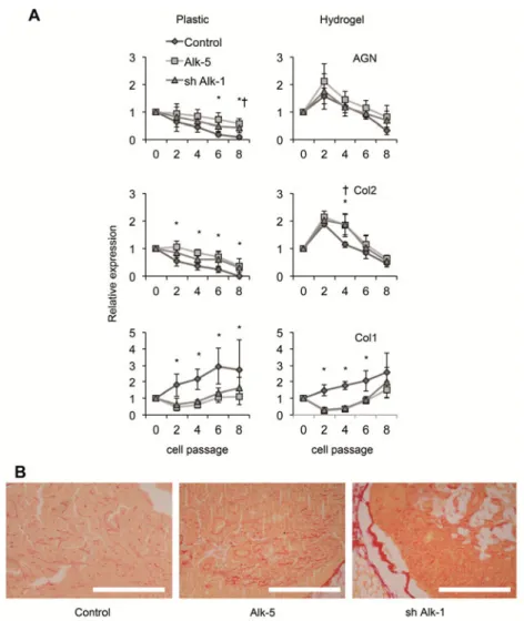
The capacity to form a matrix during the course of the experiment was analyzed by placing expanded cells in passages 0–8 in pellet cultures. In all cases (Fig. 3A), the expression of the cartilage markers aggrecan and Col2 was significantly upregulated when Alk-5 and shAlk-1 cells were assayed and referred to unmodified cells, showing a much-delayed decay for these markers. In addition, when the expansion was performed onto hydrogels, the expressions of these two markers were much increased during the first passages, showing an interesting synergy for the genetic modification and the hydrogel culture effect, which involved an improved expression of cartilage markers and confirmed previous results. In the assay of Col1, a significant decrease was observed when Alk-5 and shAlk-1 cells were used.
Fig. 3. The expression of cartilage markers in pellets obtained for cells in every passage. A – Pellets were dispersed and analyzed for the expression of markers using RT-PCR. The average ± SD for every group (n = 9) is represented for passages 0, 2, 4, 6 and 8. B – The in vivo assay was developed by injecting chondrocytes from passage 2 into the adductor muscles of nude mice. Sections were stained using Sirius red. Scale bar: 200 µm. *p < 0.05 for Alk-5 cells relative to the control cells in every passage. †p < 0.05 for Alk-5 cells
relative to the control cells in every passage.
Inhibition of TGFβ in hydrogel-cultured Alk-5 and sh-Alk-1 cells
In order to study the biological role of Alk-1 and Alk-5 receptors in
chondrocytes, we used the TGFβ inhibitors P17 and P144. Here, we report the
and shAlk-1 cells, a clear effect was observed in passage 2 cells: an increase in aggrecan and Col2 expression and a decrease in Col1 expression, which suggests increased signaling through Alk-5, an effect that would positively influence the chondrocyte phenotype. We also used hydrogel expansion for the inhibition of
TGFβ, adding P17 after the 4th passage, when we had observed an increased
level of Alk-1 [8]. At this point, the effect of inhibition was clear and significant, showing an increase in the expression of aggrecan and Col2, and a decrease in Col1 after the addition of the peptide.
DNA methylation analysis
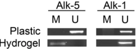
We used primers specific for selected CpG islands on the promoter of Alk-1 and Alk-5, which are capable of amplifying modified or unmodified DNA, respectively. Fig. 5 shows the presence of bands in the gel for those primers specific for unmodified DNA, indicating the presence of methylation in the CpG islands studied for the promoter of Alk-1 both in plastic and in hydrogel-cultured chondrocytes. This is an indication of the lack of regulation of the gene for Alk-1 in our model of chondrocyte culture by methylation, at least in the islands studied. For Alk-5, we could detect the appearance of a band for the primers specific for modified DNA in its promoter. This indicates that, at least in our model, the cells cultured in collagen hydrogels suffer a de-methylation of the promoter for Alk-5.
Fig. 5. Methylation analysis of CpG islands in the promoters of Alk-1 and Alk-5. Genomic DNA was transformed using the bisulfite modification and subjected to PCR using primers for the specific sequence of modified (M) or unmodified (U) DNA, respectively indicating unmethylated and methylated DNA in chondrocytes from passage 2 cultured both on plastic and on collagen hydrogels. The band pattern remains constant for Alk-1 (positive using primers specific for unmodified DNA) but changes for Alk-5 suggest a de-methylation process.
DISCUSSION
These results concur with those from our earlier research, in which we suggested that Alk-1 and Alk-5 are suitable candidates for the final effector of the response of chondrocytes to changes in substrate rigidity [8]. These two transmembrane
proteins function as opposite receptors for TGFβ [19]. In chondrocytes, Alk-5
(Fig. 3) strongly confirm these earlier findings, showing SMAD 2/3 activation
by TGFβ only when cells were expanded onto hydrogels, which had been shown
in a previous experiment to induce an improved expression for Alk-5 [8]. These two receptors have been described to have a role in the changes that age has on the reparative capabilities of cartilage. An alteration in the ratio of Alk-5 to Alk-1 to a higher expression of Alk-1 is observed in older chondrocytes [16, 20], in osteoarthritis (OA) [16] and during expansion in plastic [8]. We demonstrate here that both are relevant effectors of chondrocyte mechanosensing and modulators of chondrocyte phenotype since, by modifying their expression (increasing or silencing), we are able to modulate their chondrogenetic capacity. Expansion of cells with an overexpression of Alk-5 or a downregulation of Alk-1 mimics the effect that expansion onto hydrogels had on dedifferentiation of chondrocytes [8]. Indeed, a combination of the two factors (overexpression and silencing and hydrogel culture) could offer a synergistic effect that could be of interest in efforts to find optimal culture systems for cartilage cells (Figs 2 and 3). The data in Fig. 2 point to a regulation of Alk-1 by Alk-5 and vice versa. This coordinated regulation has not yet been demonstrated, but the opposing effects for the two receptors described here and in previous studies [16, 20] strongly support this hypothesis.
Interestingly, the methylation analysis performed showed how culture onto hydrogels produced a decrease in the methylation of the promoter of Alk-5. We suspect that this change is probably responsible for the increase in the expression of Alk-5 through the duration of the culture. We did not detect a similar behavior for Alk-1, which points to a relevant role of epigenetics in chondrocyte biology. In previous studies, epigenetic mechanisms have also been related to cartilage biology. The epigenetic state of the Sox9 promoter has been described to change during the process of OA [21]. In the same way, promoters for several metalloproteinases have been described as less methylated in OA-derived samples [22–24]. However, other studies have yielded results that could not demonstrate significant gene methylation in the aggrecan promoter of aged and OA chondrocytes [25].
inhibitors against TGFβ P17 and P144 (data not shown for the latter) [28–31]. Our results (Fig. 4) demonstrate an increased chondrocytic phenotype when
TGFβ was inhibited in Alk-1 expressing cells and, on the contrary, a lower
chondrogenic capacity when we inhibited TGFβ on cells expressing Alk-5. All
of this is consistent with the role described for TGFβ receptors. Of particular
interest are the results found for P17. When added to the culture medium at the
moment of the switch from Alk-5 to Alk-1 (around the 4th passage), P17 causes
the cells to stop or at least slows down the dedifferentiation process. If the
expansion of chondrocytes on hydrogels delays the dedifferentiation to the 4th
passage, treatment with P17 at that moment could maintain the differentiated state in passages 6 through 8.
In any case, changes in stiffness as observed in our model are caused by a reduction in the collagen content of the hydrogel. This could also lead to a reduced availability of binding sites for integrins in the soft hydrogels that may cause the cells to decrease the FA sites and, subsequently, the signal transduction intensity. In fact, it has been extensively reported that decreased rigidities have precisely this effect on the turnover of FA, since the number of FA is dependent on the rigidity of the substrate [32, 33]. All of this evidence suggests that the mechanisms evaluated in this study are valid as a model for studying the effect of changing the substrate mechanics on chondrocytes. Thus, we offer relevant data that help to increase the understanding of mechanosensing mechanisms. We demonstrate that Alk-1 and Alk-5 may be important targets for modulating chondrocyte behavior in cartilage pathologies, which include the
strategies that try to inhibit TGFβ when expression of Alk-1 cannot be silenced.
Acknowledgments. This study is funded by PIUNA (Plan de Investigación, Universidad de Navarra, 2010-40). We would like to thank Digna Biotech for
kindly providing peptides to inhibit TGFβ. Patricia Sanz-Ramos received
a Fellowship from the Asociación de Amigos de la Universidad de Navarra. The authors have no conflict of interests.
REFERENCES
1. Glowacki, J., Trepman, E. and Folkman, J. Cell shape and phenotypic
expression in chondrocytes. Proc. Soc. Exp. Biol. Med. 172 (1983) 93–98.
2. Brodkin, K.R., García, A.J. and Levenston, M.E. Chondrocyte phenotypes
on different extracellular matrix monolayers. Biomaterials 25 (2004) 5929–5938.
3. Benya, P.D. and Shaffer, J.D. Dedifferentiated chondrocytes reexpress the
differentiated collagen phenotype when cultured in agarose gels. Cell 30
(1982) 215–224.
4. Binette, F., McQuaid, D.P., Haudenschild, D.R., Yaeger, P.C., McPherson, J.M.
and Tubo, R. Expression of a stable articular cartilage phenotype without evidence of hypertrophy by adult human articular chondrocytes in vitro.
5. Darling, E.M. and Athanasiou, K.A. Rapid phenotypic changes in passaged
articular chondrocyte subpopulations. J. Orthop. Res. 23 (2005) 425–432.
6. Sanz-Ramos, P., Mora, G., Ripalda-Cemboráin, P., Vicente-Pascual, M. and
Izal-Azcárate, I. Identification of signalling pathways triggered by changes
in the mechanical environment in rat chondrocytes. Osteoarthr. Cartil. 20
(2012) 931–939.
7. Sanz-Ramos, P., Mora, G., Vicente-Pascual, M., Ochoa, I., Alcaine, C.,
Moreno, R., Doblaré, M. and Izal-Azcárate, I. Response of sheep chondrocytes to changes in stiffness in the range from 2 to 20 Pa. Effect of
cell passaging. Connect. Tissue Res. 54 (2013) 159–166.
8. Sanz-Ramos, P., Duart, J., Rodríguez-Goñi, M.V., Vicente-Pascual, M.,
Dotor, J., Mora, G. and Izal-Azcárate, I. Improved chondrogenic capacity of collagen hydrogel-expanded chondrocytes. In vitro and in vivo analysis.
J. Bone Joint Surg. Am. 96 (2013) 1109–1117.
9. Nakao, A., Imamura, T., Souchelnitskiy, S., Kawabata, M., Ishisaki, A.,
Oeda, E., Tamaki, K., Hanai, J., Heldin, C.H., Miyazono, K. and ten Dijke, P.
TGFβ receptor mediated signalling through Smad2, Smad3 and Smad4.
EMBO J. 16 (1997) 5353–5362.
10.Lin, H.Y. and Moustakas, A. TGFβ receptors: structure and function. Cell.
Mol. Biol. 40 (1996) 337–349.
11.Moustakas, A., Souchelnytskyi, S. and Heldin, C.H. Smad regulation in
TGFβ signal transduction. J. Cell. Sci. 114 (2001) 4359–4369.
12.Inman, G.J., Nicolas, F.J. and Hill, C.S. Nucleocytoplasmic shuttling of
Smads 2, 3, and 4 permits sensing of TGFβ receptor activity. Mol. Cell. 10
(2002) 283–294.
13.Roberts, A.B. TGFβ signaling from receptors to the nucleus. Microbes
Infect. 1 (1999) 1265–1273.
14.Goumans, M.J. and Mummery, C. Functional analysis of the TGFβ
receptor/Smad pathway through gene ablation in mice. Int. J. Dev. Biol. 44
(2000) 253–265.
15.Ito, H., Akiyama, H., Shigeno, C. and Nakamura, T. Noggin and bone
morphogenetic protein-4 coordinately regulate the progression of
chondrogenic differentiation in mouse clonal EC cells, ATDC5. Biochem.
Biophys. Res. Commun. 260 (1999) 240–244.
16.Blaney Davidson, E.N., Remst, D.F., Vitters, E.L., van Beuningen, H.M.,
Blom, A.B., Goumans, M.J., van der Berg, W.B. and van der Kraan, P.M. Increase in ALK1/ALK5 ratio as a cause for elevated MMP-13 expression in
osteoarthritis in humans and mice. J. Immunol. 182 (2009) 7937–7945.
17.Dell’Accio, F., De Bari, C. and Luyten, F.P. Molecular markers predictive
of the capacity of expanded human articular chondrocytes to form stable
cartilage in vivo. Arthritis Rheum. 44 (2001) 1608–1619.
18.Livak, K.J. and Schmittgen, T.D. Analysis of relative gene expression data
using real-time quantitative PCR and the 2(-Delta Delta C(T)) Method.
19.Finnson, K.W., Parker, W.L., ten Dijke, P., Thorikay, M and Philip, A. ALK1 opposes ALK5/Smad3 signaling and expression of extracellular
matrix components in human chondrocytes. J. Bone Mineral Res. 23
(2008) 896–906.
20.van der Kraan, P.M., Goumans, M.J., Blaney Davidson, E. and ten Dijke, P.
Age-dependent alteration of TGF-β signalling in osteoarthritis. Cell Tissue
Res. 347 (2012) 257–265.
21.Kim, K.I., Park, Y.S. and Im, G.I. Changes in the epigenetic status of the
SOX-9 promoter in human osteoarthritic cartilage. J. Bone Mineral Res. 28
(2013) 1050–1060.
22.Bui, C., Barter, M.J., Scott, J.L., Xu, Y., Galler, M., Reynard, L.N., Rowan, A.D.
and Young, D.A. cAMP response element-binding (CREB) recruitment following a specific CpG demethylation leads to the elevated expression of the matrix metalloproteinase 13 in human articular chondrocytes and
osteoarthritis. FASEB J. 26 (2012) 3000–3011.
23.Cheung, K.S., Hashimoto, K., Yamada, N. and Roach, H.I. Expression of
ADAMTS-4 by chondrocytes in the surface zone of human osteoarthritic
cartilage is regulated by epigenetic DNA de-methylation. Rheumatol. Int.
29 (2009) 525–534.
24.Roach, H.I., Yamada, N., Cheung, K.S., Tilley, S., Clarke, N.M., Oreffo, R.O.,
Kokubun, S. and Bronner, F. Association between the abnormal expression of matrix-degrading enzymesby human osteoarthritic chondrocytes and
demethylation of specific CpG sites in the promoter regions. Arthritis
Rheum. 52 (2005) 3110–3124.
25.Poschl, E., Fidler, A., Schmidt, B., Kallipolitou, A,. Schmid, E. and Aigner, T.
DNA methylation is not likely to be responsible for aggrecan down
regulation in aged or osteoarthritic cartilage. Ann. Rheum. Dis. 64 (2005)
477–480.
26.Choi, M.R., In, Y.H., Park, J., Park, T., Jung, K.H., Chai, J.C., Chung, M.K.,
Lee, Y.S. and Chai, Y.G. Genome-scale DNA methylation pattern profiling
of human bone marrow mesenchymal stem cells in long-term culture. Exp.
Mol. Med. 44 (2012) 503–512.
27.Zhang, Z.X., Guan, L.X., Zhang, K. Wang, S., Cao, P.C., Wang, Y.H.,
Wang, Z. and Dai, L.J. Cytogenetic analysis of human bone marrow-derived
mesenchymal stem cells passaged in vitro. Cell Biol. Int. 31 (2007) 645–648.
28.Dotor, J., López-Vázquez, A.B., Lasarte, J.J., Sarobe, P., García-Granero, M.,
Riezu-Boj, J.I., Martínez, A., Feijoó, E., López-Sagaseta, J., Hermida, J., Prieto, J. and Borrás-Cuesta, F. Identification of peptide inhibitors of transforming growth factor beta 1 using a phage-displayed peptide library.
Cytokine 39 (2007) 106–115.
29.Ezquerro, I.J., Lasarte, J.J., Dotor, J., Castilla-Cortázar, I., Bustos, M.,
transforming growth factor beta type III receptor inhibits liver fibrogenesis
in rats with carbon tetrachloride liver injury. Cytokine 22 (2003) 12–20.
30.Santiago, B., Guitierrez-Cañas, I., Dotor, J., Palao, G., Lasarte, J.J., Ruiz, J.,
Prieto, J., Borrás-Cuesta, F. and Pablos, J.L. Topical application of a peptide inhibitor of transforming growth factor-beta1 ameliorates
bleomycin-induced skin fibrosis. J. Invest. Dermatol. 125 (2005) 450–455.
31.Hermida, N., López, B., González, A., Dotor, J., Lasarte, J.J., Sarobe, P.,
Borrás-Cuesta, F. and Díez, J. A synthetic peptide from transforming growth factor-beta1 type III receptor prevents myocardial fibrosis in spontaneously
hypertensive rats. Cardiovasc. Res. 81 (2009) 601–609.
32.Bershadsky, A.D., Balaban, N.Q. and Geiger B. Adhesion-dependent cell
mechanosensitivity. Annu. Rev. Cell. Dev. Biol. 19 (2003) 677–695.
33.Pelham, R.J. Jr. and Wang, Y. Cell locomotion and focal adhesions are
regulated by substrate flexibility. Proc. Natl. Acad. Sci. USA 94 (1997)