http://www.ijorl.com pISSN 2454-5929 | eISSN 2454-5937
Original Research Article
Allergic fungal rhinosinusitis: an overview on pathogenesis, early
diagnosis and management
Neeraj Suri*, Bhavya B. M.
INTRODUCTION
Allergic fungal rhinosinusitis is a distinct clinical entity, which was first reported in 1976, characterized by the presence of allergic fungal mucin, which consists of thick tenacious eosinophilic secretion with characteristic histologic findings.1 AFRS is a form of non-invasive fungal rhinosinusitis coupled with the clinical entity of fungus ball (mycetoma). It is a unique pathologic entity which has the mucin which is similar to that found in the
lungs of allergic bronchopulmonary aspergillosis
(ABPA). This pulmonary correlation helped the researchers to understand the pathogenesis of AFRS over last decade.2 Earlier it was known as a paranasal sinus
tumor. AFRS is now believed to be an IgE mediated allergic reaction to the aerosolized environmental fungi,
commonly the dematiaceous species in an
immunocompetent host. Aspergillus was the only fungal species recovered in the cultures of AFRS due to lack of culture techniques and knowledge.
Plaignaud reported the first case in 1791. A detailed clinical description of the causative fungus – Aspergillus was given by Schubert in 1885. Katzenstein et al, described allergic aspergillus sinusitis as a newly recognized form of sinusitis.3 Robsonet al introduced the terminology allergic fungal sinusitis (AFS).4
ABSTRACT
Background: The objective of the study wasto evaluate the criteria for diagnosing allergic fungal rhinosinusitis and
to maintain permanent drainage and ventilation, while preserving the integrity of the mucosa.
Methods: This is a prospective study of 50 patients with allergic fungal sinusitis with or without polyposis all of whom were treated with endoscopic debridement. Mucous sample collection, nasal secretion culture, surgical specimen handling, and histological evaluation of surgical specimens are described. All patients treated with endoscopic sinus surgery, debridement, post-operative use of steroids and antifungal therapy.
Results: Fungal mucin was found in all 50 cases, histology and fungal cultures confirmed the diagnosis. Out of 50
patients, 29 were females and 21 were males, with a mean age of 32 years. The most common symptom was nasal discharge 41 (82%) cases, nasal obstruction in 38 (76%) cases, headache and facial pain in 32 (72%) cases, 7 (14%) patients had bronchial asthma. Symptoms of nasal obstruction and nasal discharge were improved in 46 (92%) cases. All preoperative versus postoperative changes in AFRS associated complaints reached statistical significance of p value <0.001 except in patients with asthma.
Conclusions: Comprehensive management with endoscopic sinus surgery, oral steroids and antifungals reduces the
recurrence or need for revision surgery. Long term follow up is very important.
Keywords: Allergic fungal rhinosinusitis, Type I hypersensitivity, Oral corticosteroids, Immunotherapy, Endoscopic
sinus surgery
Department of ENT, GMERS Medical College and Civil Hospital, Gandhinagar, Gujarat, India
Received: 28 February 2018
Accepted: 26 March 2018
*Correspondence: Dr. Neeraj Suri,
E-mail: singhn_16@yahoo.com
Copyright: © the author(s), publisher and licensee Medip Academy. This is an open-access article distributed under
the terms of the Creative Commons Attribution Non-Commercial License, which permits unrestricted non-commercial use, distribution, and reproduction in any medium, provided the original work is properly cited.
Approximately 6-8% of surgically treated chronic rhinosinusitis had fungal elements in their biopsied specimens. Enceet al, identified five different organisms responsible for AFRS.5 Codyet al simplified the criteria for diagnosis of AFRS which included characteristic allergic mucin, and non-invasive fungal hyphae within the collected mucin or positive fungal cultures.6 Some studies added, type I hypersensitivity reaction diagnosed by history, positive skin test or serology as other prerequisites for diagnosing AFRS.7
In 1994, Bent and Kuhn published their diagnostic criteria centered on the histologic, radiographic, and immunologic characteristics of the disease.8 Others have proposed several sets of criteria that have served to further the discussion of an investigation into this unique disease; however, the Bent and Kuhn criteria (Table 1) are largely regarded as the standard for diagnosis today. Patients must meet all the major criteria for diagnosis, while the minor criteria serve to support the diagnosis and describe individual patients but are not used to make a diagnosis. The major criteria include a history of type I hypersensitivity by history, skin testing, or in vitro
testing; nasal polyposis; characteristic computed
tomography (CT) scan/MRI findings; the presence of eosinophilic mucin without invasion; and a positive fungal stain of sinus contents removed at the time of surgery. The minor criteria include a history of asthma,
unilateral predominance of disease, radiographic
evidence of bone erosion, fungal cultures, presence of Charcot-Leyden crystals in surgical specimens, and serum eosinophilia.
Since its initial description, AFRS has been a topic of debate and controversy regarding its pathogenesis, diagnosis, classification and management.
This study was a prospective look into the cases we encountered at our centre. Our aims are:
To re-evaluate the diagnostic criteria for AFRS.
To reduce the complications by surgically removing
the inciting fungal allergic mucin and
marsupialisation of the involved sinuses.
To provide a comprehensive management for
permanent drainage and ventilation of the involved sinuses while preserving the integrity of the mucosa.
Table 1: Bent and Kuhn diagnostic criteria.
Major Minor
Type I hypersensitivity Nasal polyposis
Characteristic CT findings Eosinophilic mucin without invasion
Positive fungal stain
Asthma
Unilateral disease Bone erosion Fungal cultures
Charcot-Leyden crystals Serum Eosinophilia
METHODS
A total of 50 patients aged 15-65 years diagnosed with chronic rhinosinusitis suspected to have AFRS by clinical history and examination were included in the study. All 50 patients were treated surgically between 2013-2017. In this study 29 were females and 21 were males, with an average age of 32 years.
The essential criteria for diagnosis of AFRS were:
1. Nasal polyposis
2. Presence of thick tenacious allergic mucin.
3. Computed tomographic scan of paranasal sinuses
showing opacification of the sinus with areas of hyperattenuation.
4. Positive fungal culture of the surgical specimen.
Sample collection and culture technique
As the fungus colonises the mucus, a simple non invasive procedure to obtain as much mucin as possible should be done in all patients who present with sinus infections.
Xylocaine 10% spray was used (1-2 puffs) in each nostril to anaesthetize the nasal mucosa. After about two to three minutes, each nostril was flushed with 20cc normal saline using sterile disposable syringe and needle, during which the patient was asked to take a deep inspiratory breath and hold just before instillation of the saline and then forcefully exhale. The return fluid was collected in a sterile pan. This sample was sent to the laboratory and inoculated onto inhibitory agar mould (containing ciprofloxacin or chloramphenicol to prevent bacterial growth) or brain heart infusion agar. Fungal culture was usually seen around 30-40 days.
Surgical specimen collection
The allergic mucin was manually collected with the inflamed tissue during the surgical procedure while keeping in mind to preserve the normal mucosa. The collected sample was transferred to sterile normal saline solution/ nonstick sheet. Use of suction devices to clear the mucin was minimised to get high yield of the fungal mucin. Specimen was stained using haemotoxylin and eosin (H&E) and Gomori methanamine silver and the microbiologist was asked to look for allergic fungal mucus.
Surgical treatment
Preoperative workup
To reduce the recurrence of the disease all patients were given a short course of oral prednisolone 0.5mg-1mg/kg/day for 1 week prior to the surgery. Steroids were given to ameliorate the underlying inflammatory process, to decrease the bulk of nasal polyposis and intraoperative bleeding. Preoperative antibiotics were also given to avoid concomitant post obstructive bacterial sinusitis.
Intraoperative details
Surgery was aimed to achieve (1) Complete extirpation of the allergic mucin and fungal debris, (2) Provide permanent drainage and ventilation of the affected sinus while preserving the integrity of normal mucosa, and (3) Postoperative access to the diseased operated areas for look up for recurrence during follow up sessions.
The surgical procedure was tailored to the extent of the disease, all patients underwent endoscopic sinus surgery. The disease was seen evident as nasal polyp, hypertrophied mucosa, with necrotic avascular blackish or greenish crusts / caseous thick secretions which was debrided. The ethmoid sinuses were involved in about 48% cases. The Inter sinus septa were eroded and seen lying free embedded in the caseous mucin. The expansile nature of AFRS, provides better access for the surgeon to debride the areas which are usually difficult to reach in endoscopic approach. Most of the patients had widened nasal cavity, middle meatus, frontal recess even dehiscence of the frontal sinus due to the disease process.
Orbital extension was seen in 7 cases (14%) which was explored via endoscopic ethmoidectomy. The fungal debris was seen not breaching the orbital periosteum. 2 patients (4%) had the disease extending upto the cribriform plate but not eroding it. No neurosurgical exploration was required. 2 patients (4%) had disease extending to the sphenoid sinus which was cleared by suction and minimal manipulation.
Once the debridement was done, all the sinuses were re-inspected for any leftover disease, saline irrigation done and wide marsupialisation of the sinuses was achieved.
Figure 1: Shows thick tenacious, dirty white fungal mucin.
Figure 2: Nasal polyp in a patient with AFRS.
Postoperative care
Nasal irrigation with normal saline and antifungal solution was started in the immediate post operative period once nasal packing was removed. Antibiotics were given for 1 week. Oral steroids (prednisolone) was given for 2-4 weeks duration in tapered dosage.
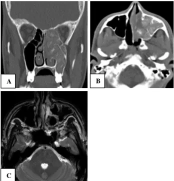
Figure 3: (A) Coronal CT shows soft tissue shadow involving left ethmoids and maxillary sinus with hetergenecity, (B) Axial non-contrast CT of same patient, (C) MRI – Axial cut showing signal void on
T2W images.
Follow-up
All patients were followed up at regular intervals, weekly for first 3 months 2 weekly for 6-12 months and monthly for 12-24 months. The average follow up period was 12 months, with maximum follow up till 36 months.
A
C
On follow up – patients were advised 1. Twice daily nasal irrigation with antifungal drops (clotrimazole) 2. Intranasal steroid spray (budesonide 2 puffs in each nostril once a day) for a mean period of 12-16 months. 3. Oral fluconazole 150 mg once daily for 7 days course. With repeat liver function tests with in normal limits oral antifungals were continued for 7 more days. All patients underwent suction clearance and debridement for retained fungal disease during weekly follow up for 3 months. All were advised for avoidance of allergens.
During follow up in the first 3-6 months we observed hypertrophied oedematous mucosa (7 cases), synechiae (3 cases), healing granulations (5 cases), retained fungal debris (8 cases).
RESULTS
Demographic data
Of the 50 patients who were included in the study, 3 patients were lost to follow up. 29 were females and 21 were males, with a mean age of 32 years. Of these, 38 patients belonged to lower socio-ecomonic status, 22 (44%) patients resided in high humid areas. Revision surgery was done in 11 (22%) cases.
Symptoms of AFRS
In our present study, the most common compliant was nasal discharge 41 (82%) cases, blood stained nasal discharge in 10 (20%) cases, nasal obstruction in 38 (76%) cases, headache and facial pain in 32 (72%) cases, 7 (14%) patients had bronchial asthma, 13 (26%) patients had history of hyposmia.
Symptoms of nasal obstruction and nasal discharge were improved in 46 (92%) cases, hyposmia and headache improved in almost all patients. All preoperative versus postoperative changes in AFRS associated complaints reached statistical significance of p<0.001 except in patients with asthma. According to the results 46 patients had improvement.
In our study, we observed recurrence of the disease in 4 (8%) cases, which was limited to maxilla and ethmoidal sinuses with no intracranial or orbital extension. This was attributed mainly to irregular follow up, noncompliance with medical and antifungal therapy, discontinuation of nasal irrigation, Non-avoidance of allergens.
Nasal airway obstruction in AFRS is a gradual process that many a times patients are unaware about the underlying disease. 7 (14%) patients had proptosis less than 2 mm, 5 (10%) patients had cheek swelling, 7 (14%) patients had eye lid swelling, while none of the patients had diplopia or visual loss.
Radiologically, patients with AFRS show high
attenuation with in the soft tissue shadow of the involved
sinuses on non-contrast CT scan.9-11 Expansion
remodelling or thinning of the sinus wall was noted commonly in our patients. Bony erosion of the sinus wall with extension into adjoining sinuses was noted in 23 (46%) cases. Maxilla and ethmoids were the most commonly affected sinus. Sphenoid was involved only in
2 (4%) patients. Lamina papyracea showed
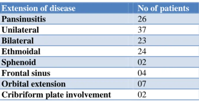
demineralisation in 8 (16%) cases. None of the patients had intracranial extension or orbital extension (Table 2).
Table 2: Extension of the disease.
Extension of disease No of patients
Pansinusitis 26
Unilateral 37
Bilateral 23
Ethmoidal 24
Sphenoid 02
Frontal sinus 04
Orbital extension 07
Cribriform plate involvement 02
DISCUSSION
Since the description of the disease in 1970, many studies have been conducted towards the pathogenesis,
symptomatology and association, diagnosis and
management of AFRS.12 Many studies evaluating the diagnostic criteria, and treatment regimen have appeared in literature.6-8,13-14 The diagnostic criteria for AFRS
includes (1) chronic rhinosinusitis confirmed
radiologically by CT scan of paranasal sinuses; (2) Presence of allergic fungal mucin with predominant eosinophils; (3) Presence of fungus confirmed by the culture of mucin or histology.15,16 Studies have reported an incidence varying from 40-65%. Relatively higher incidences are seen depending on the geographical regions like high humid areas and occupational differences and also specimen isolation techniques. All patients who were suspected to have fungal infection underwent mucus culture/histology. Chances of positive culture is high with increased volume of mucus sample collected. Suction clearance and microdebrider decreased the amount of the recovered fungal mucin.
Studies observed that some patients with AFRS clinical symptoms did not give any history of allergies. This observation led to further research in pathogenesis of
AFRS. Ponikau et al proposed an alternative
theory, which demonstrated ubiquitous presence of fungi within the nose and paranasal sinuses in 93% of patients undergoing surgery for any form of CRS.17 This study also showed that fungal-specific allergy was uncommon in these patients and concluded that most CRS is a T-cell mediated response to fungi, resulting in eosinophilic chemotaxis and activation.
fungus-specific IgE in the mucin of AFRS as well as non-AFRS patients, evidence for a local type I response entirely localized to the nose and paranasal sinuses without signs of systemic involvement.18
Pant et al in his study demonstrated the significance of humoral immunity in the pathogenesis of AFRS.19 He studied patients with eosinophilic mucin CRS, polypoid rhinosinusitis with or without fungal elements. He stated that elevated levels of fungal-specific IgG3, rather than total serum IgE, is more specific in diagnosing AFRS.
Histopathologic findings in AFRS are critical to the diagnosis is the striking number of eosinophils.
Microscopic review of mucosal specimens on
haematoxylin-eosin (H&E) staining will show typical
inflammatory infiltrate composed of eosinophils,
lymphocytes, and plasma cells.20 The presence of Charcot -Leyden crystals alone is not specific for AFRS, hence should not be used as a single diagnostic criterion. Other markers like major basic protein which is more specific needs further study. The mucosa will be hypertrophic and hyperplastic but should not have evidence of necrosis, giant cells, granulomas, or invasion into surrounding structures. Such findings would lend support to a diagnosis of a fungal process other than AFRS.
Radiologically, computed tomography shows
heterogeneous signal intensity characteristic of AFRS. Magnetic resonance imaging has a high specificity for AFRS, especially when combined with CT.21 The high protein concentration of allergic mucin (greater than 28%) results in crosslinking and slows macromolecular motion, giving rise to T1 central hypointensity and T2 central signal void (Figure 2). Both T1 and T2 series demonstrate peripheral enhancement.
Laboratory findings are also helpful in the diagnosis of AFRS. Total immunoglobulin E (IgE) levels are generally elevated, often to more than 1,000 U/mL. Mabry and colleagues.22-24 demonstrated broad sensitivity to both fungal and nonfungal antigens, emphasizing that AFRS patients are generally atopic. Interestingly, the reactions were not fungal specific, although typically only one fungus was isolated from the culture. This finding could represent a common fungal epitope to explain the broad reactivity, or possibly—as Schubert described—the presence of a superantigen that could contribute to the nonspecific reactivity of these patients.25 Manning's work has identified the dematiaceous fungi, namely Bipolaris, in the vast majority of cases.26 Correct identification of the causative organism has been accompanied by the development of multimodality treatment algorithms, with surgical therapy remaining the cornerstone for this recidivistic disease.
Oral corticosteroid and other medical therapies are used in the treatment of AFRS, increased cure rates and decreased recurrence, increased time to revision surgery,
reduction in mucosal stage of disease, and reduced systemic IgE levels.27
Immunotherapy has also been shown to be very efficacious in the treatment of AFRS since it was initiated in 1993. Mabry and colleagues have published results on the use of immunotherapy in AFRS, showing that treating reactivity to both fungal and nonfungal antigens resulted in elimination of nasal crusting and mucin deposits.23,24 He also observed there was decreased need for oral
steroid therapy in patients who underwent
immunotherapy. Therapy is initiated 4–6 weeks after surgery and is predicated on the removal of all allergic mucin at the time of surgery to reduce the antigenic load and prevent worsening of disease. The optimal length of treatment has not yet been determined.
Antifungal therapy initially started because of high rates of recurrence following surgical therapy alone but has largely debated for their efficacy since the advent of oral corticosteroids and immunotherapy. Kennedy and colleagues showed no improvement in the radiographic appearance of the disease or in symptoms in patients treated with oral terbinafine for 6 weeks. Several investigators have evaluated intranasal antifungals with mixed results.28,29 These findings emphasize the need for further work in this area and underlie the reason why antifungal therapy is not widely employed in the treatment of AFRS.
Minimally invasive endoscopic approach but complete surgical debridement of the disease with removal of polyps and marsupialisation of the involved sinuses is mandatory. Schaefer and co-workers in their study reported that an open approach is required if the disease extends to orbit or anterior cranial fossa.30 However, in our study we have used only endoscopic approach. Due to the expansile nature of disease and destruction of bone, distorted anatomy was potentially disorienting during surgery. Involved paranasal sinus acting as epicentre of fungal mucin for the spread of disease to adjacent tissues.
Systemic corticosteroids and antifungal medicines were advised during postoperative period to prevent recurrence with close follow up. Care was taken in teaching the patients for proper nasal irrigation, and avoidance of allergens. Kupferberg et al observed that in his study 19 out of 24 patients developed recurrence owing to discontinuation of steroids, and improper follow up.31,32 Bent and Kuhn emphasized the importance of follow up to prevent recurrence of the disease. A study done by Rains et al done on 139 patients, suggested that post-operative medical treatment of recurrent AFRS may avoid the need for revision surgery.33
for post-operative suction clearance to look for retained fungal debris.
In our study, with this comprehensive management 92% of patients had symptomatic improvement. None of our patients showed any complications in the form of diplopia, blindness, haemorrhage, CSF leak during follow up.
CONCLUSION
AFRS is a relatively new clinical entity; diagnosis requires a high index of suspicion. The Bent and Kuhn criteria are generally the most widely accepted diagnostic criteria in use today. Theories on pathogenesis include hypersensitivity and T-cell mediated reactions as well as a humoral immune response. A confirmatory diagnosis is made from characteristics of the fungal mucin, histological findings and CT scan findings. Mainstay of treatment is surgical debridement using endoscopic sinus surgery, along with a strong role for oral corticosteroids and an emerging role for IT. Comprehensive management with steroids and antifungals reduces the recurrence or need for revision surgery. Antifungals, both systemic and topical, currently have a limited role in treatment, although this area needs further study. Long term follow up is very important.
Funding: No funding sources Conflict of interest: None declared
Ethical approval: The study was approved by the Institutional Ethics Committee
REFERENCES
1. Safirstein B. Allergic bronchopulmonary
aspergillosis with obstruction of the upper respiratory tract. Chest. 1976;70:788–90.
2. Kuhn FA, Javer AR. Allergic fungal rhinosinusitis: our experience. Arch Otolaryngol Head Neck Surg. 1998;124(10):1179–80.
3. Katzenstein AL, Sale SR, Greenberger PA. Allergic
Apergillus sinusitis: a newly recognized form of sinusitis. J Allergy Clin Immunol. 1983;72:89-93.
4. Robson JM, Hogan PG, Benn RA, Gatenby PA.
Allergic fungal sinusitis presenting as a paranasal sinus tumour. Aust N Z J Med. 1989;19:351-3.
5. Ence BK, Gourley DS, Jorgensen NL, Shagets FW,
Parsons DS. Allergic Fungal Sinusitis. Am J Rhinol 1990;4:169-78.
6. Cody DT II, Neel HB III, Ferreiro JA, Roberts GD.
Allergic fungal sinusitis: the Mayo clinic
experience. Laryngoscope 1994;104:1074-9.
7. Morpeth JF, Rupp NT, Dolen WK, Bent JP, Kuhn
FA. Fungal sinusitis: an update. Ann Allergy Asthma Immunol. 1996;76:128-39.
8. Bent JP III, Kuhn FA. Diagnosis of allergic fungal
sinusitis. Otolaryngol Head Neck Surg
1994;111:580-8.
9. Zinreich SJ, Kennedy DW, Malat J, Curtin HD,
Epstein JI, Huff LC et.al. Fungal sinusitis: diagnosis
with CT and MR imaging. Radiology.
1988;169:439-44.
10. Manning SC, Mekel M, Kriesel K, Vuitch F, Marple
B. Computed Tomography and Magnetic Resonance diagnosis of allergic fungal sinusitis. Laryngoscope. 1997;107:170-6.
11. Mukherji SK, Tableueroa RE, Ginsberg LE, Zeifer BA, Marple BF, Alley JG et.al. Allergic fungal sinusitis: CT findings. Radiology. 1998;207:417-22.
12. Bent JP III, Kuhn FA. Allergic fungal
sinusitis/polyposis. Allergic Asthma Proc.
1996;17:259-68.
13. Bent JP III, Kuhn FA. Antifungal activity against Allergic fungal sinusitis organisms. Laryngoscope. 1996;106:1331-4.
14. Berretini S, Carabelli A, Papini M, Cianca E, Sellari FS. Allergic fungal rhinosinusitis: is this rare
disease an allergy or infection? Acta
Otorhinolaryngol Ital. 1996;16:447-54.
15. deShazo RD, Swain RE. Diagnostic criteria for
allergic fungal sinusitis. J Allergy Clin Immunol. 1995;96:24-35.
16. deShazo RD, O’Brien M, Chapin K, Soto AM,
Swain R, Lyons M, et.al. Criteria for the diagnosis of sinus mycetoma. J Allergy Clin Immunol. 1997;99:475-85.
17. Ponikau JU, Sherris DA, Kern EB, et al. The
diagnosis and incidence of allergic fungal sinusitis. Mayo Clin Proc. 1999;74(9):877–84.
18. Collins M, Nair S, Smith W, Kette F, Gillis D, Wormald PJ. Role of local immunoglobulin E production in the pathophysiology of noninvasive fungal sinusitis. Laryngoscope. 2004;114(7):1242– 6.
19. Pant H, Kette FE, Smith WB, Wormald PJ,
Macardle PJ. Fungal-specific humoral response in
eosinophilic mucus chronic rhinosinusitis.
Laryngoscope. 2005;115(4):601–6.
20. Marple BF. Allergic fungal rhinosinusitis: current theories and management strategies. Laryngoscope. 2001;111(6):1006-19.
21. Zinreich SJ, Kennedy DW, Malat J, Curtin HD,
Epstein JI, Huff LC, et al. Fungal sinusitis: diagnosis with CT and MR imaging. Radiology. 1988;169(2):439–44.
22. Mabry RL, Manning SC, Mabry CS.
Immunotherapy in the treatment of allergic fungal
sinusitis. Otolaryngol Head Neck Surg.
1997;116(1):31–5.
23. Mabry RL, Marple BF, Folker RJ, Mabry CS.
Immunotherapy for allergic fungal sinusitis: three years' experience. Otolaryngol Head Neck Surg. 1998;119(6):648–51.
24. Mabry RL, Mabry CS. Allergic fungal sinusitis: the
role of immunotherapy. Otolaryngol Clin North Am. 2000;33(2):433–40.
25. Schubert MS. A superantigen hypothesis for the
allergic fungal sinusitis, and related disorders. Ann Allergy Asthma Immunol. 2001;87(3):181–8.
26. Manning SC, Schaefer SD, Close LG, Vuitch F.
Culture-positive allergic fungal sinusitis. Arch Otolaryngol Head Neck Surg. 1991;117(2):174–8. 27. Kuhn FA, Javer AR. Allergic fungal rhinosinusitis:
our experience. Arch Otolaryngol Head Neck Surg. 1998;124(10):1179–80 .
28. Kennedy DW, Kuhn FA, Hamilos DL, Zinreich SJ,
Butler D, Warsi G, et al. Treatment of chronic rhinosinusitis with high-dose oral terbinafine: a
double blind, placebo-controlled study.
Laryngoscope. 2005;115(10):1793–9.
29. Stankiewicz JA, Musgrave BK, Scianna JM. Nasal
amphotericin irrigation in chronic rhinosinusitis.
Curr Opin Otolaryngol Head Neck Surg.
2008;16(1):44–6.
30. Schaefer SD, Liu JK, Moscatello AL, Cloudwell
WT. Neurosurgical implications of allergic fungal sinusitis. J Neurol. 2004;100:883-90.
31. Kupferberf SB, Bent JP. Allergic fungal sinusitis in
the pediatric population. Arch Otolaryngol Head Neck Surg 1996;122:1381-4.
32. Kupferberf SB, Bent JP, Kuhn FA. Prognosis for allergic fungal sinusitis. Otolarygol Head Neck Surg. 1997;117:35-41.
33. Rains BM III, Mineck CW. Treatment of allergic fungal sinusitis with high dose iatraconazole. Am J Rhinol. 2003;17(1):1-8.
Cite this article as: Suri N, Bhavya BM. Allergic fungal