Full-Field Digital Mammography for Breast Cancer Screening:
An Example of Evidence, Lost in Translation
By
Stacy Mclellan Cowherd
A Master's Paper submitted to the faculty of the University of North Carolina at Chapel Hill in partial fulfillment of the requirements for the degree of Master of
Public Health in the Public Health Leadership Program. Chapel Hill
Abstract
Screening mammography is the gold standard for early breast cancer detection and a cornerstone of preventive medicine. Traditional mammography is currently being replaced by newer, more eloquent digital technology. Clinical trials have not proven that digital mammography reduces breast cancer mortality or burden of suffering. Nevertheless, the technology has spread according to Everett Roger's "Diffusion of Innovation" pattern. The purpose of this Master's Paper is closely to examine the process by which breast cancer screening in the United States has evolved from screen film mammography to full-field digital
mammography. This is a qualitative, iterative analysis that triangulates analyses of the medical literature, elite interview responses, and media coverage to
cultivate a storyline about the development and dissemination of digital
mammography. The technology has spread because of our national hunger for computers and innovation, our eagerness to support the "war on cancer," public perceptions about the technology's theoretical advantages, and ongoing efforts of advocacy groups to maintain health care equality. Although digital
mammography is significantly more expensive than is screen film mammography, cost-effectiveness considerations have been deferred by many health care
Acknowledgements
Preface
This paper is meant to be a critical analysis, written from the perspective of a student of public health, medical literature, public policy, and social change. chose a topic about which I am passionate, which is cancer prevention, screening, and mammography. I chose to examine digital mammography because, when I began the work, I did not have a strong, predefined opinion about whether this is the appropriate direction for breast cancer screening. The reader should know my hypothesis when I began this project: The adoption and dissemination of digital mammography has followed a trajectory similar to that of other medical technologies in the United States. That is, digital screening
Table of Contents
Abstract ... ii
Acknowledgements ... iii
Preface ... iv
Table of Contents ... v
List of Figures ... vi
Introduction ... 1
Qualitative Methods: Elite Interviewing and Newspaper Analyses ... 4
Weighing the Evidence: Review of the Medical Literature ... 7
"An Idea Whose Time Has Come" ... 22
The FDA as a Gatekeeper ... 35
Perceptions with Real Consequences ... 41
Diffusion of Innovation ... 53
The Shifting Cost Burden ... 64
Lessons from Digital Mammography ... 69
Resources ... 74
Appendix 1: Coding Summary of Interview Responses ... 80
Appendix 2: Coding Summary of Media Review ... 82
Appendix 3: Digital Mammography Interview Questions ... 88
Appendix 4: Interview Respondents and Positions ... 91
List of Figures
1. Summary of Trials Comparing Film and Digital Mammography ... 11 2. Surveillance, Epidemiology, and End Results (SEER) Incidence of Invasive Breast Cancer by Age ... 19 3. SEER 5-Year Survival Rates of Invasive Breast Cancer by Age ... 19 4. National Screen Film Mammography Facility Count... ... 25 5. Important Dates in the History of Breast Cancer Screening & Digital
Mammography ... 28 6. Keyword Frequency: Computerrrechnology ... 29 7. Newspaper Portrayal of DMIST Results ... 44 8. Best Characteristics of Digital Mammography as Identified by Interview
Introduction
.. . this has sort of been an interesting issue around evidence-based medicine. It's sort of what happens, and particularly in an environment where you have
a
strong, significant advocacy group ... the public has driven this, and it sort of went right by where the evidence was.-- Carl Ravin, MD, Chairman, Department of Radiology, Duke University Medical Center (DUMC)
In September 2007, the University of North Carolina at Chapel Hill (UNC) became the first hospital in North Carolina to transition completely from screen film to all-digital mammography. These two screening modalities both use X-rays to obtain breast images. Screen film images are captured on film, but digital images are deposited in computer software. Thus, digital images may be manipulated to adjust contrast, brightness, or magnification. We have been eager to implement digital mammography because of its potential to improve the diagnostic accuracy of traditional, screen film mammography. This potential, if realized, would be enormously positive for the fields of preventive medicine and oncology.
Many of our current medical technologies were also introduced into the health care system because of their potential to improve public health. Unfortunately, too many of these have turned out to be extremely expensive without contributing significantly to improved clinical outcomes. The medical profession has recently, however, become more attuned to using evidence as a guide for individual practices, and a method for improving both effectiveness and cost-effectiveness of interventions. In the case of digital mammography, several large clinical trials have compared the technology with screen film mammography. Overall, these trials have not demonstrated considerable diagnostic advantage of digital
The purpose of this analysis is to conduct a critical examination of digital mammography by identifying the variables most likely to explain its emergence and rapid adoption. Popular culture encourages Americans to buy the most expensive and innovative things on the market, whether they are electronics, automobiles, or health care devices. Doctors may choose to adopt uncertainly supported practices because of available resources, personal experiences, or even political loyalties. At the health system level, adoption of new technologies is fragmented because separate parties are responsible for developing,
approving, funding, marketing, and distributing products. In this disorganized process, no one has the advantage of a completely balanced, "big picture" understanding of new medical interventions. Moreover, no entity has the authority to regulate technologies using this "big picture."
Above and beyond these baseline forces that seem to impel the adoption of technology, digital screening mammography has been introduced into the system against a backdrop of ongoing national discussions about breast cancer
screening and prevention. Participants in this discussion include government organizations, cancer and women's advocacy groups, innovators of digital technology, breast imaging specialists, private vendors of digital equipment, and third-party payers. All these interests have had stakes in digital technology's dissemination. In this complex process, the evidence has been muffled,
manipulated, misunderstood, and even completely forgotten by both groups and health care leaders.
Nowhere can these various perspectives be better explored than through interviews with elite stakeholders and analyses of media coverage of digital mammography. Therefore, this qualitative analysis is iterative, relying on a triangulation of analyses of medical literature, respondent interviews, and media accounts to draw conclusions about influences on the adoption of digital
digital mammography, which provides one account of its development and introduction.
Qualitative Methods: Elite Interviewing and Newspaper Analyses
Two standard methods of gathering social science data are through analyses of interviews and documents.1-3 I extracted data from interviews and newspaper articles using a dynamic analytical process in which I generated, tested, and refined hypotheses about the most significant social factors affecting the
adoption and dissemination of digital mammography. I used the well-recognized social research process of coding to both develop and support hypotheses from my field work2· 3 I acquired skill in document coding through intensive training at the Odum Institute for Research in Social Science (University of North Carolina at Chapel Hill)3 and by reading three reference books about qualitative research: Bogdan's Introduction to Qualitative Research Methods, Bailey's Methods of Social Research, and Berg's Qualitative Research Methods for the Social
Sciences.1• 2· 4
Briefly, I coded both interview responses and newspaper articles using the following parameters: (1) most important topics, as determined by length of interview discussion or word counts in newspaper articles, (2) areas of apparent consensus, for example agreement among interviewed radiologists about the similar subjective experience of women receiving a digital versus film
mammogram, and (3) areas of disagreement or contradiction, such as different opinions about the role scientific evidence in the adoption of digital
mammography. I also coded each newspaper article according to its publication date, words used in the title, key words and/or phrases used in the article's text, presence of medical evidence citations, and overall tone of the article.
Summaries of the coding schema for the interviews and newspaper articles are found in Appendices 1 & 2. Ideally, the codes I developed for data extraction should be reviewed and verified by another social science researcher to bolster their validity and reliability. Due to limited resources and time constraints,
The purpose of interviewing elite stakeholders is to gather information and perspectives that cannot be obtained from surveys or other public documents. Elite interviewing focuses on how leaders frame their views and how they make connections or reveal disjunctions between their opinions. In her work with elite interviewing, Hochschild says the purpose of such interviews is to contextualize issues from the perspectives of those involved in shaping them. 5 In the case of mammography, elite interviewing is appropriate because leaders from various fields have essentially spearheaded the national push for digital screening in the absence of mandates compelling such a transition. Understanding their views of the process is, in significant ways, to understand the process itself.
Over the course of one month, I conducted a total of ten interviews with twelve elites in the fields of radiology, breast cancer screening, hospital administration, and public policy. I received permission from UNC's Institutional Review Board (IRB) to conduct these interviews, and obtained verbal consent from each participant to record, transcribe, and include their responses in my analysis. transcribed all interviews, which varied in length between 15 and 45 minutes, in the "naturalist" style including notations of pauses, incomplete statements, and other non-verbal communication. The IRB-approved interview questions and a list of respondents are located in Appendices 3 & 4.
about digital technology to the public and framing the public's perception of the technology.
My media search included the top twenty-five circulating United States
newspapers, according to A. C. Nielsen's spring 2007 assessment (Appendix 5). Although newspaper readership among the U.S. lay public has declined with our increased preference for television and internet news, policy makers and medical elites continue to read and contribute to newspaper articles. Also, the results of newspaper searches are very reliable indicators of media coverage, since the choice of stories in other media is reflected, and often even stimulated by, what newspapers choose to cover.
I accessed the majority of newspapers electronically through the UNC Libraries website; however, USA Today, The Wall Street Journal, and The L.A. Times were not directly available through the website. I used LexisNexis® to access USA Today articles and Procite® to access The Wall Street Journal and The L.A.
Times. I searched each newspaper database using key terms "digital
mammography" and excluded reprints, brief financial statements, articles that did not contain the words "digital mammography" together in context, and articles that only mentioned digital mammography as a peripheral subject (ie. in
Weighing the Evidence: Review of the Medical Literature
Approach to Reviewing the Literature
The strongest evidence-based approach to evaluating digital mammography would involve answering the following question: "Does digital mammography screening decrease breast cancer mortality rates more than does screen film mammography?" This fundamental question of comparative effectiveness should be addressed prior to adopting any new screening tool. In addition, it is critical to review the literature regarding relative harms and costs of digital as compared to film mammography. This chapter addresses both the comparative effectiveness and cost-effectiveness of digital mammography. A subsequent chapter, entitled Perceptions with Real Consequences, investigates the potential hazards of adopting digital mammography as the standard breast cancer
screening method.
The purpose of any screening test is not simply to detect disease, but to help people Jive longer Jives with higher quality because they are treated for the
disease intended to be detected at an early stage by screening. Thus, screening tests are only effective if they detect diseases that can be successfully treated.
In the case of digital mammography, researchers have conducted randomized and paired-comparison trials to study the diagnostic accuracy of digital versus film screening for breast cancer lesions. These types of studies are necessary to ensure that digital mammography does not miss breast cancers that would have been detected by screen film mammography. The inherent problem with these studies, however, is that they cannot demonstrate which modality is a more effective screen for relevant breast cancers, or those that can be treated to enable women live longer lives with less morbidity.
because they remain in the pre-symptomatic, screening-detectable phase of disease for a longer time. The natural history of indolent breast cancers, such as low-grade ductal carcinoma in situ (DC IS), is variable. Although digital
mammography may enable more breast cancer diagnoses, these diagnoses could include a significant number of indolent breast cancers. These cancers may be detected earlier on full-field digital mammography, but could have been detected later on film mammography with no change in clinical outcome for the woman. Alternatively, many digitally detected breast cancers may never have become clinically apparent. Women with these sub-clinical cancers may be exposed to painful and unnecessary treatments, such as surgery and
chemoradiation. In scenarios such as this, digital screening mammography does not serve its intended purpose of helping women live longer and/or more
healthfully.
The best way to evaluate the value of digital versus film mammography for breast cancer screening would involve a randomized controlled trial (RCT). The
investigators should randomize women to receive either digital or film
mammography, and follow them over years (or even decades) to determine which group experienced lower breast cancer mortality rates and also which group had the greatest number of false positive and/or negative screens.
Although having data from such an RCT would be ideal from the perspective of evidence-based medicine, designing the trial would be difficult for several reasons. First, the relationship between screen film mammography and
decreased breast cancer mortality is already well-documented in the literature. Restarting mortality trials with the digital "upgrade" would require tremendous funding and other resources to monitor patients over time.
sophisticated therapies on breast cancer mortality. To explore this phenomenon, Berry and associates used mathematical models to estimate the distinct effects of screening, chemotherapy, and tamoxifen on breast cancer mortality reduction since 19758 The models provide estimates of how much mortality reduction is attributable to cancer screening versus therapy. The models attribute breast cancer screening with reducing breast cancer mortality by a range of 7.5% to 22.7%, with a median value 15.3%, over the past 25 years. The proportion of the overall decreased breast cancer mortality attributable to screening, as compared to chemotherapy and/or tamoxifen, ranged between 28% and 65% with a median value of 46% since 1990.
As becomes clear, systematically reviewing the literature for effectiveness
evidence about digital screening mammography is complicated by several factors. Studies examining the influence of digital screening on mortality are not available, and studies examining the accuracy of digital screening are subject to length-time bias. Despite these limitations, I proceeded with a systematic literature review to answer the following question: "Among women seeking breast cancer screening, does digital mammography have greater diagnostic accuracy than film mammography?" Studies which evaluate the diagnostic accuracy of digital and screen film mammography must be rigorously analyzed for high-quality,
transparent reporting of methods and results as outlined in the Standards for Reporting of Diagnostic Accuracy (STARD) criteria9 Thus, I only reviewed articles that included calculations of sensitivity/specificity of the screening tests, described the study population and participant recruitment, outlined the data collection process, explained the expertise of radiologists reading mammograms, and explicitly reported statistical methods.
Systematic Review
Controlled Clinical Trial. This initial search yielded 103 articles. I only reviewed studies of asymptomatic women undergoing breast cancer screening. The study interventions were full-field digital mammograms, the comparisons were screen-film mammograms, and the outcome of interest was breast cancer detection. For purposes of this review, breast cancer includes both DC IS and invasive disease. Because my focus is on the real-world effectiveness of digital mammography as a tool to improve breast cancer outcomes in the population, I specifically
excluded studies that considered breast tissue calcification(s) a "positive"
screening test, because detection of only breast macro- and micro-calcifications is often an ambiguous indicator for a breast neoplasm. I also excluded studies using biopsy specimens or phantom images for comparison between digital and film modalities, and studies that relied primarily on computer-aided detection (CAD) devices for diagnosis or computed radiography (CR) systems for image acquisition.
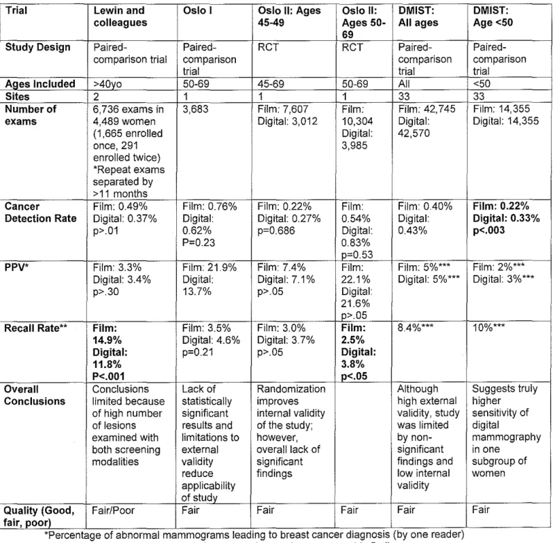
Four major studies met these inclusion/exclusion criteria: (1) Lewin and
Figure 1: Summary of Clinical Screening Trials Comparing Film and Digital Mammography
Statistically significant findings appear in bold
Trial Lewin and Oslo I Oslo II: Ages Oslo II: DMIST:
colleagues 45-49 Ages 50- All ages
69
Study Design Paired- Paired- RCT RCT
Paired-comparison trial comparison comparison
trial trial
Ages Included >40yo 50-69 45-69 50-69 All
Sites 2 1 1 1 33
Number of 6,736 exams in 3,683 Film: 7,607 Film: Film: 42,745 exams 4,489 women Digital: 3,012 10,304 Digital:
(1 ,665 enrolled Digital: 42,570
once, 291 3,985
enrolled twice) *Repeat exams separated by >11 months
Cancer Film: 0.49% Film: 0.76% Film: 0.22% Film: Film: 040% Detection Rate Digital: 0.37% Digital: Digital: 0.27% 0.54% Digital:
p>.01 0.62% p=0.686 Digital: 043%
P=0.23 0.83%
p=0.53
PPV* Film: 3.3% Film: 21.9% Film: 74% Film: Film: 5%*** Digital: 34% Digital: Digital: 7.1% 22.1% Digital: 5%*** p>.30 13.7% p>.05 Digital:
21.6% p>.05
Recall Rate** Film: Film: 3.5% Film: 3.0% Film: 8.4%*** 14.9% Digital: 4.6% Digital: 3.7% 2.5%
Digital: p=0.21 p>.05 Digital:
11.8% 3.8%
P<.001 p<.05
Overall Conclusions Lack of Randomization Although Conclusions limited because statistically improves high external
of high number significant internal validity validity, study of lesions results and of the study; was limited examined with limitations to however, by non-both screening external overall lack of significant modalities validity significant findings and
reduce findings low internal
applicability validity
of study
Quality (Good, Fair/Poor Fair Fair Fair Fair fair, poor)
*Percentage of abnormal mammograms leading to breast cancer d1agnos1s (by one reader) **Recall after consensus meeting, based on abnormal mammographic findings
***Values were extrapolated from data presented by DMIST authors
DMIST: Age <50 Paired-comparison trial <50 33
Film: 14,355 Digital: 14,355
Film: 0.22% Digital: 0.33% p<.003
Film: 2%*** Digital: 3%***
10%*** Suggests truly higher sensitivity of digital mammography in one
subgroup of women
Lewin and associates, 2001:10• 11This was the first large study comparing the
effectiveness of film versus digital mammography for screening purposes. Women over forty years who presented at one of two mammography centers were included, for a total of 6,736 paired examinations of film and digital mammography. Overall, the study found no significant difference between the modalities in terms of cancer detection rates or positive predictive values (PPVs). The authors did not design this analysis for hypothesis generation, and could not conduct statistically meaningful subgroup analyses to compare accuracies of each modality among different age groups.
The major, statistically significant result discussed in this study was lower recall rates for patients receiving digital (11.8%) than for patients who had had film mammography (14.9%). The authors attributed these lower recall rates to fewer false positive results with digital mammography. Interpreting radiologists were not masked, though, and could therefore have been more meticulous about recalling film than digital cases.
Oslo 1:12 The Oslo I trial, conducted by Skaane and associates, was published two years after the study by Lewin and colleagues. Oslo I took place at one site, and was designed as a paired-comparison study with 3,683 women aged 50-69 years receiving both film and digital mammograms. The study found no
significant difference in the two modalities' rates of cancer detection, PPV, or recall. Since the Oslo I did not include women in their forties, the trial's findings are not applicable to this subgroup of women.
Despite not finding significant differences, the Oslo authors did improve upon the experimental design of Lewin's study. Each image was reviewed by two
independent radiologists, who were not given previous screen film images for comparison. The radiologists discussed indeterminate breast lesions in teams, but the teams were only allowed to use original images to make final diagnoses.
Oslo 11:13 The main differences between the Oslo I and II studies were that Oslo II had a larger study population of 25,263 women, it included women aged 45-49 years, and it randomized participants to either film or digital mammography. The authors found no overall, significant difference in cancer detection rate or PPV between screening modalities. Notably, both Oslo I and II found higher recall rates for digital mammography than for screen film mammography. These results reached statistical significance in the Oslo II study. The higher recall rate for digital imaging is the obverse of the trial results by Lewin and associates, who found a significantly lower recall rate for digital mammography. This difference may be partially attributable to more frequent mammography recalls in the United States as compared to Europe.14
women participated in both Oslo studies. This population is more homogeneous than the population in the United States, where women of different races,
ethnicities, and even socio-economic classes have distinct screening patterns and presentations of disease. Finally, the Oslo studies only used one type of digital machine (General Electric's Senographe 20000®). The authors therefore could not explore different manufacturers, which may influence cancer detection rates.
One major contribution from both Oslo studies was the documentation of cancer types detected by each screening modality. This information is important, as histopathology of cancer estimates of the cancer's clinical aggressiveness. Histopathology can serve as an intermediate outcome, or marker for cancer mortality. Unfortunately, the Oslo I study only detected 31 malignancies, so the authors could not draw any significant conclusions about the diagnostic
performance of either screening modality. A total of 120 cancers were detected during the Oslo II study; however, the study was not powered to find very small differences in detection of DC IS versus invasive cancer. Therefore, although the Oslo studies measured histopathology, neither study was able to draw significant conclusions about which screening modality detected more invasive and/or curable cancers.
Overall, the DMIST found no significant difference between digital and screen film mammography. The authors did, however, find significantly higher Area Under the Curve (AUC), sensitivity, and PPV of digital mammography among women younger than 50 years old who are pre- or peri-menopausal with dense breastsu This subgroup included 7,315 women, or 17.1% of the DMIST study population. The AUC among this subgroup of women was 0. 791 for digital mammography and 0.544 for screen film mammography [p=.0015]. The sensitivities were 0.591 for digital mammography and 0.273 for film
mammography [p=.0013]. Finally, the PPV of digital mammography was 0.033 and the PPV of film mammography was 0.015 [p=.0005] among these younger, dense-breasted women. It should be noted that the specificities of the screening modalities were not significantly different within this subgroup of women.
One major concern about the DMIST, raised in editorials in The New England Journal of Medicine, was that digital mammography may not perform as well as
film mammography for women older than 50 years. This concern was addressed in the DMIST follow-up analyses of subgroups. Among women older than 65 years with less dense breasts, screen film mammography was more accurate than digital mammography in terms of AUC, sensitivity, and PPV. The AUC for film mammography 0.877 and for digital mammography was 0.705 in this
subgroup of women [p=.0025]. The sensitivity of film mammography was 0.694, versus sensitivity of 0.532 for digital mammography [p=.031]. Because of the Bonferroni correction, these results just barely missed statistical significance.17 Nevertheless, the near-significantly better diagnostic accuracy of screen film mammography than digital mammography among older women without dense breasts raises the possibility that digital mammography could actually be more harmful than film mammography mammography in this large subgroup of women.
on either a digital or a film image, (s)he was permitted to evaluate images from both modalities during the patient's work-up. Thus, Lewin's study and the DMIST introduced similar image interpretation biases.
Like the Oslo I and II authors, the DMIST authors also recorded histopathology of cancers detected by each screening modality. The DMIST, however, did not detect enough cancers to make statistically sound conclusions about which screening modality was more accurate for detecting specific types of breast lesions. The authors conclude the "histologic findings and stage of the breast cancers detected by the two methods were similar.16"
The DMIST was designed to have superb external validity and generalizability, as the trial was the largest and most widespread comparison of digital and
screen film mammography to date. The trial results do suggest better diagnostic performance of digital mammography among one subgroup of women. These results must be interpreted with caution, given the trial's threats to internal validity and inability to measure mortality endpoints.
Cost-Effectiveness of Full-Field Digital Mammography
not offer sufficient health gains to warrant its increased cost unless its use is limited to younger women. Specifically, age-targeted digital screening for women in their forties cost $26,500 per quality-adjusted life year (QAL Y) gained, and age- and density-targeted digital screening for women in their forties cost $84,500 per QAL Y gained. Among women over 65 years, density-targeted digital screening cost $97,000 per OAL Y gained. Overall, the cost for all-digital mammography relative to all-film mammography was $331,000 per QAL Y gained.
The primary limitation of this cost-effectiveness study was its reliance on models. Although the models were externally validated, they were based on predictions of breast cancer risk and treatment patterns, which change significantly over time with new medical developments. In addition, Tosteson and colleagues do not include costs of anxiety and fear that women experience when receiving mammograms or being called back for work-up of suspicious breast lesions. These transient hardships are difficult to capture in a cost-effectiveness analysis, but the authors do consider a "personal visit time" cost for breast cancer
screening. These allowances, which range from $3 for a woman who has a true-negative exam to $106 for a woman with a true-positive exam, may be gross underestimations of what women truly pay during breast cancer screening.19
Conclusions: Digital Mammography for Population Screening
In the best of worlds, three questions would be answered with a "yes" before we implemented any wide-scale prevention strategy: "Can it work? Does it work? Is it worth it?" In theory, digital mammography can work as a population screening tool for breast cancer. The clinical trials indicate all-digital screening is certainly plausible, and is diagnostically comparable to screen film mammography.
earlier detection of aggressive, treatable disease. The literature does not provide evidence that digital mammography "works" in this sense. The DMIST does show higher sensitivity of digital mammography in women less than 50 years old, who are pre- or peri-menopausal with dense breasts. Therefore, it can be
approximated that more breast cancers could be detected, and burden of suffering could decrease, by using digital screening in just this subpopulation.
The recent trend toward all-digital mammography makes it appropriate to examine the population-wide burden of breast cancer suffering that could be relieved with digital technology. This will shed light onto the final question: "Is it worth it?" Burden of suffering is estimated by considering (1) how many people in a given population are affected by a disease, and (2) how severe a disease is among those who have it.
Figure 2: SEER Incidence of Invasive Breast Cancer by Age21
I
i
300
250
8
2008
... 150~
100i
50Age
Source: SEER database, National Cancer Institute, Cancer Statistics Branch
Five-year survival rates provide an indicator of disease lethality, which is the second burden of suffering component. These survival rates, by age at breast cancer diagnosis, are presented in Figure 2. The graph shows that women in their forties may have slightly lower, but not significantly lower, 5-year breast cancer survival rates than do older women.
Figure 3: SEER 5-Year Survival Rates of Invasive Breast Cancer by Age21
Figures 1 and 2 make clear that women in their forties are not the majority of breast cancer patients, and they do not appear to have significantly more lethal forms of breast cancer. Screening only this population with digital
mammography could be cost-effective, at a theoretical $26,500 per QAL Y gained. However, it is inappropriate and not cost-effective to use digital screening
mammography for the entire population.
The research community cites expense and resources as major obstacles to comparing the long-term outcomes of digital versus screen film mammography. The Principal Investigator of the DMIST said in her interview:
... the reason why we did what we did exactly was because we only had so much money. If we had more money, then we could have followed
women. We could have gotten more years of data collection and had more information about annual screening mammography, and that would have been a stronger study. With infinite resources, which you don't have, we might have studied mortality as an endpoint. But, those things are impractical. I mean, it's just too expensive. We had one year of data collection, we had one year of imaging results ... and then a year to
follow-up. So, we had a total study length of three years and it still cost us twenty-six and a half million dollars.
"An Idea Whose Time Has Come"
.. . I say openly, to the world, that we're not doing well. Still 40,000 women are dying a year. It's not criminal, but it's not low enough. There are still too many people dying. We have to acknowledge that we're finding breast cancer in some women when it's too late. We're doing the best we can. People get all defensive about it ... Some of the radiologists, they get all defensive about it: "How dare you attack us? We're doing the best we can; we're working our asses off; we're really working hard on this." Yes we are, but our tools aren't good enough. -- Etta Pisano, MD, Principal Investigator of DMIST
Digital mammography was born into a time of great potential for any new breast cancer screening technology. In the late 1990's, our "war on cancer" was fully waged, with advocacy groups acting as commanders of the siege. The
effectiveness of breast cancer screening is, however, limited because of the imperfect sensitivity and specificity of mammography. While the breast cancer lobby has been very successful in summoning enthusiasm and infrastructure for their cause, we have remained frustrated with breast cancer screening because, as Dr. Pisano stated, "our tools aren't good enough." Therefore, we may
consider digital mammography an "idea whose time has come." John Kingdon states, in American politics, "an idea whose time has come is so powerful that it pushes aside everything that might stand in its path."6
The War On Cancer
Deborah Stone argues that "war" is ingrained into policy language because it invokes our desire to survive.23 Despite decreasing incidence and mortality rates, breast cancer remains the most commonly diagnosed cancer among women. General apprehension among healthy women about developing breast cancer contributes significantly to its burden of suffering. The anxiety-provoking nature of breast cancer screening was described by the Chairman of Duke's Radiology Department:
everybody unhappy ... or two, you're OK this year, we'll see you. It's not like "you're cured, you're healthy, go away." And all the women are
familiar with it, and everybody's got at least friends who have had biopsies done. So they're very anxious, and because they're anxious and healthy, they turn out to be a more difficult population.
In addition to the ambiguous nature of screening, breast cancer is also worrisome because it is not strongly associated with individual choices and habits. Popular knowledge teaches women to feel safe from lung cancer if they are non-smokers and from cirrhosis if they are non-drinkers. The tendency for breast cancer to invade a woman's life without warning or clemency perhaps contributed to the development of the "war on cancer."
Advocacy groups, however, have been instrumental in popularizing this "war." Breast cancer advocacy is a relatively new political movement, as Betty Ford was one of the first women to speak openly about her disease in 197 4. This
encouraged other women with breast cancer to discuss what had previously been an utterly private and even shameful diagnosis24 Betty Ford and others opened the floodgates of popular awareness and advocacy, and it was not difficult to tie breast cancer awareness and research to other goals espoused by the Women's Movement at the time. The Susan G. Kamen Foundation was founded in 1982, and other advocacy groups, including the National Breast Cancer Coalition and Breast Cancer Action, were founded in the mid 1990's.
These groups have been influential agenda-setters for Congress, which
increased funding for breast cancer research from $40 million per year to over $200 million per year through the 1990's.25 In the year 2007, funding for breast cancer included $572.4 million from the National Cancer Institute and $127.5 million from the Department of Defense, for a total of about $700 million for breast cancer research through federal organizations.26• 27 Breast cancer
Mammography Day" to encourage women to make yearly mammography appointments.29 A senior policy analyst at the Susan G. Komen Foundation summarized the role of advocacy groups in placing breast cancer screening on the national agenda when he stated:
I think the greatest success in screening is the ... availability of mammograms and the education effort that's taking place ... and the partnerships between federal and state governments and private
organizations like ours, the Susan G. Komen Foundation for the Cure, and others ... the American Cancer Society and others to get the word out that mammography is the gold standard in screening for breast cancer, and that you really have your chances of survival increase by having breast cancer detected early ...
Advocacy groups have been so successful in their efforts to "get the word out" about the "war on cancer" that women have historically felt overly vulnerable to the disease. In her book Gender and American Politics, Sue Tolleson-Rinehart found women in the mid 1990's overestimated their risk of breast cancer by up to twenty-fold 24
The public's tendency to be excessively nervous about breast cancer was
Figure 4: National Screen Film Mammography Facility Count
9400,---,
9200
9000
8800 8600
8400
Oct, 2002
Oct, 2003
Oct, 2004
Oct, 2005
Oct, 2006
Oct, 2007
Feb, 2008
Source: MQSA National Statistic Archive, FDA Center for Devices and Radiological Health
The media were responsible for amplifying the perceived mammography shortage, as reports of women crowded into mammography centers appeared especially in the northeastern newspapers. The Boston Globe published an article in October 2000 headlined "Radiologists are quitting, making women wait longer to find out if they have breast cancer."30 The article cautioned readers about an upcoming mammography shortage, since the number of women seeking mammograms doubled between the years 1985 and 2000. In 2002, a Newsday article headlined "A Long Wait for Mammograms" warned New York
women they would wait between one and three months to receive mammograms.
At the same time the media were popularizing a mammography shortage, they were also maintaining the "war on cancer" through digital mammography
coverage. Of 215 digital mammography articles analyzed, 44 used the following war-related terms in either their headlines or opening paragraphs: weapon, war, struggle, fight. Among interview respondents, six out of twelve used the terms "fighter," "war," and/or "save lives," with four respondents repeating these words multiple times over the course of the interview.
"Medicine from Space," "CIA lets loose of technology for health use," and "From bombs to breast cancer aid" appeared in papers across the country.31-33 In October 1993, the Sacramento Bee described digital mammography as "a real-life example of turning swords into plowshares."34 Several articles referenced the Regan Administration's "Star Wars" Defensive Initiative, stating technologies from this campaign would be used to enhance digital mammography.35-37 In 1994, the government created the "Missiles to Mammograms" program to foster cooperation between digital mammography innovators and the intelligence community.38 The expectation for "Missiles to Mammograms" was that "space, defense and intelligence technology allow the unprecedented mapping of the landscape of the breast."
These initial descriptions of digital mammography could appeal to the public on several different levels. First, the media accounts attract our sense of fantasy and "the unknown" in a very real way, as digital mammography was connected with outer space and even the Search for Extra-Terrestrial Intelligence (SETI)a2 In addition, the notion of combining wartime technologies with a potentially life-saving technology could be pleasing for both peace-lovers and ironic satire-lovers. Professor JoAnne Earp believes these original accounts could have been purposefully marketed to adorn screening mammography, a uniquely female experience, with more masculine qualities:
When you invest innovation with verbiage that surrounds it with tougher, more manly kind of sales aura ... such as war, or technology, or space ... then I think as an ad person, you think that's going to sell your sort of softer, "breast, women, and lower status" ... kind of take it away from that kind of softer, more feminized, gendered image to a more masculine, more "war toys," technology, masculinized image ... where the money has been and the technology has been and the resources have been and the control has been. So I think it probably was no coincidence that it was sold that way.
The State of Breast Cancer Screening
Figure 5: Important Dates in the History of Breast Cancer Screening & Digital Mammography
1895 -Wilhelm Roentgen develops the X-ray process39
1960-Dr. Robert Egan adapts high-resolution industrial film for mammography, allowing simple and
reproducible mammograms with improved image detail40
1963 -Initiation of the first randomized controlled trial of breast cancer screening by the Health Insurance
Plan of New York. Data from 18 years of follow-up indicates 25 percent reduction in breast cancer mortality
among women over 50 years who received screening mammography40· 41
1974-Betty Ford speaks openly about her breast cancer diagnosis"
1982-Susan G. Kamen Foundation for the Cure founded25
1986-American Cancer Society and American College of Radiology develop breast cancer screening
accreditation program40
1992-Mammography Quality Standards Act requires all mammography facilities to be licensed by the
FDA28
*NCI designates digital mammography "the imaging technology with the highest potential" for detecting and
diagnosing breast cancer42
1993-Suzanne Fletcher and colleagues publish evidence review, "Report of the International Workshop on
Screening for Breast Cancer," which acknowledges unknown benefit of screening for women aged 40-4943 *President Bill Clinton designates October 21" as National Mammography Day29
1994-"Missiles to Mammograms" program launched by Public Health Service's Office on Women's Health,
with the pur;pose of digital technology sharing between the intelligence community and mammography
innovators3 · 44' 45
1997-NIH Consensus Development Conference on Breast Cancer Screening for Women Ages 40 to 49
does not recommend universal mammography for women in their forties; decision met with great resistance from breast cancer screening enthusiasts and politicians46
1998-NEJM article reports cumulative risk of false positive mammogram at 49.1% after 10
mammograms47' 48
*FDA approves lmageChecker for mammography double-check49· 50
2000-FDA approves G. E.'s digital mammography machine, the Senographe 2000D®51
2001 - National Cancer Institute awards team of investigators, headed by Dr. Etta Pisano, $26 million grant
to study digital mammography52
*Medicare coverage for digital mammography increases to 150 percent of traditional mammography
reimbursement rates53
2002-Tommy Thompson announces federal guidelines, which strongly recommend mammography for
women aged 40-50 years28
*BiueCross, BlueShield North Carolina begins reimbursing for digital mammography at the same rate as
screen film mammographl4
2003-FDA starts providing accreditation to mammography facilities which only use digital technolog/5
2005-DMIST results show digital mammography more effective for younger, dense-breasted women
Other radiology tools, such as CT, MRI, and PET scans, have been digital since their inception. These sophisticated technologies have paved the way for popular concepts like "the digital revolution" and "wireless medicine." For example, a January 1998 article in the Dallas Morning News reported, "More radiologists and hospital administrators seem to be dreaming in digital these days because a new generation of digital technology promises to enhance clarity, reduce costs and improve convenience."57 Film mammography, which requires a dark room for image development and a view box for analysis, seems archaic compared to other radiology technologies. Duke's Director of Breast Imaging described the state of breast cancer screening in somewhat hyperbolic terms:
The entire rest of the radiology department has been on soft copy display -that means CAT scans, MRI, even X-rays of big toes ... for many, many years. So, as radiologists we've had experience using soft copy displays when we get our call.
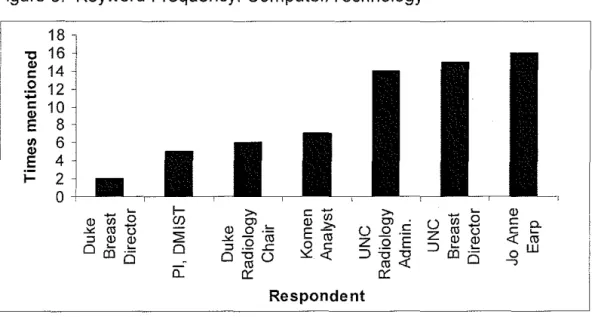
The importance of digital mammography as "technology" was demonstrated in other interviews, as the following respondents mentioned either "computers" or "technology" several times throughout our encounter:
Figure 6: Keyword Frequency: Computerrrechnology 18
"0 16
~ 14
,g
12~ 10
E 8
UJ 6
"'
E 41- 2
0
~
Q) 1i5 0
~rot> ::J Q) Q)
0
cD
.!::::0 1-(f) 2 0 0.. c
-Ql UJ
E~
~~
>. Cl . 0 .2 .!:
z o E
=>:01.:1 rn<C 0::: Respondent ~ u1il£ Z "' Q) u Q)
::>
00
.!::::0
Q) c a. c ~
<( "' ow
--,
frequently than did respondents affiliated with Duke University. The trend is telling: UNC converted to all-digital technology early, and has a stake in
embracing the technology in which it has made a substantial early investment. Duke, on the other hand, is only now overhauling its screen film systems and does not evince the language of early adopters.
The media have also been attuned to the innovative aspect of digital
mammography, and have used concrete, simple metaphors to help explain the technology to the public. These include comparing digital mammography to word processors rather than type-writers, fax machines rather than snail mail, and digital cameras rather than traditional cameras. In fact, 10% of reviewed articles likened digital mammography to digital cameras, and about two-thirds of articles that explained the mechanics of digital mammography used the analogy of either a digital camera or a word processor. A Newsday article published in September 2005 provided one such typical description of digital mammography: "Both digital and film mammograms require the use of X-rays to produce an image. But digital mammography, which is similar to taking pictures with a digital camera, allows doctors to view the mammogram on a computer and to change the contrast on their screens to view suspicious areas."58 The digital camera metaphor, which is even being used by clinicians, makes digital mammography very palatable for the average woman and also draws on our fondness for the newest and most convenient technologies.
Breast imaging specialists also like new technology, especially when it may lead to higher status and more reimbursement. Breast specialists have perceived themselves to be underpaid and subject to higher rates of lawsuits, compared to other radiologists. The Director of Breast Imaging at UNC described the inherent humility associated with reading mammograms as follows:
high-paying reimbursement. .. by them (digital mammograms) having a little bit higher reimbursement, it's just starting to make the tables ... in my opinion, from my side of the fence ... a little bit better. The reimbursement for mammography ... for what people pay ... it's just so low.
Media have also reported on the relatively lower compensation and higher liability for breast imaging. 59 Numerous articles have described mammography as a money-loser, and the New York Times even quoted a renowned breast imaging specialist who described fellowships in mammography as a "lousy career move."60 Other articles have described breast imaging specialists as the "most highly sued radiologists."
Between August 1985 and 1995, the Physician Insurers Association of America (PIAA) tracked over 125,000 medical malpractice claims for missed breast cancer diagnoses, and found breast cancer-related claims yielded higher settlements than did any other malpractice suits except for those arising from cases of brain-damaged infants. These findings led the American College of Radiology (ACR) to support regular screening mammography for women in their forties, supposedly to decrease the number of missed breast cancers.61
The perception of higher numbers of lawsuits against radiologists who read mammograms was reaffirmed by several interview respondents, including Dr. Earp, who commented:
Breast cancer is the first. .. among those diseases that cause lawsuits. mean, there are papers out there that show, of the suits that happen ... breast cancer was the first cause of lawsuits.
Partially in response to these widespread malpractice concerns, the St.
Petersburg Times conducted research into malpractice history for breast imaging specialists in Florida, and found no significant difference in number of lawsuits or malpractice insurance rates for radiologists between 1991 and 2002.62
than evidence about the actual amount of litigation, may be considered a litmus test for the political strength of breast imagers and screening advocates. It is not surprising that these groups were very influential in launching the adoption and widespread distribution of digital mammography.
In addition to digital mammography's potential to advance what might be thought of as the "social status" of breast imaging, the technology also brought great hope for more accurate diagnoses. Digital mammography came to market during a time of intense debate, among professional organizations and cancer
specialists, about the true value of breast cancer screening. In October 1993, the Journal of the National Cancer Institute published the "Report of the
International Workshop on Screening for Breast Cancer." Investigators reviewed eight major randomized controlled trials of breast cancer screening, and found a one-third reduction in mortality for screening women ages 50-69, but no benefit for screening women in their forties43 In 1994, the American Cancer Society (ACS) and the National Cancer Institute (NCI) engaged in a fierce discussion about mammograms for women in their forties, with the ACS supporting screening and the NCI concerned about potential harms offalse results.63 In April 1998, even more public attention was drawn to the potential for false positives when the New England Journal of Medicine published an analysis estimating the cumulative risk of a false positive result at 49.1% over the course of 10 years, or 10 mammograms47 False negative screenings have also drawn media attention, as an article published in the Houston Chronicle explained: "It (mammography) misses 20 percent to 25 percent of cancers in women under 50 and 8 percent to 10 percent in women over 50 ... 64"
So the number of false positives ... if you look at the data on how many biopsies and things we do, that turn out to be normal. .. The patients don't usually get horrendously upset, in the sense that, in the world of women, having a breast biopsy is fairly commonplace. In a way, doctors always say ... "three quarters of these are going to be normal, so don't get yourself too excited." The other side of that though, is if only three-quarters of those are normal, couldn't we do a better job about figuring that out before you have to worry for days? ... and needles ... and all that kind of stuff. We should be doing a better job.
The uncertain analyses of screening mammography have been accented by several definitive, though not necessarily evidence-based, statements by public health officials. For example, in March 2000, the United States Preventive Services Task Force (USPSTF) developed a guideline for screening
mammography in women over 40 years old. The USPSTF gave their guideline a "B" recommendation, indicating fair evidence in favor of screening
mammography. Despite the "B" recommendation, Tommy Thompson emphatically announced to American women that benefits of screening mammography outweigh potential harms. Thompson said the federal
government was sending a "powerful and clear" message to women: "If you are 40 or older, get screened for breast cancer with mammography every one to two years."65
Despite Tommy Thompson's declaration, confusion and debate have continued to surround breast cancer screening. One reporter for the Chicago Tribune perhaps phrased it best in October 2004 when she wrote: "Many women are confused about mammography, and it's no wonder. For years, doctors have been debating and changing breast cancer screening guidelines. It seems like our bras last longer than mammography recommendations do."66
Conclusion
Digital mammography, "an idea whose time has come," entered health care as a technology that could potentially relieve the uncertainties and frustrations
advocacy groups' "war on cancer," update the mammography process, boost the prestige and profitability of breast imaging, and provide greater diagnostic
accuracy in screening mammography. The New York Times described these popular hopes for digital technology in a January 2001 article entitled
"Mammography's Next Step is Assessed:"
The FDA as a Gatekeeper
We are
a
"gatekeeper" ... and we need to assure that things on the market are not going to hurt people. Once we can assure ourselves of that, then I think we need to allow technologies on the market with the understanding that we will get more information as time goes on.-- Daniel Schultz, MD, FDA Director, Center for Devices and Radiological Health
General Electric's Senographe 2000D® full-field digital mammography unit was approved by the United States FDA in January 2000. As with other medical technologies, FDA approval was crucial for distribution of digital mammography because it allowed General Electric, and other private vendors like Hologic, Fischer, and Fuji, to begin widespread marketing to patients and clinicians. In addition, FDA approval was a necessary step before the federal government could increase its Medicare reimbursement of digital mammography to 150 percent of screen film mammography reimbursement. 53
The FDA's approval happened at an ambiguous time in the accrual of
effectiveness data for digital mammography. In January 2000, none of the major trials comparing digital with screen film mammography had been published. The study by Lewin and associates was not published until March 2001 and the Oslo I and II studies were published in 2003 and 2004, respectively. The DMIST did not even have funding until one year after the FDA approved digital
mammography. Dr. Schultz summarized the approval process as follows: ... as with many technologies, we were able to provide reasonable assurance of safety and effectiveness, which is the standard that we use and that Congress has given us to approve new medical devices. So, what that means is that in many cases we don't know everything that there is to know and we fully expect that additional information will be gathered over time.
Only a patchwork picture of the approval process may be discerned from media accounts and interview respondents affiliated with the FDA and/or digital
Administrative Director of UNC's Radiology Department described Dr. Pisano's cooperation with both private companies and the FDA:
Etta helped some companies achieve their FDA clearance. She's also had a role in advising the FDA on what was reasonable, to assess the turnover ... what was acceptable for the FDA to approve digital imaging. Given the critical roles of innovators and private companies in technology development, it is not accurate to consider them "external entities" trying to impose their agendas on the FDA. Nor is it appropriate to expect FDA staff of Advisory Committee members to evaluate new medical devices without input from these innovators and private companies.
In the case of digital mammography, both breast imaging specialists and technology development companies were eager for rapid approval. UNC's Administrative Director of Radiology described the environment prior to buying digital equipment:
Now, when it gets closer to, you know, hitting the market and the vendors were so excited about the eminent FDA approval. .. and then postponed, postponed, postponed. The vendors were trying to stage the equipment. In some ways, we were willing to purchase, but if it's not FDA approved ... we don't go for it. .. but we will continue clinical research work on the fringe, until it is approved. And then the vendors got to the point where they were so anxious to sell. .. that they got a little bit reluctant, because they thought FDA was right around the corner. As soon as they got FDA, they'd be able to sell.
The administrator's language in this passage, particularly his use of the words "excitement," "anxious," and his repetition of "postponed," captures the great anticipation with which private companies must have awaited FDA approval. Private companies, however, may not have been as eager for approval as were breast imaging specialists. Dr. Pisano expressed frustration with the process from her perspective as an innovator:
dealing with breast cancer. My generation, your generation ... So I feel really a huge pressure to try and move things forward and get things done more quickly. It really bothers me that it takes so long ... and that we're making such slow progress. I wish the breast cancer community would focus on that, because the FDA. .. it's not just the FDA, it's the companies, it's radiologists, it's everybody.
Dr. Pisano's dissatisfaction with the approval process, which almost took on the tone of a personal mission in this excerpt, has been echoed by other groups of breast imaging specialists. One group of radiologists wrote a statement of
concern about the digital mammography approval process for Diagnostic Imaging in December 1999. Their statement was also published in a USA Today article entitled "Going digital Proponents say it's time the FDA updates mammography system."68 The group sent the following joint message to the federal
government: "We believe that the review process, which has extended for more than three years, is unduly prolonged and complicated."
Dr. Schultz acknowledged pressures from private vendors and breast specialists during his interview: "Um, there's always pressure from companies to approve their products as quickly as possible. I mean, that is a given in what we do." Dr. Schultz also noted that in the case of digital mammography, pressure from technology developers was so great that the agency needed to "take a stand:"
There was a lot of pressure, and a lot of it actually was coming from very prominent clinicians, and even academic clinicians, who felt that the mere fact that the image was ... appeared to be ... a higher-quality image ... should in fact be enough for us to approve the technology, as we had for other types of digital technologies. And I think this was a case where we really needed to sort of take a stand and say that, you know ... the fact that it's a better picture, or what appears to be a better picture, is not enough. The digital mammography approval process was thus caught between the
Conflicting interests even spilled into the FDA's internal discussions about the technology. While certain FDA members pushed for rapid approval, others called for more extensive, head-to-head comparisons of digital and film
mammography. A USA Today article reported specific information about dueling perspectives of the agency members, certain top FDA officials, and the advisory committee.68 The article reads:
The agency wants manufacturers to conduct screening trials that would compare digital and film mammography head-to-head. Such a trial would need thousands of patients and cost millions of dollars.
"This is not an industry that's used to those kinds of costs," says David Feigal, top medical devices official at the FDA.
But the advisory committee, fearing that patients bear the cost of a screening trial, urged the FDA not to require one of G.E.
The end result of these conflicts, both between the FDA and "outside" groups, and among FDA members themselves, was a compromise. After a three-year review process, the FDA had sufficient evidence to convince itself of digital mammography's safety and effectiveness. Dr. Schultz described the compromise:
It was one of those situations, I think, where nobody was totally happy with us. So I think we were probably in close to the right place.
In retrospect, the FDA was actually "close to the right place," because clinical trials have now demonstrated similar diagnostic accuracies of digital and film mammography. Data collection for the actual approval process, however, appears to have been far messier. Specifics are unclear about which data were collected, and what evidence was gathered, before the Senographe 2000D® was approved in 2000. In our April 2008 interview, Dr. Schultz gave one account of the FDA's protocol:
affected and still get that kind of clinical information, without the large trials that were originally proposed.
The Dallas Morning News reported slightly different data collection in February 2000, although Dr. Schultz was cited as the newspaper's key informant. The article stated: "In studies of 625 women, a printout of the mammogram from the digital machine was as effective in detecting breast cancer as standard film mammograms."
These descriptions by Dr. Schultz and the Dallas Morning News are vague, particularly with regard to the number of investigational cases used to compare screen film and digital mammograms. Uncertainties were mirrored in press reports, as the Dallas Morning News article warned readers: "Digital
mammograms appear as good as - but not better than - regular mammograms in detecting breast cancer ... "
Ultimately, digital mammography won FDA approval on the basis of these uncertain data. The FDA's approval legitimized the subsequent increase in Medicare reimbursement for digital mammograms, which undoubtedly catalyzed the spread of the technology. Higher reimbursement rates made the upgrade from film to digital technology invisible, from a cost perspective, for many medical practices. In addition, the more generous Medicare reimbursement set a new standard for private insurance companies to increase their payments for digital mammography. Details about the justifications and reasoning behind higher Medicare payments for digital mammography are uncertain. At the time of this writing, no Medicare officials were available to comment on funding
appropriations for digital mammography. The 2001 increase in Medicare funding for digital mammography, however, was certainly dependent on the technology's FDA approval.
entirely determining the fate of the technology. Also because of compromises, however, providers did not have rigorous evidence to offer a convincing
Perceptions with Real Consequences
So, not to my surprise, but a little bit to my surprise, we get rid of all-film screen and we go to all-digital when there's not data to show that's effective. But I understand why because one of my most favorite of all aphorisms, sentences ... I attribute it to WI. Thomas, sociologist in the 1920's, who said:
"Things that are perceived as real are real in their consequences." -- Professor JoAnne Earp, SeD
As with many of our new technologies, the widespread distribution of digital mammography has been catalyzed by anecdotal success stories and even misinterpretation of facts by leaders in health care. This phenomenon is likely because, as Deborah Stone writes, " ... facts do not exist independent of
interpretive lenses, and they come clothed in words and numbers." Facts about digital mammography have been disseminated in a disorganized way, with the media and even key stakeholders in breast cancer screening receiving their information second- or third-hand rather than directly from the DMIST results section. Certain attributes of digital mammography have been inappropriately magnified, leading some decision-makers to justify their actions with incomplete data.
The Fate of the Evidence
Two popular perceptions about digital mammography have led the general public and radiologists to accept its superiority over screen film mammography. First, positive findings from the DMIST and other trials have been overstated by the medical literature, digital mammography enthusiasts, and the media. Second, people have exaggerated the theoretical and logistical advantages of digital mammography.
Both these perceptions can be traced to the presentation of digital