CLAIRE CAUGHEY MOST. Metabolite Accumulation during
m-Cresol Degradation by Aquatic Bacteria. (Under the
Direction of DR. FREDERIC K. PFAENDER)
The accumulation of metabolites in the aqueous phase
was measured during studies of m-cresol biodegradation with
natural water samples. 14C-m-cresol was separated from
radiolabeled products remaining in the aqueous filtrates
using HPLC. In heterotrophic activity studies, which
involve short-term incubations (< 24 h), metabolite
accumulation accounted for only a small fraction of the
radiolabeled m-cresol originally added. After longer
incubations characteristic of die-away studies, metabolite
accumulation accounted for as much as 30% of the label
originally added. The nature of metabolite accumulation
varied, and appeared to reflect differences in the initial
environmental samples. Sometimes no metabolite
accumulation was detected. One time, a few products
accumulated and they were less polar than m-cresol. At
other times, many different products accumulated and they
were more polar than m-cresol. When metabolite
accumulation occurred, it was greater in samples receiving
960 ug/1 m-cresol than 120 ug/1 m-cresol. No one
metabolite accumulated consistantly to a level greater than
TABLE OF CONTENTS
Page
LIST OF TABLES...iv
LIST OF FIGURES...v
ACKNOWLEDGEMENTS...vii
INTRODUCTION... . .___1 LITERATURE REVIEW...4
Microbial Degradation...4
Measures of Biodegradation...8
General Features of Aromatic Degradation...12
Metabolism of m-Cresol...14
Pure Culture Studies, the Natural Environment, and Metabolite Accumulation...22
MATERIALS AND METHODS...27
Sample Source and Collection...27
Sample Preparation, Incubation, and Termination...28
HPLC Methodology...31
Instrumentation...31
Separation Procedure forr Cresol Disappearance Experiment...32
Separation Procedure for Metabolite Accumulation Experiments...33
Sep-Pak Concentration Procedure...36
RESULTS AND DISCUSSION...37
Cresol Disappearance Experiments...37
Cresol-Metabolite Separation Experiments...42
Metabolite Accumulation Experiments...43
CONCLUSIONS... 70
REFERENCES...72
LIST OF TABLES
Page
Table 1: HPLC Detention Times and Fraction Numbers
of m-Cresol and Metabolite Standards...45
Table 2: Recovery in Aqueous Filtrates of the Radioactive Label Originally Injected.
September, 1984...49
Table 3: Recovery Efficiencies of Radioactive Label
in Sample Filtrates when Eluted with an
Acetonitrile Mobile Phase. September, 1984....50
Table 4: Recovery in Aqueous Filtrates of the
Radioactive Label Originally Injected.
November, 1984...54
Table 5: Recovery in Aqueous Filtrates of the
Radioactive Label Originally Injected after Different Periods of Incubation. January,
Figure 1: Degradation of m-cresol to ring-fission
substrates...16
Figure 2: The ortho fission pathway of protocatechuic
acid...19
Figure 3: The meta fission pathways of protocatechuic
acid and 3-methylcatechol...20
Figure 4: The gentisic acid pathway...21
Figure 5: Calibration Curve...34
Figure 6: Percent of m-Cresol and Radioactivity
Remaining in Samples vs. Time...39
Figure 7: Percent of m-Cresol and Radioactivity
Remaining in Controls vs. Time...41
Figure 8: Sample Chromatogram of m-Cresol and Aromatic
Metabolites...44
Figure 9: HPLC Fractionations of Sample Filtrates.
September, 1984...48
Figure 10: HPLC Fractionation of One Sample Filtrate
under Extended Gradient Elution Conditions.
September, 1984...52
Figure 11: HPLC Fractionation of Sample Filtrates.
November, 1984...55
Figure 12: Proportion of Radioactive Label Remaining in
the Sample Filtrates Which is Recovered as Metabolites vs. Incubation Time. January,
Figure 13: HPLC Fractionations of 960 ug/1 Sample
Filtrates at 0 hours, 3 days, 8 days and 16
days. January, 1985...60
Figure 14: HPLC Fractionations of 120 ug/1 Sample
Filtrates at 0 hours, 3 days, 8 days and 16
days. January, 1985...61
Figure 15: HPLC Fractionation when Collect 1 ml Fractions
instead of 2 ml Fractions. 960 ug/1 Sample
ACKNOWLEDGEMENTS
I would like to thank my advisor. Dr. Fred Pfaender,
for his time, advice and support during the past couple years. I would also like to thank Dr. Sobsey and Dr.
Turner for serving on my committee and for occasionally lending their advice. Thanks to Marj Aelion for her personal support through some hard times, and special thanks to my husband. Bob, for his support throughout it
all.
This research was supported by a grant from the U.S.
Hazard assessments for chemicals released into the
environment involve two types of studies: toxicity studies and environmental fate studies (Gledhill and Saeger, 1979; Lee and Jones, 1980). Environmental fate studies are an integral part of any hazard assessment scheme because they may reveal the ultimate modes and concentrations of
exposure to the compound. The transport and persistance of any compound will be influenced by a complex web of
physical, chemical and biological factors. One factor especially important in the disappearance of compounds in the environment is microbial degradation (Gledhill and Saeger, 1979; Alexander, 1980).
Several methods are available for assessing the
biodegradation of potentially hazardous chemicals in the environment. The techniques available have been reviewed
by Larson (1984). One of the newer and most sensitive
methods, the heterotrophic bacterial activity method, involves the use of radiolabeled substrates to assess
biodegradation kinetics. Experimental conditions are
designed to simulate those existing in the natural
environment as much as possible. Natural water samples are
amended with low concentrations of radiolabeled substrate
and allowed to incubate until a small proportion of the
sample. Following sample incubation, the radioactivity
evolved as carbon dioxide and/or incorporated into cells is
measured. This radioactivity is then used to compute rates
of biodegradation, and when appropriate, analyze the data
according to an enzyme-saturation kinetic model.
Heterotrophic activity studies are concerned with only
two possible metabolic fates of the radioactive label, uptake into cells and production of carbon dioxide. One
potential limitation of these studies is that they do not
measure a third possible metabolic fate, the release of metabolites of the parent compound from cells back into the
surrounding water. If such metabolites were to accumulate
in the aqueous phase, then the heterotrophic activity
method would underestimate the extent of biodegradation.
Little is known about which products in a metabolic
pathway are excreted and accumulated outside the microbial cell (Alexander, 1980; Fletcher, 1979). Furthermore, most
metabolic pathways are established under experimental
conditions so different from those in the natural
environment that it is difficult to extrapolate from the
results of these studies to the natural environment. For
example, degradation pathways are typically established
using pure cultures of microorganisms grown in artificial
media with the substrate of interest present at high
natural water medium. The effect of these conditions on
the mechanisms of degradation and the accumulation of
metabolites is largely unknown and difficult to predict (Alexander, 1980; Bollag, 1974).
In this project, metabolite accumulation was explored using m-cresol, which has been identified as a priority pollutant under the Toxic Substances Control Act. The compound is produced from the high-temperature
carbonization of coal and as a by-product in the petroleum
and petrochemical industries (Chapman, 1972). It is used
in the manufacture of disinfectants, fumigants,
photographic developers, and explosives (Merck Index,
1983). m-Cresol was chosen for this particular project because it has been studied extensively in this laboratory using the radiolabel method described above. The projectobjectives were threefold: (1) to determine the extent of
metabolite accumulation in biodegradation studies using
short term heterotrophic uptake assays, (2) to look at
Microbial Degradation
When an organic chemical is released into the
environment, biological, chemical and physical factors will influence its fate. Major structural changes in the
compound, however, are mainly accomplished by
microorganisms (Alexander, 1980). Two types of microbial metabolism are involved in the biodegradation of organic compounds (Harder, 1981). In the first form of metabolism, the compound serves as a substrate for growth and energy. Microbial enzymes degrade the compound into other
structures involved in intermediary metabolism. Some of the compound's carbon may then be converted into cell
constituents, while the rest may be channeled into
oxidative cycles. Complete oxidation produces carbon
dioxide and water, while providing energy for biosynthesis
(Chapman, 1979). Thus, this form of metabolism may lead to
the mineralization of an organic substrate into inorganic
products (Bollag, 1974; Rubin et_al., 1982; Subba-Rao et
al. , 1982). In a recent study, Chesney et al. (1985) found that microorganisms in freshwater samples completely
removed phenol present at trace concentrations. About
eighty percent of the parent phenol was mineralized to
polysaccharides). This study suggests that when a
hazardous compound serves as a substrate for growth and
energy, microbial metabolism may be instrumental in its complete removal from the environment.
In the second form of metabolism, a chemical is
transformed by a microorganism without deriving carbon or
energy from that transformation to support growth (Horvath, 1972). This phenomenon is termed cometabolisra and has been
demonstrated in the laboratory for compounds such as DDT, 2,3,6-trichlorobenzoate (2,3,6-TBA),
2,4,5-trichlorophenoxyacetate (2,4,5-T) and 3-methylcatechol
(Horvath, 1972). From an environmental standpoint, one of
the major features of such cometabolic transformations is
that they only lead to partial degradation of the parent
compound. Cometabolisra of 2,3,6-TBA by a Brevibacterium
sp. results in the accumulation of its transformation
product 3,5-dichlorocatechol (Horvath, 1971). Similarly,
p,p-dichlorodiphenyl methane accumulates from the
cometabolisra of DDT by Aerobacter aerogenes (Wedemeyer, 1967). Such cometabolic products are often structurally
similar to the parent compound and can be less toxic, as
toxic, or even more toxic than the parent compound
(Alexander, 1980; Bollag, 1974). Thus, partial degradation
by cometabolisra may not remove the hazard posed by the
compound differently. The insecticide carbaryl, for
example, is subject to hydrolysis by some organisms and to hydroxylation by others ( Bollag, 1979). Moreover,
hydroxylation of carbaryl by Aspergillus terreus produces
1-Naphthyl N-hydroxy-methylcarbamate as the major metabolic product. Hydroxylation by Penicillium sp., in contrast, produces 4-hydroxy 1-napthyl methyl carbamate as the major metabolic product (Bollag, 1979). Formation of the former involves a side chain hydroxylation, whereas the latter
involves a ring hydroxylation. Hence, even a similar
initial reaction can produce products differing in the
subsequent transformations required and in the products which ultimately accumulate.
A further complication are population interactions
among microbes living in a community. Gunner and Zuckerman (1968) isolated from soil an Arthrobacter sp. and a
Streptomyces sp. which together could synergistically degrade the ring structure of the organophosphate insecticide diazinon. When incubated alone, neither
organism could degrade the ring structure. Similarly, Beam
and Perry (1974) reported a commensal relationship involved
in the degradation of cyclohexane. Mycobacterium vaccae cometabolicly degraded cyclohexane to cyclohexanone while
growing on propane. A second organism, which was unable to
compounds, especially those only partially degraded or cometabolized. Negative interactions such as competition, predation, or parasitism may also influence compound
degradation.
A host of physical and chemical factors affect
biodegradation in the natural environment. Factors of
importance in freshwater habitats include temperature, pH, dissolved oxygen, and the presence of organic and inorganic compounds (Atlas and Bartha, 1981; Fewson, 1981). Such
environmental factors tend to influence overall microbial
metabolic activity rather than biodegradation specifically
(Scow, 1982). These factors also may determine which
microbial populations persist within an ecological niche or are active at particular times. Consequently, a chemical
susceptible to microbial attack under one set of conditions
may not be degraded under other environmental conditions. Substrate concentration is one factor that may
specifically influence the rate and extent of microbial
attack. Some studies suggest that at least some organisms
may have dual enzyme systems, one which functions at low
lakes, "the rate of phenol mineralization as a function of concentration was linear below 1 ug/ml, fell off between 1 and 100 ug/ml, and was again high at levels above 100
ug/ml." They hypothesized that this reflected the activity
of oligotrophs at the lower concentrations and eutrophs at
the higher concentrations. In a more recent study, Wang et al. (1984) found that the microbial community in lake
water samples mineralized N-phenylcarbamate (IPC) at a
concentration of 400 pg/ml. At the higher concentration of 1 ug/ml, however, IPC was not mineralized but was converted
to organic products. The partial degradation at the higher
concentration may reflect the activity of different
organisms or enzyme systems. All together, the above studies suggest that concentration may influence the mode,
extent and kinetics of biodegradation, and thus, the
persistance of a chemical and/or its metabolites in the
environment.
Measures of Biodegradation
A wide variety of methods are used to study
biodegradation. Methods typically include three parts: the analytical test, the chemical to be studied, and the
Lee, 1979). Consequently, the methods chosen will depend
on the nature of chemical and where it is released into the
environment.
Most analyses follow the accumulation of biomass, the
disappearance of reactants, or the appearance of degradation products (Raymond, 1979). Some of these
analyses only follow biodegradation indirectly, as in the
measurement of oxygen uptake and carbon dioxide evolution.
These analyses tend to be simple and inexpensive because a
specific analytical technique does not have to be developed
for each chemical tested (Gilbert and Lee, 1979). They are
typically limited to screening tests, however, because of
their low sensitivity and limiting test conditions (Gilbert and Lee, 1979; Gledhill et_al, 1979).
Biodegradation is followed directly by analyses which
monitor the disappearance of parent compound. Common
techniques include gas chromatography, high pressure liquid
chromatograpy, and ultraviolet spectrophotometry. In
contrast to the nonspecific analyses discussed above,
direct methods are applicable under a wide range of test
conditions (Gibert and Lee, 1979). Complete degradation is
not distinguished from partial degradation, however, unless degradation products are also monitored. The development
of such procedures to detect parent compound and all
because the identity of products is rarely known (Gilbert
and Lee, 1979).
The most reliable methods for measuring biodegradation
involve radiolabeled compounds (Johnson, 1979). These
compounds not only provide a sensitive and specific means
for assessing degradative activity, but they are applicable to nearly all types of tests (Brock, 1979; Gilbert and Lee, 1979). The overall fate of the compound may be established
by monitoring the radioactivity present as the parent compound, metabolites or in different environmental
compartments. Marinucci and Bartha (1979), for example,
used radioactive labeling to follow the biodegradation of
1,2,3- and 1,2,4-trichlorobenzene in soil. They measured
the radioactivity evolved as carbon dioxide, volatilized
products, and other products, as well as the radioactivity
bound to and extracted from soil. The primary disadvantage
of such studies is the expense of the radiolabeled
compound, but the development of specific analytical
techniques to monitor the fate of the compound may be more
expensive and more complicated (Lee and Jones, 1979).
In the aquatic environment, radiolabeled compounds are
frequently used in two types of biodegradation studies:
(1) river die-away studies, and (2) heterotrophic bacterial
activity studies. In an effort to simulate environmental
conditions as much as possible, both methods often use
natural water samples which are amended with low
environmentally relevant conditions. The methods differ,
however, in their length of incubation and in the kinetic
models usually applied to the generated data.
In die-away studies, the disappearance of compound is
monitored as a function of time. These studies typically
involve long incubations because they continue until a
significant fraction of the compound has disappeared
(Larson, 1984; Scow, 1982). Radioactivity evolved as carbon dioxide, incorporated into cells, and/or removed from solution may be monitored as an indication of compound
disappearance. Lee and Ryan (1979), for example, monitored
only 14C02 production in a die-away study of surface waters
amended with radiolabeled benzene, phenol, chlorobenzene,
and other organochlorine compounds. They found that the
mineralization of many of the compounds could be described by first-order kinetics i.e. the rate of biodegradation was
directly proportional to the concentration of the chemical
present (Larson, 1984). The first-order kinetic model has been applied in many other die-away studies (Larson, 1984;
Larson and Payne, 1981; Paris et al. 1981). Other
investigators have used a second-order kinetic model (Paris et al. , 1981), which includes a term for biomass.
Heterotrophic activity studies, in contrast, are based
on the idea introduced by Parsons and Strickland (1962)
that radiolabeled compounds can be used to measure
substrate uptake by heterotrophic bacteria in natural
followed enzyme saturation kinetics and could be analyzed
by the Michaelis-Menten kinetic model (Wright, 1974). The Parsons and Strickland method has subsequently been
modified by other investigators (Hobbie and Crawford, 1969;
Wright and Hobbie, 1965), and more recently, has been
applied to the biodegradation of pollutants in natural
waters (Bartholomew and Pfaender, 1983; Larson, 1984;
Pfaender and Bartholomew, 1982; Shimp and Pfaender, 1985
a,b). Pfaender and Bartholomew (1982), for example,
measured radioactivity mineralized and incorporated into
cells in natural water samples amended with m-cresol,
chlorobenzene, nitrilotriacetic acid, and
1,2,4-trichlorobenzene. They observed saturation kinetics with
each pollutant tested. Because of assumptions inherent to the kinetic model, these studies involve short incubations
which are stopped after a very small proportion of the
label has been metabolized (Larson, 1984; Wright, 1973).
Short term incubations minimize the effect of bottle
confinement on the community composition and metabolic activity of aquatic samples (Ferguson at al, 1984). Consequently, the biodegradation rates observed in
heterotrophic activity studies may more closely approximate
rates which would occur in the natural environment.
General Features of Aromatic Degradation
been studied extensively using pure culture methods.
Fewson (1981) suggests that the degradation of an aromatic may be divided into five phases. In the first phase, an aromatic compound must gain entry to the microbial cell.
Some compounds enter by diffusion, whereas others enter by
facilitated diffusion or active transport processes. In
the second phase, the aromatic compound is modified to
form a substrate appropriate for ring fission. This
modification may involve the manipulation of side chains and the introduction of hydroxyl groups into the ring. A ring fission substrate must have at least two hydroxyl
groups located ortho or - para to each other, and the
placement of these groups will determine the one pathway
used by the organism to degrade the compound (Dagley,
1978).
The third phase of degradation involves enzymatic
cleavage of the modified substrate ring. The enzymes
responsible for ring cleavage are dioxygenases, and in the process of ring fission, these enzymes incorporate both atoms of molecular oxygen into the substrate molecule
(Fewson, 1981; Dagley, 1978). When the substrate contains
hydroxyl groups on adjacent carbon atoms, ring cleavage may
occur either between the two carbon atoms bearing the hydroxyl groups or between only one carbon bearing a
hydroxyl group and a different carbon. The former mode of
In the fourth phase of degradation, the products of
ring fission are converted to tricarboxylic acid (TCA) cycle intermediates. This conversion occurs by one of
several catabolic pathways, each involving separate suites of enzymes (Dagley, 1978). The enzymes include hydrolases, hydrolyases, aldolases, and thiolases. Although the
enzymes in different pathways catalyze similar reactions, they are specific for their substrates and usually do not attack the metabolites of other pathways (Dagley, 1978).
Final products of the pathways include TCA intermediates
such as pyruvate, fumarate, and oxaloacetate, and other
structurally related compounds (Fewson, 1981).
In the fifth phase of aromatic degradation, these
final products are catabolized as a source of energy, used
as building blocks for other compounds, or excreted. How the compound is used will depend on the current environment
within the cell i.e. endogenous metabolite pools and
alternate substrates, enzyme induction and repression, and
other factors governing the integration of metabolism
within the organism. Thus, the exact fate of the end
products of aromatic degradation pathways may be complex to
determine and variable. The likelihood that the ultimate
catabolic or anabolic products would pose an environmental hazard, however, is small.
M-cresol is metabolized by three known pathways. In
each pathway, the m-cresol is modified to form a different substrate for ring fission (Figure 1). While all three
ring fission substrates appear to be central intermediates in the degradation of aromatic compounds, the particular substrate formed from m-cresol depends on the genus or
species of the organism involved. A study by Bayly et al.
(1966) revealed that a fluorescent Pseudomonas modified
m-cresol to form 3-methylcatechol. They found that (1) whole
cells of the organism oxidized 3-raethylcatechol when grown
on m-cresol, (2) heat-treated extracts of the organism
oxidized 3-methylcatechol to a ring-fission product, which
they characterized by spectrophotometry, and (3) this same
ring-fission product was isolated from whole cells grown on m-cresol. Hence, they concluded that 3-methylcatechol was
the ring fission substrate for that Pseudomonas species.
Hopper and Chapman (1970), in contrast, found that
some pseudomonads first oxidize the methyl group of
m-cresol and then introduce a hydroxl group to form the
ring-fission substrate. When m-cresol was metabolized in the
presence of an inhibitor of bacterial oxygenases, the
intermediates formed prior to ring cleavage accumulated in
the culture medium. This enabled them to isolate
m-hydroxybenzoic acid, the oxidized intermediate, by
thin-layer chromatography.
Previous investigators (Yano and Arima, 1958; Wheelis
CH.
OH m-Cresol
CH,
COOH
OH
3-Methylcatechol m-Hydroxybenzoic Acid
/
Protocatechuate
\
COO-OH
Gentisate
hydroxylated at two different locations, thus forming two
different ring-fission substrates. Yano and Arima (1958) isolated numerous bacterial strains from soil and sewage,
and found that some strains metabolized m-hydroxybenzoic
acid through the gentisic acid pathway and others through
the protocatechuic acid pathway. They extracted and partially characterized the m-hydroxybenzoate hydrolases
from strains using different pathways. One hydrolase introduces a hydroxl group at the C-4 position, thus
forming protocatechuic acid, while the other introduces a hydroxyl at the C-6 position, thus forming gentisic acid.
Gentisic acid, protocatechuic acid, and
3-methylcatechol are each cleaved by dioxygenases. The mode
of cleavage determines which pathway is used to metabolize
the fission product to TCA intermediates or related
compounds (Bayly and Barbour, 1984; Dagley, 1971). In the case of protocatechuic acid, whose hydroxyl groups are
located on adjacent carbon atoms, an ortho or a meta
cleavage may occur. The subsequent pathways of metabolism
are thus termed the ortho-fission pathway and the
meta-fission pathway, respectively. The latter pathway is also
involved in the metabolism of 3-methylcatechol; although
the specific enzymes involved are different, the sequence
of reactions are the same (Stanier and Ornston, 1973). In
the case of gentisic acid, whose hydroxyl groups are para
to one another, cleavage occurs between the carbon bearing
group, and the subsequent pathway of metabolism is termed
the gentisate pathway (Bayly and Barbour, 1984).
Metabolism of protocatechuic acid by the ortho-fission
pathway is shown in Figure 2 (Stanier and Ornston, 1973). Ring fission by protocatechuate 3,4-dioxygenase produces
beta-carboxy-cis,cis-muconate, and subsequent metabolism
yields the two end products of the pathway, succinate and
acetyl-CoA. Metabolism of protocatechuic acid and 3-methyl catechol by the meta-fission pathway is shown in Figure 3 (Chapman, 1972; Stanier and Ornston, 1973; Bayly and
Barbour, 1984). The meta cleavage of protocatechuate by protocatechuate 4,5-dioxygenase produces
alpha-hydroxy-gamma-carboxymuconic semialdehyde. Subsequent metabolism
produces two molecules of pyruvate and one molecule of formate. Cleavage of 3-raethylcatechol by catechol
2,3-dioxygenase produces 2-hydroxy-6-ketohepta-2,4-dienoate.
End products of this pathway are acetate, acetaldehyde and
pyruvate.
The gentisate pathway is shown in Figure 4 (Bayly and
Barbour, 1984). Gentisic acid is cleaved by gentisate
1,2-dioxygenase to form maleylpyruvate. In some organisms
maleylpyruvate is isomerized to form fumarylpyruvate, which
is then hydrolysed to form fumarate and pyruvate.
Hydration of fumarate produces L-malate, which is an
intermediate in the TCA cycle. In other organisms, the
isomerization step is omitted. Maleylpyruvate is directly
"OjC
OjC
^co.
C02 c—o
CO C02
>3-Carboxy-cis,cis-muconate
y-Carboxy-muconolactone
>6-Ketoadipate enol-lactone
2' /3-Ketoadipyl-Coi
o
H
C-SCoA
CO,
Succinate and Acetyl-CoA
/<3-Ketoadipate
Figure 2: The ortho fission pathway of protocatechuic acid
"•
ͣ
•
ͣ
a
a
rr
CH, cor
HC02 Formate
CO,
c=o
CH,
CH.
CO,
CH3CO2 Acetate
u
CH3 CO
HO
Pyruvate Pyruvate
CH,
I
C=0
I
H
Acetaldehyde
CO2
I
C
I
CH,Pyruvate
Figure 3: The meta fission pathways of protocatechuic acid
and 3-methylcatechol (Chapman, 1972; Stanier and
cocoo
CH3COCOO
coo-HO-C-H HC-OH
coo-
coo-COCOO"
COO"
0-Malate
L-Malate
raaleic acid produces D-malate, which cannot enter directly
into the TCA cycle. The latter form of metabolism has been
observed in the degradation of m-cresol by two species of
Pseudomonas (Hopper and Chapman, 1970).
As was discussed in the previous section, the end
products of the ortho, meta and gentisate pathways may be
excreted, further oxidized, or channeled into anabolic
pathways. It must be emphasized, however, that ATP is not
generated from m-cresol during the couse of its degradation
via the ortho, meta or gentisate pathways. ATP is
generated by the oxidation of their respective end products
to CO2 through the TCA cycle, and the re-oxidation of NADH
in the electron-transport system.
Pure Culture Studies, the Natural Environment, and
Metabolite Accumulation
The mechanisms of m-cresol degradation have been
revealed by studies using pure cultures of microoganisras.
These cultures are usually isolated by their ability to
grow in artificial media with the substrate of interest
present at high concentration and as the sole source of
carbon (Alexander, 1980; Dagley, 1975). The strains,
mutants of the strains, or isolated enzymes are then used
in an effort to discern the mechanism of interest. Though
these methods have been valuable in the elucidation of
different from those in the natural environment that it is
difficult to extrapolate from the results of these studies
to the natural environment (Alexander, 1980).
The effects of mixed populations, low substrate
concentrations, alternate carbon sources, and physical
conditions on degrading mechanisms are all difficult to
predict (Alexander, 1980). Little is known about which
pathways predominate in the environment, whether the
organisms isolated by enrichment cultures contribute to a
compound's degradation in nature, or whether other
degradation pathways exist (Alexander, 1980; Bollag, 1974).
Furthermore, little is known about which products in a
metabolic pathway are excreted and accumulated outside the
microbial cell (Alexander, 1980). Even if a product is not
directly toxic to humans, its accumulation may inhibit
microbial activity and prevent further degradation of the
toxic primary substrate.
In the case of m-cresol, studies with pure cultures
suggest that few of the metabolites formed after
ring-fission would accumulate because of the regulation of
enzyme induction. In two Pseudomonas putida isolates, for
example, m-cresol induces simultaneously all the enzymes of
the meta-cleavage pathway (Bayly and Harbour, 1984).
beta-ketoadipate, whereas in Acinetobacter calcoaceticus, nearly
all enzymes of the pathway are coincldently induced by the
substrate, protocatechuate (Stanier and Ornston, 1973).
Thus, in the organisms studied so far, few metabolites in
the pathway have an inductive function, and with coincident
enzyme induction, most metabolites along the sequence will
not accumulate within the cell. Furthermore, most of these
metabolites are chemically unstable in aqueous solution and
are usually unable to permeate the membrane of intact cells
(Dagley, 1975; Stanier and Ornston, 1973). These factors
suggest that the intermediary metabolites are unlikely to
accumulate outside the cell.
In contrast, pure culture studies suggest that the
aromatic metabolites (before ring fission), end products,
and intermediates of central oxidative pathways may
accumulate. Ring-fission substrates, for example, have
been detected in culture fluids (Dagley and Chapman, 1971).
Dagley (1978) suggests that when the supply of dissoved
oxygen is limited, the "rate of formation of a dihydric
phenol may sometimes exceed its rate of oxidation", thus
leading to the accumulation and excretion of the
ring-fission substrate.Classic microbial and biochemical studies with pure
cultures have often revealed that overall metabolism is afinely tuned process exhibiting such phenomena as feedback
inhibition and allosteric control mechanisms. Batchclosely balanced with the rate of glucose metabolism.
Similarly, feedback inhibition of meta pathway enzymes by the end product acetate has been observed in Pseudomonas putida (Bayly and Barbour, 1984). In this case, the
continued accumulation of acetate and other end products
wouldnotbeexpected.
Neijssel and Tempest (1979) argue that metabolic processes are not always so finely tuned, and that
overproduction of metabolites can and does occur. In
chemostat cultures of Klebsiella aerogenes, these
investigators studied the effect of nutrient limitation on
the use of four carbon substrates: glucose, glycerol,
manitol and lactate (Tempest and Neijssel, 1978; Neijssel and Tempest, 1979). They found that when the carbon
substrate was the limiting nutrient the only growth products were organisms and carbon dioxide. When a
different nutrient was limiting, however, the non-limiting
carbon substrate was used less efficiently and certain
intermediary metabolites were excreted into the culture
fluids.
Nearly all these metabolites were intermediates of
central oxidative pathways. They included the TCA
similar phenomenon was observed with the organisms
Escherichia coli and Bacillus subtilis. Thus, this study
suggests that the overproduction and excretion of central metabolites may occur in response to stressful
environmental conditions. Whether such excretion and accumulation would occur in batch studies with a mixed
Sample Source and Collection
Natural water samples were collected from two water
sources: the B. Everett Jordan Lake and the Haw River.
Surface water from Jordan Lake was used in the first phase
of the research project. Jordan Lake is a eutrophic
reservoir located in the Piedmont region of North Carolina.
The sampling site was located on a bridge where State Road
1008 crosses the reservoir. The water temperature was recorded at the time of collection. Samples were thenstored in polyethylene containers, returned to the
laboratory, and processed for experiments within two hours
ofcollection.Degradative activity in Jordan Lake water had been
studied in numerous short-term kinetic studies and in three
long-term die-away studies during the past two years. A
kinetic study on May 24, 1984 found unusually low
degradation rates for that time of year. Similarly, the
first cresol disappearance study (May 29) was terminated
on its seventh day because almost no degradation had
occurred. In the previous die-away studies at least 45
percent of the m-cresol was degraded by the seventh day.
Comparable activity was observed in the second cresol
this experiment suggested that at these levels of activity,
metabolites did accumulate in the water. In the first two
metabolite accumulation experiments, however, little
degradative activity and no metabolite accumulation was
observed. It became apparent that the Jordan Lake
environment had changed, precipitating changes in microbial
activities and/or community composition.
In view of time constraints, a scarce supply of
radioactive m-cresol, a desire to have degradative activity
comparable to the levels observed in the time course
studies over the last two years, and the need to have
m-cresol degraded in order to study whether its metabolites
accumulate in the aqueous phase, we decided to try a
different water source, the Haw River. The Haw River is a
polluted, nutrient-rich river located in the Piedmont
region of North Carolina and which flows into Jordan Lake.
Samples were collected from a bridge where State Road 1545
crosses the river about 10 miles southeast of Saxapahaw,
North Carolina. Samples were collected in the same manner
as those from Jordan Lake. Because sufficient degradative
activity was consistently observed, this water source was
used for the rest of the project.
Sample Preparation, Incubation and Termination
Although certain details varied depending on the
basic method used in the die-away studies is outlined
below.25 ml aliquots of the collected water sample were
dispensed into autoclaved 250 ml flint-glass bottles.
Typically, three bottles were used for each concentration
and time period. Two served as sample replicates and the
other received 50 ul of 6.8% mercuric chloride or was
autoclaved, and served as an abiotic control. After
allowing the autoclaved bottles to cool, the appropriate
volumes of 12c m-cresol and l^C m-cresol delivery solutions
were added to each bottle to give the desired final
concentration (120 - 1000 ug/1) and level of radioactivity
(typically 1 uCi/bottle). Delivery solutions were prepared
just before the experiment from the stock 12c and l^C
m-cresol solutions. Diluent for the 12c was distilled water,
and for the l-^C, 50% EtOH. Duplicate injections of the
radioactive delivery solutions were added directly to
scintillation vials. In this way, the radioactivity added
initially to each bottle could be determined.
All bottles were then capped with screw caps and
silicone septa lined with Teflon (Pierce Chemical Co.,
Rockford, XL) and incubated. In some experiments,
evolution of 14C02 was measured. In this case, duplicate
sets of samples and controls were inoculated at each
concentration. Suspended from each septumof the
respiration vials was a small plastic cup. After capping,
*C). The incubation period depended on the experiment and
ranged from 0 days to 16 days. The typical incubation
period was 8 days.
For the respiration samples, incubation was stopped by
the injection of 50 ul of 2 N H2SO4 through the septum and
into the water sample. Then 150 ul of 1 N KOH, a CO2
trapping agent, was injected into the suspended cup.
Bottles were placed on an rotary platform shaker for 5-6
hours. The caps were subsequently removed and a folded
piece of filter paper (Whatman #1) was used to absorb the
KOH from the trap. Filter papers were then added to
scintillation vials containing cocktail, and subsequently
counted for radioactivity. Measured respiration wascorrected for the CO2 recovery efficiency, as determined by
the radioactive 14C02 recovered from a 25 ml water sample
amended with Bal4c03 (New England Nuclear) and processed
identically to the other respiration samples.
For the uptake/filtrate samples, incubation was stopped
by filtering the water sample through a 47 mm, 0.45 um
polycarbonate filter (Nuclepore Corp., Pleasanton, CA).
The vacuum apparatus included a glass funnel and a glass
filter holder equipped with a stainless steel screen
(Millipore Corp., Bedford, MA). This apparatus was
autoclaved prior to each experiment. After each
filtration, the apparatus was rinsed with 300-400 ml of
sterile distilled, deionized water.
equipped with a loop injector (Waters Associates, Model
U6K), a 254 nm fixed wavelength UV absorbance detector
(Waters Associates, Model 440), a fluorescence
spectrophotometer (Perkin-Elmer, Model 650-lOS), a recorder
(Linear Instruments, Model 300), and a prepacked
uBondapack-Ci8 column (Waters Associates; 3.9 cm x 30 cm). Fractions
were collected with a LKB 2112 RediRac fraction collector(LKB-Prodkter AB, Bromma, Sweden). An Uptight precolumn
32
(Upchurch Scientific, Inc.) packed with C^g material was
placed between the injector and analytical column. Another
precolumn (identical to the analytical column) was placed
between pumps to clean up the water mobile phase (Randy
Goodman, personal communication). This column was cleaned
following each day of use by backflushing with
acetonitrile.The mobile phase consisted of two solvents, A and B.
Solvent A was water or water with 0.2% (v/v) glacial acetic
acid (J.T. Baker Chemical Co., Phillipsburg, N.J., 99.9%
pure). The water had been purified with a Corning
Mega-Pure system. Solvent B was acetonitrile (Fisher Scientific
Co., Fair Lawn, N.J., HPLC grade). Each solvent was
degassed prior to use. Though the flow rate was almost
always 2 ml/min, the elution conditions depended on the
particular experiment.
Separation Procedure for Cresol Disappearance Experiment:
In this experiment, samples were eluted isocraticly
using a 20% acetonitrile/80% water mobile phase. The
injection volume for all samples and standards was 10 ul.
m-Cresol was detected using the fluorescence detector with
emission and excitation wavelengths set as follows:
excitation A = 275 nm, slit width = 5 nm; emission A =
310 nm, slit width = 5 nm. Neither the UV detector nor the
fraction collector were used.
Under these conditions, m-cresol eluted in 5.25 minutes
sample mixture was achieved. Detection was monitored by
the UV detector set at 254 nm and a sensitivity of 0,1.
The sample mixture was an aqueous solution of m-cresol,
protocatechuic acid, m-hydroxybenzoic acid,
4-methylcatechol, and when available, gentisic acid. The
m-cresol was prepared from the stock 12c solution, whereas
aqueous solutions of the other compounds were all prepared
fresh the day of the experiment. Typically 2 ug of each
34 z o Ui X v 3 -ͣ S -7 -X X y S -•'' 5
y^
4. - ,/
X
3
-2 - /'D
-1
-^/
0
<z^
1 \ 1 1 1 120 4.0 SO ao
m-CRESOL INJECTED (ng)
too 120
Figure 5: Calibration Curve,
aliquots of each sample were also injected directly into
scintillation vials so that the recovery efficiency could
be determined. All scintillation vials were counted on a
Packard Tri-Carb 300CD scintillation counter.
Sep-Pak Concentration Procedure
In the cresol disappearance study, reverse-phase
Sep-Pak C^g cartridges (Waters Associates) were used to
concentrate m-cresol from the aqueous samples. The
cartridge was first prepared by passing 2 ml of
acetonitrile through the cartridge, followed by 5 ml of
water (the same as that used for the HPLC mobile phase).
Then 20 ml of sample filtrate were passed through the
cartridge at a flow rate of 6 ml/min. Sample elution was
with 2 ml of acetonitrile, and the exact eluent volume was
measured and recorded.
A preliminary study was performed using the same water
source and initial concentration as the subsequent cresol
disappearance study. Four replicate samples were passed
through Sep-Pak cartridges as described above and then
analyzed by HPLC. The average m-cresol recovery efficiency
was 89%, with a standard deviation of 2%. HPLC data in the
Cresol Disappearance Experiments
In the Environmental Microbiology laboratory of the UNC Department of Environmental Science and Engineering, the biodegradation of m-cresol has been studied in numerous heterotrophic activity experiments and in several die-away
experiments during the past two years. In these
experiments, water samples were amended with radiolabeled
m-cresol and biodegradation was measured by the evolution of
14C02 and the uptake of l^C into cells. When radioactivity
in the aqueous phase was monitored, it was assumed that allradioactivity remaining in this phase was in the form of the unmetabolized parent compound, m-cresol. In other
words, the phenomenom of metabolite excretion was assumed
to be insignificant in the degradation of m-cresol. Two
questions were thus posed: (1) With regard to the past experimental methods, has this assumption been valid?, and
(2) if metabolites are formed, what conditions favor their
production, and what might they be? The cresol
disappearance experiment was designed to test the validity of this assumption and to determine whether there was
metabolite accumulation to study in future experiments. This experiment was set up just like a typical die-away
For each incubation period, however, two sample sets were
inoculated. One set received a m-cresol solution
containing 14C m-cresol, whereas the other received a
solution containing only 12C m-cresol. The final
concentration of both sets was 1000 ug/1. The sample set
containing l^C m-cresol was used to follow the
disappearance in radioactive label in sample filtrates.
The 12c samples, in contrast, were used to specifically
follow the disappearance of m-cresol in the sample
filtrates. m-Cresol in these filtrates was concentrated
using a Cis Sep-Pak cartridge and then analysed by HPLC.
The concentration chosen for this experiment was about
2x-4x higher than the maximum concentration typically used in
earlier experiments, but was chosen so metabolism observed
over the experiment would be within the detection limits of
the procedure.
Results from this experiment are shown in Figure 6.
After 48 and 96 hours of incubation, the percent of
radioactivity and m-cresol remaining in the aqueous phase
is very similar. Thus, most radioactivity remaining in solution is in the form of the parent compound, m-cresol, rather than its metabolites. After longer incubation periods, a discrepancy appears between the percent of
radioactivity remaining and the percent of m-cresol
remaining. This discrepancy suggests that radioactivity remains in solution in the form of compounds other than
1Q0S"
z
2= 60"
ui 4&
a:
0"
li
^ CRESOL
a RADIOACTIVITV
H—h
V
H---i---i---i---1---i-4S 144 249
'-!'-idt.
0 96 192 2Sy
TIME (HiDLIRS>
Figure 6: Percent of m-Cresol and Radioactivity Remaining in
These results have obvious implications for the
previous studies with m-cresol. For the heterotrophic
activity experiments, which involve short incubationperiods (less than 24 h.)» the results support the
assumption that metabolite accumulation (in the watermedium) accounts for only a small fraction of degradative
activity. For the die-away studies, which involve muchlonger incubations and are similar to most procedures used
to assess pollutant biodegradation, the results do not
support the assumption of no product formation. Rather,
the method underestimates the extent of degradation by 15%
at 168 hours (7 days) and nearly 50% at 336 hours (14
days) .
These results also have implications for future
studies. They suggest that there may indeed be metabolites accumulating during the incubation, and that they begin to
appear after approximately four days of incubation. Future
experiments were designed to confirm the results obtained
in this study while yielding more information about the quantity of metabolites.
An appreciable amount of degradation is apparent in the
HgCl2 controls, particularly after the longer incubation
periods (Figure 7). This activity could be due to abiotic
removal mechanisms, biodegradation by resistant
microorganisms, or microbial contamination. In subsequent experiments, all controls were autoclaved, and almost no
I90ar
CD S0
Z
c
Ld
>^
set
40..
20"
0'
^ CRESOL
D RADIOnCTIVITV
V
-I—I—i—i—i—!—I—i—I—1—i—i—\—h
4S 144 249 336
0 96 192 288
TIME (HOURS;
Figure 7: Percent of m-Cresol and Radioactivity Remaining in
Cresol-Metabolite Separation Experiments
The cresol disappearance experiment suggested that
metabolites may accumulate in the aqueous phase under
certain conditions. The next step was to develop a
procedure to separate m-cresol from its metabolites. The
number, quantity and identity of metabolites generated
under such conditions was unknown. Therefore, the first
step was to develop a separation procedure using some known
metabolites. This procedure could then be refined later,
as needed.The metabolites used in the procedure development
experiments were m-hydroxybenzoic acid, protocatechuic
acid, 4-methylcatechol and gentisic acid. These compounds
were chosen because (1) they were all detectable with the
UV detector at 254 nra, (2) they were readily available, and
(3) the latter three are ring-fission substrates, which
have been known to accumulate in some pure culture fluids
(Dagley and Chapman, 1971). 4-methylcatechol was used as a
substitute for 3-methylcatechol, which was unavailable. It
was subsequently discovered that the 4-methylcatechol was
impure, and thus, of little value as a substitute anyway.
Other than the ring-fission substrates, the compounds
most likely to accumulate were the end products of the
compounds, and being very polar, are likly to elute before
the aromatic metabolites on a reverse-phase column.
Therefore, the primary objective was to find elution
conditions that would separate m-cresol from the
ring-fission substrates, the ring-ring-fission substrates from more
polar compounds and, as much as possible, the ring-fission
substrates from each other.
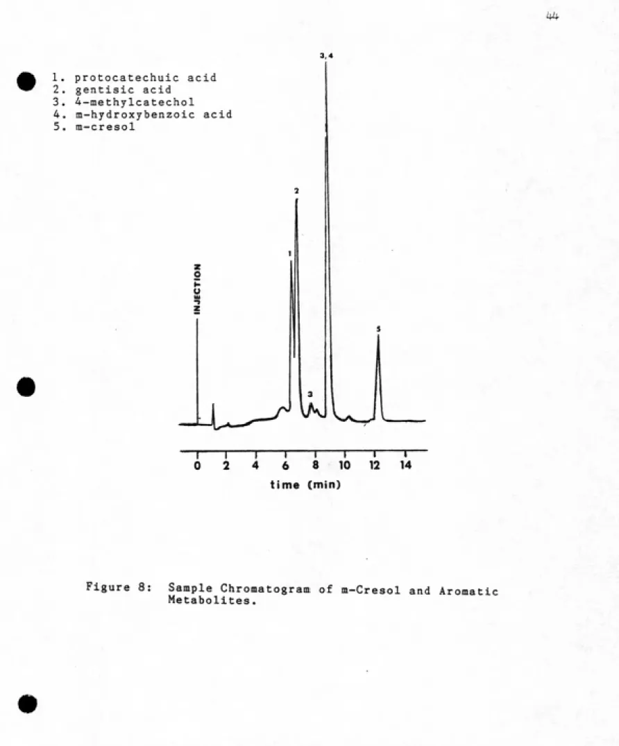
The final elution conditions are described in Materials
and Methods. The separation achieved under such conditions
is shown in Figure 8. The 4-methylcatechol eluted in two
small peaks, one of which coeluted with m-hydroxybenzoic
acid. Because the 4-methylcatechol was only being used as
a substitute for 3-methylcatechol (and would not
necessarily have the same elution characteristics as that
compound anyway), the impurity problem was not pursued
further.The detention time of each compound under the final
elution conditions is shown in Table 1. The detention
times were corrected for the 0.6 minute time lag between
the UV detector and fraction collector. From the time of
arrival at the fraction collector, the fraction containing
the compound peak maximum was estimated. In this way,
peaks detected with the UV detector could be correlated
with radioactive peaks detected in the metabolite
accumulation experiments.
1. protocatechuic acid 2. gentisic acid
3. 4-methylcatechol
4. m-hydroxybenzoic acid
5. m-cresol
o
u
T
0 6
-r
8
-r 10
"T"
12
T-14
4
time (min)
Figure 8: Sample Chromatogram of m-Cresol and Aromatic
and Metabolite Standards.
Compound Detention Time (min) Time at Collector F-'ostul ated Fraction Mo,
protocatechuic acid
gentisic acid 4 -- m e t h y 1 c a t e c h o 1
m-hydroxyben2oic acid
m--cresol
6,32 0,07
6.62 0.08 7. 6 0.04 8. 71 0.08 8. 68 0.05 12.22 0. 04
(m i n ) 1 min 0 5 min
6.92 7 14
7.22 8 15
8.2 9 17
9.31 10 19
9.28 10 19
12.82 13 26
After development of the metabolite separation
procedure, the next step was to perform a series of
die-away studies. The focus of these die-die-away studies was not
measuring mineralization, radioactive uptake, or
degradation rates, but assessing the number and relative
proportions of metabolites present in the aqueous phase. A
rigorous attempt to identify a particular metabolite would
only proceed if it were produced in significant quantities
(greater then 5% of the label).The first two experiments were performed with Jordan
Lake water, and as for two other degradation studies that
summer, unusually low degradative activity was observed.
The percent m-cresol metabolized by the seventh day was
about 1/6 the level observed in previous die-away studies,
and under these conditions, no metabolite accumulation was
detected. Because of the low levels of activity, we
decided to try a different water source, the Haw River.
In light of the problems with the Jordan Lake samples,
the first metabolite accumulation experiment with Haw River
water was a small study. Samples were incubated for 8 days
at two m-cresol concentrations, 120 ug/1 and 960 ug/1. No
uptake or respiration measurements were made; only the
radioactivity remaining in the sample filtrate was
monitored.and the radioactivity per fraction is expressed as a
percent of the radioactivity added to the water sample at
day 0. The peak eluting at fractions 26, 27 and 28 is
m-cresol. Most samples show little radioactivity coming off
before the m-cresol peak. The exception is one 960 ug/1
sample, which has radioactivity eluting in fractions 5-10
and 16-17. Together these peaks represent 16% of the
radioactivity originally added (Table 2). Only a trace of
radioactivity is apparent in these same fractions in the
other samples.
Further examination of the data, however, revealed that
the fractionation procedure failed to recover all the
radioactivity present in the sample filtrates (Table 2).
Only 41%, 58%, 81% and 76% of the injected label was
recovered from the 960#2, 960#1, 120#1, and 120#2 samples,
respectively. This suggested two possibilities: (1) that
the radioactivity was binding irreversibly to the HPLC
column, or (2) that it was eluting after, rather than
before, m-cresol.
To test these possibilities, the recovery efficiency of
each sample was determined when eluted with acetonitrile
(100%). Under these conditions, radioactive compounds
eluting after m-cresol should be recovered, whereas those
binding irreversibly to the column should not. The high
recovery efficiencies (Table 3) suggest that most of the
"missing" label was eluting after the m-cresol. This was
a ^=^1 30 Ci
§ 20-ͣ
JL
CD o
ͣ
71
3:
10-a o
rrrrrw:STsrST•' \ i 'ͣ' ->-'' { ' •
1
<.'e 5 10 15 2Q 2S 30
FRACTION NUMBER R 2!5' § 20--- 15f o z 10--O 5-:
0 ryy«-'i!!-*~
10 15 20 25 FRACTION NUMBER
25 HAW . 9^S-^ . SD . 9iSQ**l
§ 20--CD O s 10--a. a
u. 5f
o
0: 55
0 5 10 15 2© 25 30
FRACTION NUMBER U a a <r Or: a. u. o X 25
HAW . 9/34 . SD . 9oa**2
5 IS 15 20 25 FRACTION NUMBER
Ct 25
Ui Q
° 20t
CD Or 10-:
Q
5+
HAW. gy'S4 . 9D . ISaCONTROl.
0 • I II il'i' i"iii^w*' >•_ :
'0 5 10 15 20 25 3S
FRACTION NUMBER
Q 25
a cz 20+ o
" 15+
o
z l0t
o
5+
HAW . 9/S-4 . SD . 960CQN TROL
=r5s»—„_=i 0 5 10 15 20 25 3e
FRACTION NUMBER
Figure 9: HPLC Fractionations of Sample Filtrates. September.
Table 2: Recovery in Aqueous Filtrates of the Radioactive Label
Originally Injected. September, 1984.
Concentration Replicate (u.g/1) No.,
% Label % m-Cresol % Other
Remaininq Recovered Recovered
120 120 120
1
control
960 960 960
1 2 control
or ͣ!
104
66 44 101
41
31
87
o
6
6
Table 3: Recovery Efficiencies of Radioactive Label in Sample
Filtrates when Eluted with an Acetonitrile Mobile
Phase. September, 1984.
Samp1e Repli cate Nd, R e c Q V e r y E t f i c i e n c y
120 ug/1 120 ug/1 120 uq/i 960 ug/l 960 ug/1 960 ug/1
control
1
control
94 99 102
93 82 98
m-cresol standard 100
gradient was increased from 15 minutes to 45 minutes, 3
radioactive peaks eluted after the m-cresol (Figure 10).
To accomodate such a long gradient, however, fractions of 1
minute had to be collected. Consequently, in this figure, m-cresol elutes in fractions 13 and 14.The identity of peaks a, b, and c is unknown, but their
elution after m-cresol indicates that they are less polar
compounds than m-cresol, and that they are not metabolites
in its degradation pathway. One possibility is that thecompounds are growth products. In this case, m-cresol is
degraded to pathway end products (Figures 2-4) which enter
the central pathways of metabolism. Intermediates in the
central pathways may enter a diverse and diverging seriesof biosynthetic pathways to form proteins, polysaccharides,
lipids, nucleic acids, or their respective precursors.
Thus, if peak a,b, and c are in fact growth products, they
could be many different compounds.Another possibility is that peak a, b and c are
polymerization products of m-cresol. Bollag et al. (1979)
isolated an extracellular enzyme from the soil fungusRhizoctonia practicola that catalyzed the formation of
polymerized products from phenolic compounds. The enzyme
polymerized p-methylphenol to a dimer, o-methoxyphenol to
dimers and trimers, and phenol to diraers, trimers and968 UG/L GRADIENT i
2500+
2000"
J 1500"
P
1000+
500"
0-m-cresol
c
X
b
V
11111111 11 111111 III 11II1111'lTiTtlTpffi'pi^'S
5 15 25 35 45
0 10 20 30 40
FRACTION NUMBER
Figure 10: HPLC Fractionation of One Sample Filtrate under
Extended Gradient Elution Conditions (see text).
recent rains caused such a fungus to be carried with soil
runoff into the river. This possibility is further
supported by the fact that peaks a, b, and c were not
observed in any subsequent or previous experiments. Thus, their production may reflect a change in the microbial
populations present.
One peak appeared to elute about 55 minutes after an
injection, leading to an extraneous peak in the following chromatogram (see peak with "?" in Figure 10). To correct this problem and to accomodate the possibility of peaks
eluting after m-cresol, the gradients in all subsequent
experiments were 45 minutes, followed by a 15 minute wash with acetonitrile, and 1 minute fractions were collected.
In all other respects, the gradients were identical.
A second metabolite accumulation experiment was
initiated in November. This experiment was a repeat of the
September metabolite accumulation experiment.
Fractionation results are shown in Figure 11 and Table 4.
Clearly, these results are entirely different from those
observed in the September metabolite accumulation study.
Nearly all radioactivity was recovered with the expanded
fractionation procedure, but aside from the m-cresol
impurity which eluted in fractions 20 and 21, most
radioactivity eluted before m-cresol (which elutes in
fractions 13 and 14). Thus, the products which accumulate in this experiment may be metabolites in the degradation
Table 4: Recovery in Aqueous Filtrates of the Radioactive Label
Originally Injected. November, 1984.
Concentration Replicate '/. Label "'. m-Uresol 7. Other <ug/l) No. Remaining Recovered Recovered
120 1 120 2 120 control
960 1
960 2
960 control
17 16 102
49 42 101
ͣͣ
: 1
HAW . il/S-4 . SD . i2Q«l HAW . 11/S-4.aD. 960441 Q UJ O O <r CD o X 0. u. o 25 20+ 15--10-:
5 ͣ
0-C=---1 ͣͣ I I I I I s:d« r>pmS»faSim :
0 10 20 3ti -40
FRACTION NUMBER
UJ 1=1 (St O
10 "^ 20 "^ 30 4 0
FRACTION NUMBER
HAW , 11./3-4 . SD . ISBCONTRaU. O 25-t a § 20 r: 15+ X 10-^ 5-O
-^/rrrrrr^^..'
5 ^^ 15 25 35
10 20 30 40 FRACTION NUMBER
-45
HAW. il/S'*.8D. 96 0CONTRai_
Cj
251---il'
CTiI I r .1 rTTi...-rttTT«T. ^,r»','i'fn i if
^ 5 15 25 35 -4
0 10 20 3 0 -4 0
FRACTION NUMBER
identity will follow later.
Concentration differences were also clearly apparent. In the 120 ug/1 samples, only 17% of the radioactivity
initially injected remained in the aqueous phase after
eight days, and almost none of this was present as
m-cresol. In contrast, about 45% of the initial
radioactivity remained in the 960 ug/1 samples, but about
60% of this was present as m-cresol. Curiously, the %
metabolites of label initially added was about the same at
both concentrations. This is in distinct contrast from the
September experiment, when the label theoretically present as metabolites at 960- ug/1 was twice that at 120 ug/1.
Given that the type of metabolites which accumulate in the two experiments are so different, the factors which govern
their formation and release are bound to differ. Different
enzyme systems, and possibly, different organisms are
involved. Therefore, they would not be expected to respond
to concentration in the same way.
Given the conflicting results of the September and November experiments, a third metabolite accumulation
experiment was initiated in January, 1985. The objectives
of this experiment were twofold: (1) to confirm the
results of one, or both, of the previous experiments, and (2) to follow cresol disappearance and metabolite
accumulation in the filtrate at several different periods.
The experiment was similar to the two previous studies,
3, 8 and 16 days. Sample uptake and respiration were also
measured.
Time study results are shown in Table 5. m-Cresol
disappears more quickly from the aqueous phase than the radioactive label. Moreover, as the incubation time increases, the recovered metabolites account for an
increasing proportion of the radioactive label remaining in
the aqueous phase (Figure 12). These results confirm those
observed in the cresol disappearance experiment and
demonstrate that metabolite accumulation does occur with
longer incubations.
Metabolite recovery and fractionation results at day 8 are nearly identical to those observed in the November
metabolite accumulation experiment (Table 5, Figure 13-14).
A similar fractionation pattern is observed at each time
and concentration. As the percent of label in the form of
metabolites increases, the radioactivity in most fractions
eluting before m-cresol also increases. The increase
occurs after and including fraction 7, where aromatic
metabolites would elute, and before fraction 7, when
nonaromatic metabolites would elute. Thus, both aromatic
metabolites and other products may accumulate.
No one fraction accounts for a significant percentage
(greater than 5%) of the label initially added, but small
radioactive peaks do consistantly appear in fraction 7 and
fractions 3-5. Fraction 7 may be tentatively identified as
Incubation. January, 1985.
120 UG/L BAHPLES 960 UG/L SAMPLES
Incubation "/. Label "/. m...Cresol "/. Other 7„ Label 'L m-Cresol "/. Other
Time (days) Remaining F-Jecovered Recovered Remaining Recovered Recovered
0 (20 min)* 91 82 9 85 76 S
3 sample* 33 14 15 69 53 13
control 109 97 9 104 95 8
8 sample* 22 3 15 54 36 IB
control 101 86 11 102 B9 10
1.6 sample* 21 1 19 21 1 ib
control 103 91 12 100 Qg \\
I
0 jd
I
120 ug/l
960 ug/
10 ^^
ttme (dnys)
Figure 12: Proportion of Radioactive Label Remaining in the
Sample Filtrates Which is Recovered as Metabolites
a: o a. a o sw«»
5 15 25 35 -45 0 10 20 30 40
FRACTION NUMBER CD I—I o a. a t-C^"-^
5 15 25 35 45 0 10 20 30 40
FRACTION NUMBER a UJ a a o I—4 a: o a. o 25
HAW. 1-^S5 . 9D .9S0#2
-1
5 15 25 35 45 0 10 20 30 40
FRACTION NUMBER a U O a <E CO a: o a. 13
25 HAW . 1X05 . ISD . 9Se+»l
10+
«>««««»,„
5 15 25 35 4; 0 10 29 30 40
FRACTION NUMBER