Abstract
Prenatal alcohol exposure (PAE) can result in a range of mental disabilities and physical
defects. Because adults affected by PAE demonstrate increased susceptibility to alcohol
dependence, it was hypothesized that PAE leads to the loss of inhibitory GABAergic
interneurons in corticostriatal circuits that regulate reward perception. Offspring of mice that
experienced acute ethanol exposure on gestation day 7 (GD 7) were used as an animal model.
Intracranial self-stimulation studies showed a marked difference in threshold for non-exposed
controls and PAE mice during the first 15 minutes following morphine injection, but no
significant difference was shown with alcohol or cocaine. Immunohistochemistry against
parvalbumin was performed to visualize the GABAergic interneurons of the CNS in the nucleus
accumbens, medial prefrontal cortex, dorsal striatum, and medial septum. A preliminary count of
the interneurons in the nucleus accumbens showed no difference between control and PAE mice.
In the future, further quantification of GABAergic interneurons in all four regions of interest may
conclude that maternal ethanol ingestion on GD 7 causes significant damage to the normal
distribution of inhibitory interneurons in CNS structures associated with reward perception.
Introduction
Fetal alcohol syndrome (FAS) results from severe ethanol exposure during fetal
development and is characterized by stunted fetal growth, distinctive craniofacial abnormalities,
and damage to the central nervous system (CNS) that can lead to a spectrum of intellectual,
psychological, and social disabilities. Fetal alcohol spectrum disorders (FASD), an umbrella term
exposure (PAE), represent the most common non-inherited cause of birth defects in the western
world (Clarke and Gibbard, 2003). The efficacy of animal models in the study of PAE has been
demonstrated (eg. Driscoll et al., 1990; Wilson, 2011) and subsequent studies have employed
diverse methodologies to better understand the effects of FAS in humans.
Studies in PAE have been successful in utilizing neuroimaging techniques such as
magnetic resonance imaging (MRI) to observe anatomical malformations of brain structures in
both humans and animal models (Bookstein et al., 2006; Lipinski et al., 2011). It was concluded
that anatomical malformations of facial features and brain structures during fetal development
are variable and stage-dependent in PAE mice. In both humans and mice, the malformations
observed depended on the day of gestation ethanol exposure occurred. Furthermore, magnetic
resonance imaging of mouse models for PAE correlated to imaging of humans affected by PAE
(O’Leary-Moore et al., 2011).
Quantifiable evidence of damage to the central nervous system (CNS) due to PAE is
lacking. Based on previous observations of increased alcohol dependence in adults affected by
PAE (eg. Yates, 1998), we hypothesized that maternal ethanol ingestion during gestation leads to
neuronal loss in corticostriatal circuits involved in regulating reward perception and that this
damage may enhance both the positive and negative effects of alcohol and other drugs.
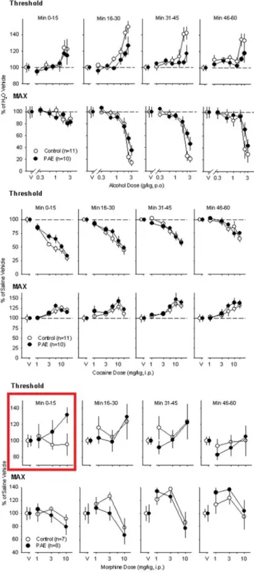
Preliminary findings utilizing intracranial self-stimulation (ICSS) as a means of delivering direct
stimulation to the brain’s reward circuitry showed no significant differences in brain stimulation
reward (BSR) between PAE mice and non-exposed controls following acute exposure to ethanol
(Figure 2A). This result did not support the initial hypothesis that PAE mice would exhibit
increased alcohol intake and alcohol reward relative to control mice. Similarly, no significant
alcohol and drugs such as cocaine would have an enhanced effect on reward perception in PAE
mice relative to the control (Figure 2B). However, a significant difference was found between
the two groups in the potentiating effects of morphine (Figure 2C). PAE mice exhibited greater
sensitivity to morphine and experienced a greater potentiation effect in comparison to
non-exposed controls. This result supports our hypothesis, but past studies have shown that PAE’s
effect on pituitary-adrenal function can lead to enhanced responses to footshock stress and
morphine amongst other stress related responses (Nelson et al., 1986). Therefore, further studies
are required to analyze whether a change in reward perception was involved in the dissimilar
response to morphine that was observed in the PAE and control mice.
Investigation of GABAergic interneuronal loss in structures of the brain underlying
reward perception was the next step in understanding the effects of PAE on the CNS. GABA is
the chief inhibitory neurotransmitter in the CNS. Studies have demonstrated that imbalances in
neuroexcitatory and neuroinhibitory amino acids, such as glutamate and GABA respectively, can
result from long-term ethanol exposure and are responsible for behavioral susceptibility during
ethanol withdraw (De Witte, 2004). Because insufficient expression of GABA in the CNS can
lead to an imbalance in inhibition and excitation, these conclusions support the hypothesis that
loss of GABAergic interneurons due to PAE may lead to increased alcohol dependency.
The process of performing a morphological analysis of GABAergic interneurons in the
nucleus accumbens, medial prefrontal cortex, dorsal striatum, and medial septum of both PAE
and non-exposed control mice was started using immunohistochemistry to visualize the
interneurons of interest and was followed by a manual count. Design-based stereology will be
utilized to quantify the interneurons in an unbiased manner in future counts.
in GABAergic interneurons, required a new protocol for effective staining due to a lower density
of inhibitory interneurons in the nucleus accumbens relative to the other three regions of interest.
Previous work done by the lab has achieved proper staining of the medial prefrontal cortex,
dorsal striatum, and medial septum. The optimal protocol for staining in the nucleus accumbens
was determined by testing multiple primary antibody ratios and incubation periods and assessing
them with brightfield microscopy (Table 1 and 2).
In the near future, the samples that have undergone staining for parvalbumin will be
assessed using design-based stereology that allows for systematic random sampling (Figure 3a).
The availability of design-based stereology in quantitative morphology of the CNS has allowed
for neuronal counting in entire brain structures of interest without bias due to the size, shape,
spatial orientation, or spatial distribution of cells (Schmitz and Hof, 2005). Quantifying the
extent of neuronal loss or maldistribution in FAS mice may indicate the circuits and routes of
neuronal migration damaged by PAE to provide insight into the origin of defects in reward
Figure 1. The craniofacial abnormalities of FAS individuals correlated with magnetic resonance
imaging of malformations in facial features and brain structures of PAE mice exposed on
gestation day 7 (GD7) and gestation day 8.5 (GD8.5). The malformations in the mouse models
Figure 2A. No difference was shown in threshold or max response between PAE and
Figure 2B. No difference was shown in threshold or max response between PAE and
non-exposed control mice following administration of cocaine.
Figure 2C. A significant difference was shown in threshold between PAE and non-exposed
control mice during the first 15 minutes following administration of morphine. PAE mice
experienced a marked attenuation of ICSS not observed in the control mice.
Figure 3a. The systematic-random sampling made possible by design-based stereology to yield
an unbiased count of interneurons of the mouse nucleus accumbens (Courtesy of Malanga, C. J.).
Figure 3b. An unbiased counting frame generated by the stereological probe (Courtesy of
Materials and Methods
Mice:
The investigation utilized a mouse model of FAS produced by C57BL/6J dams given
acute ethanol exposure (X mg/kg, p.o.) on GD 7. The offspring of these dams were grown to
young adulthood (PXX) prior to anesthetization and transcardial perfusion with 0.1 M
phosphate-buffered saline (PBS) and 4% paraformaldehyde. A non-exposed control group given
no ethanol exposure during gestation was also used. All work leading up to the fixation of the
brains in 4% paraformaldehyde and PBS was performed by other members of the lab.
Immunohistochemistry:
Histology was performed using coronal slices on a sliding microtome to acquire an
exhaustive sample of 50 μm thick sections that encompass the entirety of all 4 brain regions of
interest: the nucleus accumbens (NAc), medial prefrontal cortex (mPFC), dorsal striatum, and
medial septum. These sections were placed in cryoprotectant (1.0% w/v polyvinylpyrrolidione,
30% w/v sucrose, and 30% v/v ethylene glycol in 0.1 M PBS) and stored at 0oC until use.
Sections representative of entire regions of interest were removed from cryoprotectant and rinsed
with 0.1 M PBS before undergoing free-floating immunohistochemistry against parvalbumin.
The sections were treated with 0.05% H2O2 followed by a 0.5% triton with 3.0% normal goat
serum solution before incubation with goat anti-parvalbumin primary antibody of varying
primary antibody for 72 hours and 48 hours respectively. In both trials, 2 sections were stained
in 2 separate wells at each primary antibody concentration. All samples were incubated in a
rabbit anti-goat secondary antibody with a 1:250 concentration for 4 hours and visualized with a
DAB peroxidase substrate kit. The sections were mounted on gelatin-subbed slides to be
counterstained and coverslipped prior to assessment with light microscopy.
Nissl Counterstain:
A Nissl counterstain was performed on half of the sections from the second
immunohistochemistry trial against parvalbumin. The protocol for Nissl counterstaining will be
tested and modified in future immunohistochemistry trials meant to confirm the concentrations
of primary antibody to be used for optimal visualization of parvalbumin in each of the brain
structures of interest.
Manual Counts:
A preliminary count of interneurons in the nucleus accumbens of PAE and non-exposed
control mice was performed with brightfield microscopy and an outline of the region of interest
from a mouse brain atlas.
In the near future, a quantitative analysis of GABAergic interneurons in the nucleus
accumbens, medial prefrontal cortex, dorsal striatum, and medial septum of the PAE and control
brain sections will be performed with design-based stereology. The optimal counting frame area,
dissector height, and guar frame width for stereological analysis of each brain region will be
determined through practice trials prior to counting (Figure 3B).
Results:
Sections from trials 1 and 2 of immunohistochemistry against parvalbumin were
incubated in goat anti-parvalbumin primary antibody for 72 hours and 48 hours respectively.
The 1:1500 concentration was used as a control because this ratio was successful in
immunostaining all regions of interest other than the NAc in past trials. Regardless of the
concentration of primary antibody used, trial 1 resulted in sections that were too dark to
accurately quantify the number of GABAergic interneurons in neither the NAc nor any of the
other three structures of interest (Table 1). In contrast, trial 2 resulted in variable success with
staining of inhibitory interneurons in the NAc depending on the concentration of primary
antibody used. Sections that underwent incubation in a 1:690 concentration of primary antibody
in trial 2 yielded the best staining for visualizing interneurons of the NAc. In trial 3, the
concentrations of primary antibody used in trial 2 were repeated to better determine an
intermediate protocol that visualizes interneurons in all 4 regions of interest simultaneously. A
1:1230 concentration of primary antibody was selected as the optimal protocol for our
interneurons in the nucleus accumbens without over-staining in other regions of greater
interneuron density (Figure 4).
Nissl counterstaining with cresyl violet was performed on 1 of the sections at each
primary antibody concentration in trial 2. Only patchy staining was observed in most sections
and those that were mounted towards the end of the slide showed no sign of Nissl staining (Table
2). There were insufficient amounts of Nissl staining to determine whether the white or dark
matter of the sections was more heavily stained. Areas of the sections that were Nissl stained
were not indicative of increased staining in any specific biological structures of the brain.
However, some counterstained areas demonstrated success in providing contrast to better
visualize the parvalbumin immunostaining when compared to the non-counterstained sections
from trial 2 that were incubated at the same concentration of primary antibody.
Manual counts of interneurons in the NAc of the PAE and non-exposed mice showed no
significant difference between the two groups in the number of interneurons present in the NAc
(Figure 5). This result is contrary to our hypothesis because we expected to observe a lower
number of interneurons in structures of the brain associated with reward perception in the PAE
mice due to damage to the CNS during fetal development. However, a conclusion cannot be
reached until the remaining brains are accounted for and a more reliable method of quantification
is used to minimize sources of bias during counting. In the future, the GABAergic interneurons
of the immunostained sections from both GD 7 PAE mice and non-exposed controls will be
quantified using design-based stereology in the four regions of interest. Estimates of the total
number of inhibitory interneurons in the nucleus accumbens, medial prefrontal cortext, dorsal
striatum, and medial septum will be reported using the optical fractionator method with
1o Antibody Concentration Incubation Time (hr) Staining in NAc
Staining in mPFC, Medial Septum, and Dorsal Striatum
1:1500 72 Too Dark Too Dark
1:1230 72 Too Dark Too Dark
1:960 72 Too Dark Too Dark
1:690 72 Too Dark Too Dark
1:420 72 Too Dark Too Dark
1:150 72 Too Dark Too Dark
Table 1. Immunohistochemistry trial 1 for determining the optimal goat anti-parv primary
antibody concentration demonstrated that an incubation time of 72 hours yields overly dark
staining
1o Antibody
Concentration IncubationTime (hr) Staining inNAc Medial Septum, andStaining in mPFC, Dorsal Striatum
Staining of Nissl Bodies
1:1500 48 None Good Patchy
1:1230 48 Present Present Patchy
1:960 48 Present Present None
1:690 48 Good Too Dark Patchy
1:420 48 Too Dark Too Dark Patchy
1:150 48 Too Dark Too Dark None
Table 2. The second immunohistochemistry trial showed variable staining in the nucleus
accumbens depending on the primary antibody concentration. A 1:690 concentration with 48
hours of incubation time (highlighted) yielded the best results for staining in the nucleus
accumbens. A 1:1230 concentration was selected as optimal for staining in all 4 regions of
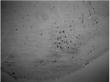
Figure 4. Staining in the nucleus accumbens and surrounding structures using a 1:1230
concentration of primary antibody. The interneurons are easily visualized and there is no
Figure 5. A preliminary count of GABAergic interneurons in the NAc of PAE and non-exposed
control mice (n=2). There was no significant difference between the two groups thus far.
Discussion
At this point in the study, proper protocols for immunostaining of inhibitory GABAergic
interneurons of the NAc, mPFC, dorsal striatum, and medial septum were established. The
immunohistochemistry protocol for the latter 3 structures was determined by the work of
previous lab members. Because parvalbumin immunostaining was not present in the NAc of
these past trials, they demonstrated that a separate protocol was necessary. It was concluded that
the NAc had a lower density of GABAergic interneurons relative to the other regions of interest.
A higher primary antibody concentration, a longer incubation time with the primary antibody, or
some combination of the two was necessary to properly immunostain the inhibitory interneurons
in the NAc.
The three trials used to determine a protocol suitable for staining in the nucleus
accumbens led to several conclusions regarding immunohistochemistry against parvalbumin.
The failure of trial 1 in properly staining the GABAergic interneurons of any of the regions of
interest demonstrated the significant effect that primary antibody incubation time has on
immunostaining. Specifically, it was concluded that increasing incubation time leads to a
decrease in specific binding by the primary antibody. Once all of the parvalbumin was bound,
the remaining antibody eventually bound to other areas of the mouse brain and yielded sections
In contrast to sections from trial 1, trial 2 demonstrated variable staining of GABAergic
interneurons depending on what concentration of primary antibody was used. As originally
predicted, trial 2 showed that increasing the primary antibody concentration increases staining
for parvalbumin. However, the sections from trial 2 with the two highest concentrations (1:840
and 1:300) demonstrated that increasing the primary antibody concentration also increases the
prevalence of non-specific binding. It was concluded that using an overly high concentration
causes excess primary antibody to bind to non-specific targets in the brain after all of the
parvalbumin has been bound.
Immunohistochemistry trial 3 showed that an intermediate protocol between the
concentrations of 1:1500 (good staining in mPFC, medial septum, and dorsal striatum) and 1:690
(good staining in NAc) could be used to stain for all four regions of interest on the same brain
section. At a primary antibody concentration of 1:1230, interneurons in all structures of interest
were properly stained. This concentration was utilized as the optimal protocol for our
investigation.
The group of sections from trial 2 that underwent counterstaining led to several
conclusions on how to improve the protocol for Nissl staining using cresyl violet. The lack of
Nissl body staining in the sections suggests that the slides were treated with the acetic acid
solution for too long following staining with cresyl violet. Interestingly, the sections mounted
towards the end of the slide that made contact with the counterstaining solutions first exhibited
much less Nissl staining relative to those that were mounted towards the opposite end. It was
concluded that the submergence of slides in wells during Nissl staining must be done using quick
dipping motions to minimize disparities in the length of time that different sections have contact
towards the end of the slide that contacts the solutions first to be submerged for longer. The
results of the counterstain trial demonstrated that this difference in time can result in significant
differences in Nissl staining.
The manual count of GABAergic interneurons in the NAc of the PAE and non-exposed
control mice have shown no significant difference thus far. However, the quantification of
interneurons in the mPFC, medial septum, and dorsal striatum has yet to be done, and further
counting in the NAc is also necessary. Therefore, it is too early to make any conclusions about
our hypothesis that PAE causes damage to interneurons in structures of the brain associated with
reward perception. In the future, the quantification of GABAergic interneurons in the brain
regions of interest will be performed using design-based stereology. Another future goal may be
to study PAE’s effect on the adrenal-pituitary response and whether it has any connection to a
greater propensity for alcohol and drug addiction.
Literature Cited
McBroom, J. A., and Streissguth, A. P. (2005). Preliminary evidence that prenatal alcohol
damage may be visible in averaged ultrasound images of the neonatal human corpus
callosum. Alcohol 36, 151-160.
Clarke, M. E. and Gibbard, W. B. (2003). Overview of Fetal Alcohol Spectrum Disorders for
Mental Health Professionals. Canadian Academy of Child and Adolescent Psychiatry 12,
57-63.
De Witte, P. (2004). Imbalance between neuroexcitatory and neuroinhibitory amino acids causes
craving for ethanol. Addictive Behavior 29, 1325-1339.
Driscoll, C. D., Streissguth, A. P., and Riley, E. P. (1990). Prenatal alcohol exposure:
comparability of effects in humans and animal models.
Guerri, C., Bazinet, A., and Riley E. P. (2009). Foetal Alcohol Spectrum Disorders and
alterations in brain and behavior. Alcohol and alcoholism 44, 108-114.
Lipinski, R. J., Hammond, P., Ament, J.J., Pecevich, S.J., Jiang, Y., Dehart, D.B., Johnson,
G.A., and Sulik, K. K (2011). Magnetic resonance microscopy-based 3D face-brain
correlations in an FASD mouse model. Alcohol Clin Exper Res 35
Nelson, L. R., Taylor, A. N., Lewis, J. W., Poland, R. E., Redei, E., and Branch B. J. (1986).
Pituitary-adrenal responses to morphine and footshock stress are enhanced following
prenatal alcohol exposure. Alcohol Clin Exper Res. 10, 397-402.
Resonance-based imaging in animal models of Fetal Alcohol Spectrum Disorder.
Neuropsychology Review 21, 167-185.
Schmitz, C. and Hof, P. R. (2005). Design-based stereology in neuroscience. Neuroscience 130,
813-831.
Wilson, S. E. and Cudd, T. A. (2011). The Use Of Animal Models For The Study Of Fetal
Alcohol Spectrum Disorders. Alcohol Research & Health 34, 92-98.
Yates, W. R., Cadoret, R. J., Troughton, E. P., Stewart, M., and Giunta, T. S. (1998).