Introduction:
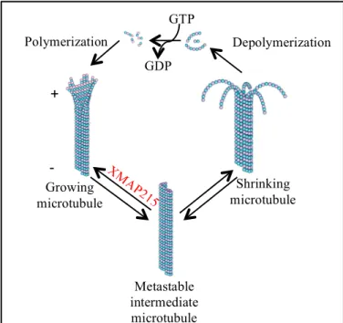
Microtubules (MTs) are dynamic cytoskeletal polymers composed of αβ-tubulin that define cell structure, establish platforms for the transportation of intracellular cargos, and form the mitotic spindle to facilitate chromosomal segregation. These hollow tube-like polymers are formed by the GTP hydrolysis-dependent head-to-tail polymerization of α- and β-tubulin heterodimers (1-3). The polarity of the heterodimer is reflected in the polymer; β-tubulin is exposed at the MT plus end where polymerization mainly occurs and α-tubulin is exposed at the minus end (1, 2) (Fig. 1). The dynamic
properties of MTs are readily displayed at the interphase-mitosis transition. During interphase, MTs are much less dynamic and organize into a radial array; whereas, in mitosis, MTs are much more dynamic and assemble into the dense bipolar mitotic spindle (1, 2, 5, 6). The remarkable diversity present within the dynamics of the interphase MT array versus the mitotic spindle proposes the existence of proteins that
regulate MT dynamics (5, 6). One type of such proteins are MT-associated proteins (MAPs), which are crucial for the regulation of MT polymerization and depolymerization, as well as intracellular organization (7). Included among MAPs is the XMAP215 family, which was originally discovered in Xenopus egg extracts (4). XMAP215 family proteins serve as conserved
GTP
GDP
Polymerization Depolymerization
XM AP
215
Growing microtubule
Metastable intermediate microtubule
Shrinking microtubule
Figure 1.Polymerization (assembly) and depolymerization
(disassembly) of MTs is driven by the binding, hydrolysis, and exchange of a guanine nucleotide on the β-tubulin monomer. The presence of the equilibrium between MT growing, shrinking and paused states confers microtubule dynamic instability. Crucial for the regulation of rapid MT assembly are XMAP215 family proteins.
regulatory factors to catalyze rapid MT elongation (8). All of the proteins within the XMAP215 family consist of a C-terminal domain that promotes XMAP215 localization via its interaction with other MAPs and an array of N-terminal TOG (tumor overexpressed gene) domains that interact with αβ-tubulin in order to facilitate MT growth, promote the formation of the mitotic spindle, and enhance MT dynamics (1, 2, 9-11). The number of TOG domains within the TOG domain array differs across XMAP215 family members (Fig. 2). Yeast homologs have only two
TOG domains that comprise a dimeric structure, whereas higher eukaryotes are monomeric and contain a pentameric TOG domain array (4, 9, 15). In juxtaposition to the TOG arrays observed in yeast and higher eukaryotes is the ZYG-9 TOG array, which is trimeric (12, 17). Connecting the TOG domains are linkers ranging from 60-100 amino acids. Although these linkers obtain no projected secondary structure, they do contain basic residues that are important for interacting with microtubules (16).
Across all TOG arrays, TOG domains share conserved features. TOG domains are defined by six (A-F) HEAT (Huntingtin, elongation factor 2, protein phosphatase 2A, target of rapamycin 1) repeats (HRs) that align to form an oblong framework (17, 18) (Fig. 3A). The arrangement of intra-HEAT loops on one face of the TOG domain provides a conserved surface for proper tubulin binding (13) (Fig. 3B). How the XMAP215 TOG domain array utilizes conserved tubulin binding residues to bind and incorporate αβ-tubulin into MT plus ends is a
Figure 2. The XMAP215 family proteins share a conserved architecture, consisting of
a C-Terminal domain or coiled-coil that associate with other MAPs and an N-terminal TOG array that interacts with αβ-tubulin. XMAP215 family TOG domain
long-standing question in the cytoskeletal field. Currently, there are two models to explain this phenomenon. The first is the
wrap-around model, which suggests that XMAP215 proteins utilize their TOG array(s) to surround a tubulin heterodimer and transport it to the MT plus end. On the other
hand, the templating model assumes that each TOG domain within the array uses a similar mechanism to interact in a concerted manner with multiple tubulin heterodimers (8, 13, 18). To differentiate between these two proposed models, it is important to uncover the architecture and MT polymerization behaviors of each TOG domain within the array. Structure function
investigations show that TOG domains differentially promote MT
polymerization and spindle structure. Specifically, studies indicate that TOGs 1-2 have a much greater affect on regulating MT dynamics than TOGs 3-5 (9, 12).
This differential ability to
promote MT polymerization and spindle structure may be in part justified by structural deviations among TOG
Figure 3. A. Stu2 TOG1 conserved tubulin-binding surface. B. Stu2 TOG1
bound to a curved αβ-tubulin heterodimer.
90°
90°
A. B.
β
α +
-
A B C D E F
HR
Msps TOG4
Msps TOG2 Msps TOG3 Msps TOG5
ZYG-9 TOG3 ch-TOG TOG4
Stu2 TOG1 Stu2 TOG2
Figure 4. TOG domain architectures currently solved, featuring
domains. Presently, there have been eight TOG domain structures determined by X-ray crystallography that have provided substantial insight into the mechanism of XMAP215-mediated MT polymerization. These
structures include Saccharomyces cerevisiae Stu2 TOG1 and TOG2, Drosophilia melanogaster Mini spindles (Msps) TOG2, TOG3, TOG4, and TOG5, Caenorhabditis elegans ZYG-9 TOG3, and Homo
sapiens ch-TOG TOG4 (12, 13, 17-20) (Fig. 4). Studies show that TOG1, TOG2, and TOG3 have conserved architectures, which contrast to the divergent structure of TOG4 (12, 20). In particular, TOGs 1-3 contain a
similar tubulin-binding surface to facilitate complimentary interactions with tubulin
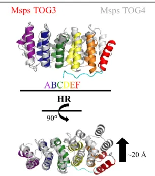
heterodimers (Fig. 5). Conversely, studies report that TOG4 transiently binds tubulin, perhaps due to the differential positioning of the HR D-F triad (Fig. 6). The diverse tubulin-binding mechanism of TOG domains 1-3 versus TOG4 provides evidence for the contribution of a polarized arrangement of XMAP215 to promote MT dynamics (12, 18-20). It has further been discovered that the ultimate TOG domain in ZYG-9 (TOG3) and Msps (TOG5) have
A B C D E F HR
Stu2TOG1 MspsTOG3
2.4 Å rmsd
Figure 5. Msps TOG3 is most similar to Stu2
TOG1, with an rmsd of 2.4 Å. The structural conservation between Msps TOG3 and Stu2 TOG1 provides evidence that TOGs1-3 share a conserved tubulin-binding mechanism.
90°
ABCDEF HR
90°
~20 Å
Figure 6. TOG4 is structurally divergent. Msps TOG4
and Msps TOG3 aligned pairwise results in a differential arrangement of HRs D-F. Specifically, alignment yields an ~20 Å shift in HR F. These structural deviations suggest that TOG4 has an alternative tubulin-binding mechanism.
similar domain architectures that deviate from other solved domain structures in the TOG array (19) (Fig. 6). Specifically, these domains have a characteristic seventh N-terminal HEAT repeat (HR) that binds orthogonal to HRs A-C. It is suspected that the N-terminal HR facilitates lateral tubulin interactions; however, its function is still at large (16). In addition, these domains have a conserved phenylalanine residue in intra-HR loop A, which contrasts with the tryptophan in the same position in TOG domains 1-4 (12). These conserved aromatic residues are exposed on the surface of the protein and have been shown to be of value in securing the interaction between the TOG domain and tubulin (15, 19).
The inherent differences in TOG domain architectures and their affect on MT dynamics provoke two separate questions. The first is in regards to whether the TOG domains are similar or different in their functionality, and the second concerns the type of tubulin-binding mechanism utilized by the TOG domain array. I hypothesize that TOG domain architectures comprising XMAP215 TOG domain arrays are
Figure 7. A. Msps TOG5 displays noteworthy architectural characteristics, including an
extra N-term HR and a conserved phenylalanine. B. Instead of a tryptophan, Msps TOG5 has an equally aromatic residue, phenylalanine, which is positioned on the surface to promote tubulin binding. C-E. Residues R1157, E1151, and W1169 are hypothesized to be of significance in stabilizing the N-terminal HR. Point mutations were introduced in order to disrupt the extra HR to gain a better understanding of how this domain aids in overall TOG domain stabilization.
D. 90° N-terminal HR
A.
W1169 E1161 C.
D.
E. C. E.
B.
R1157 B.
arranged in a specific orientation to distinguish between different tubulin structural states, which may play an important mechanistic role in XMAP215-mediated MT polymerization. To study this hypothesis, I am going to determine if TOG domain structures are conserved between XMAP215 family members, test the role of different TOG domain features in promoting domain stabilization, and understand how different TOG architectures affect MT polymerization. These studies will be achieved by (1) structurally characterizing TOG domains 3 and 5 from species across the XMAP215 family by X-ray crystallography, (2) completing the Msps TOG array by crystallizing Msps TOG1 (3) probing the role of TOG5’s extra N-terminal HR in promoting secondary structure and domain stability by creating mutant constructs and performing
comparative circular dichroism studies, and (4) examining the effects of intra-HR loop A WE (TOG3) and FE (TOG5) mutations on the catalysis of MT polymerization by these TOG domains in a light scattering assay. Collectively, these experiments will provide further comprehension of the mechanism of XMAP215 proteins and will provide insight into how the architecture and structural elements of arrayed TOG domains dictate their functionality and differential contribution to MT dynamics.
Methods:
A. DNA Cloning
PCR protocol was followed to amplify the desired DNA sequence of the Msps TOG1, ch-TOG TOG3 and TOG5 constructs. The Msps TOG1, ch-TOG TOG3 and TOG5 PCR products were digested with NdeI and BamHI restriction enzymes at 37 °C for 1 hour and 30 minutes. The purified Msps TOG1, ch-TOG TOG3 and TOG5 DNA products were then ligated into a precut pET28-p vector, a Kanamycin resistant bacterial expression vector that introduces an N-terminal His-tag, using T4 DNA ligase for an hour at 25 °C. For each construct, approximately 5 µL of the resulting DNA plasmid was transformed into 75 µL XL1B E. coli high copy cells and placed on ice for 20 minutes. The transformed bacteria was heat shocked at 37 °C for one minute and then placed on ice for an additional two minutes. Luria broth (LB) was added and the cells were incubated at 37 °C for approximately an hour. The bacteria was concentrated via centrifugation at 8,000 x g for four minutes and plated on kanamycin agar plates for selection. The transformed bacteria was allowed to incubate at 37 °C overnight.
In order to ensure that the DNA sequences of Msps TOG1, ch-TOG TOG3 and TOG5 were successfully ligated into pET-28-p, colony PCR was performed using a T7 forward primer and BamHI reverse primer. The positive colonies were placed in a culture tube containing 5 mL LB and 5 µL Kanamycin to amplify the plasmid DNA. The plasmid DNA from the 5 mL growths was purified and confirmed by sequencing.
colonies were grown up in 5 mL culture tubes at 37 °C under kanamycin selection. The amplified plasmid DNA was purified and the sequences of the constructs were verified. B. Protein Expression and Purification
The desired constructs were transformed into BL21 DE3 E. coli high expression cells using the transformation protocol described above. Cultures containing 6 L LB were grown at 37 °C under kanamycin selection. Once an optical density of 0.6 was reached, protein expression was induced with 0.1 mM ispropyl-1-thio-β-D-galactopyranoside and incubated at 18 °C
overnight. The cells were pelleted via centrifugation, resuspended in buffer A (25 mM Tris, 300 mM NaCl, 10 mM Imidazol, 0.1% beta-mercaptoethanol (βME), pH 8.0), and lysed using sonication. The lysed cells were centrifuged for 45 minutes at 15,000 x g (4°C) to separate the cell debris from the proteins in solution. The lysate containing the expressed constructs were loaded onto a Ni2+-NTA column by gravity flow. The protein was eluted using an imidazole gradient ending with a concentration of 25 mM Tris, 300 mM NaCl, 300 mM Imidazole, and 0.1% βME, pH 8.0. For the mutagenic Msps TOG3 construct, the His-tag was cleaved using Bovine α –Thrombin; whereas, for the Msps TOG1, ch-TOG and Msps TOG5 F1204E constructs, the His-tag was cleaved using PreScission Protease. To increase stabilization for CD experiments, the His-tag was not cleaved for wild type Msps TOG5 nor for the other mutagenic Msps TOG5 constructs. The thrombin was removed using a 0.5 mL benzamidine column. For all constructs, excluding those retaining a His-tag, the protein was passed over the Ni2+-NTA
column a second time to remove any uncleaved His-tagged protein. The protein was
and TOG5 F1204E constructs were concentrated to 8 mg/mL and 13 mg/mL respectively. The wild type and mutagenic Msps TOG5 constructs were concentrated to approximately 10 mg/mL. C. Crystallization
Hampton crystal screens were set up for ch-TOG TOGs3 and 5 and MspsTOG3W606E and MspsTOG5F1204E using 2 µL of the concentrated protein and 2 µL of a 500 µL well solution. To optimize crystals of ch-TOG TOG3 and ch-TOG TOG5, streak-seeding and micro-seeding methods were utilized. First, Msps TOG3 and TOG5 crystals were harvested. The crystals were then extracted and diluted into a larger volume of stabilizing solution to be used as seed stock in order to facilitate crystallization of the human homologs. For the streak-seeding method, a clean cat whisker was dipped into the seed stock and ran through the experimental drops containing ch-TOG TOG3 or ch-TOG TOG5. As for the micro-seeding protocol, approximately 1 µL of the seed stock was transferred to a drop containing 1 µL of the
concentrated protein and 2 µL of a stabilizing solution. Both methods were implemented using Msps TOG3 and TOG5 crystals derived from a variety of stabilizing solutions in order to enhance crystallization.
Results:
A. Protein Purification
expressed, purified, and concentrated, they can now be used for further functional analysis and characterization experiments to provide a more complete picture of the XMAP215-mediated MT polymerization mechanism.
B. Structural Analysis
Despite efforts to facilitate crystallization using micro and streak seeding methods, no crystals were
observed for either TOG domain from ch-TOG. Upon analyzing the crystallized structure of Msps TOG5, it appears that the C-terminal tail is slightly unorganized. The presence of this disorder suggests that the ch-TOG homolog is destabilized, which may explain the lack of crystallization.
Msps TOG3 W606àE
Sup.
Msps TOG3 W606àE
Pellet
Msps TOG5 F1204àE
Pellet Msps TOG5
F1204àE Sup. ~100 kDa ~10 kDa ~15 kDa ~25 kDa ~35 kDa ~55 kDa ~70 kDa ~130 kDa ~250 kDa
Figure 9. SDS-polyacrylamide coomassie stained gel
showing the solubility of Msps TOG3 (aa. 582-825) W606àE and Msps TOG5 (aa. 1141-1411) F1204àE. Expected MW for Msps TOG3 is ~27 kD and that for Msps
Figure 8. SDS-polyacrylamide coomassie stained gel depicting the proteins that were
successfully cloned and purified. Expected MW for Msps TOG1 = ~24 kD, ch-TOG TOG3 = ~26 kD, ch-TOG TOG5 = ~31 kD, Msps TOG5 = ~31 kD, and Msps TOG3 = ~27 kD.
Msps TOG1 aa. 6-234 ch-TOG TOG3 aa. 593-828 ch-TOG TOG5 aa. 1155-1422 ch-TOG TOG5 aa. 1155-1425
Msps TOG3 W606àE Msps TOG5
F1205àE Msps TOG5
For the mutagenic Msps TOG domains, the low concentrations of the proteins, along with the lack of potential crystallization observed prompted solubility tests to be performed. Both Msps TOG3 (W606E) and TOG5 (F1204E) were expressed in larger quantities for solubility trials. For both proteins, more than 95% was expressed in the pellet (Fig. 10). Thus, these tests support the hypothesis that the introduced mutations destabilize the protein, making it insoluble in solution.
Discussion:
The purification and crystallization of ch-TOG TOG3 (aa 593-828) and TOG5 (aa 1155-1425) proved to be unsuccessful, perhaps due to the lack of stabilization of the constructs. Currently, I am in the process of crystallizing a new TOG5 construct (aa 1155-1422). I hypothesize this construct to be more stable than the latter, as some of the residues of the C-terminal tail are not included within the sequence. Since there is no information to date regarding the structures of ch-TOG TOG3 and TOG5, the crystallization of these proteins will provide novel information about the structure and will lead to a better understanding of its importance in MT dynamics. Furthermore, the crystallization of ch-TOG TOG3 and TOG5 will also confirm whether the design of the TOG domains is conserved across domains and various species. In addition, I am also working on crystallizing Msps TOG1 to complete the Msps TOG array.
References:
1. Desai, A., and Mitchison, T. J. (1997) Microtubule polymerization dynamics. Annu. Rev. Cell Dev. Biol. 13, 83–117 2.
2. Gelfand, V. I., and Bershadsky, A. D. (1991) Microtubule dynamics: mechanism, regulation, and function. Annu. Rev. Cell Biol. 7, 93-1163.
3. Wang, H.-W., and Nogales, E. (2005) Nucleotide-dependent bending flexibility of tubulin regulates microtubule assembly. Nature 435, 911–915
4. Gard DL, Kirschner MW. (1987) A microtubule-associated protein from Xenopus eggs that specifically promotes assembly at the plusend. J. Cell Biol. 105, 2203–2215
5. Syred, H. M., Welburn J., Rappsilber, J., Ohkura, H. (2013) Cell Cycle Regulation of Microtubule Interactomes: Multi-layered Regulation Is Critical for the Interphase/Mitosis Transition. Mol. Cell. Proteomics 12, 3135-3147.
6. Kline-Smith, S. L., Walczak, C. E. (2004) Mitotic Spindle Assembly and Chromosome Segragation: Refocusing on Microtubule Dynamics. Molecular Cell. 15, 317-327.
7. Galjart, N. (2010) Plus-End-Tracking Proteins and Their Interactions at Microtubule Ends. Current Biology 20, R528–R537
8. Brouhard, G. J., Stear, J. H., Noetzel, T. L., Al-Bassam, J., Kinoshita, K., Harrison, S. C., Howard, J., and Hyman, A. A. (2008). XMAP215 Is a Processive Microtubule Polymerase. Cell
132, 79–88
9. Widlund, P. O., Stear, J. H., Pozniakovsky, A., Zanic, M., Reber, S., Brouhard, G. J., Hyman, A. A., and Howard, J. (2011) XMAP215 polymerase activity is built by combining multiple tubulin-binding TOG domains and a basic lattice-binding region. Proc. Natl. Acad. Sci. U.S.A.
10. Popov, A. V., Pozniakovsky, A. Popov, A. V., Pozniakovsky, A., Arnal, I., Antony, C., Ashford, A. J., Kinoshita, K., Tournebize, R., Hyman, A. A., Karsenti, E. (2001) XMAP215
regulates microtubule dynamics through two distinct domains. The EMBO Journal. 20, 397–410
11. Cassimeris, L., Gard, D., Tran, P. T., and Erickson, H. P. (2001) XMAP215 is a long thin molecule that does not increase microtubule stiffness. J. Cell Sci. 114, 3025-3033
12. Fox, J. C., Howard, A. E., Currie, J. D., Rogers, S. L, Slep, K. C. (2014) The XMAP215 family drives microtubule polymerization using a structurally diverse TOG array. Molecular Biology of the Cell 25, 2375-2392.
13. Ayaz, P., Ye, X., Huddleston, P., Brautigam, C. A., Rice, L. M. (2012) A TOG: αβ-tubulin Complex Structure Reveals Confromation-Based Mechanisms for a Microtubule Polymerase. Science 337, 857-860.
14. Maccioni, R.B., Cambiazo, V. (1995) Role of microtubule-associated proteins in the control of microtubule assembly. American Physiological Society 75, 835-864.
15. Slep, K. C. (2009) The role of TOG domains in microtubule plus end dynamics. Biochemical Society Transactions 37, 1002-1006.
16. Currie, J. D., Stewman, S., Schimizzi G., Slep, K. C., Rogers, S. L. (2011) The microtubule lattice and plus-end association of Drosophila Mini spindles is spatially regulated to fine-tune microtubule dynamics. Molecular Biology of the Cell 22, 4343-4361.
17. Al-Bassam, J., Larsen, N. A., Hyman, A. A., Harrison, S. C. (2007) Crystal structure of a TOG domain: conserved features of XMAP215/Dis1-family TOG domains and implications for tubulin binding. Structure 15, 355-362.
19. Al-Bassam, J., Kim, H., Flor-Parra, I., Lal, N., Velji, H., Chang, F. (2012) Fission yeast Alp14 is a dose-dependent plus end-tracking microtubule polymerase. Molecular Biology of the Cell 23, 2878-2890.