1 Early Repolarization: A Marker of Increased Risk of Sudden Cardiac Death?
By
Kristoff Olson
A Master’s Paper submitted to the faculty of the University of North Carolina at Chapel Hill
in partial fulfillment of the requirements for the degree of Master of Public Health in
the Public Health Leadership Program.
Chapel Hill
2010
_________________________________
Advisor:
_________________________________
Date
_________________________________ Second Reader:
_________________________________
2 Table of contents
I. Literature Review……….3
II. Original Manuscript………. 29
III. Appendix 1: Manuscript Tables………... 46
Abstract
This Master’s Paper is composed of two principal parts. The first portion provides a brief introduction on the problem of sudden cardiac death and its possible association with the early repolarization pattern seen on electrocardiogram. This is followed by a systematic review of the existing literature on the topic. The second portion of the paper is an original research
3
Early Repolarization: A Marker of Increased Risk of Ventricular Arrhythmia and Sudden Cardiac Death?
A Systematic Literature Review
INTRODUCTION
Sudden cardiac death (SCD) accounts for more than half of all deaths from cardiovascular
disease in the United States.1-3 SCD is typically caused by sudden cardiac arrest from ventricular
arrhythmia. Most individuals who suffer SCD have underlying coronary heart disease, but
5-10% of those dying of SCD have structurally normal but electrically abnormal hearts.1, 8 In
individuals who are at risk for SCD due to cardiac electrical abnormalities, the electrocardiogram
(ECG) is one of the best tools that clinicians have at their disposal for detecting abnormal
electrical patterns.
The electrocardiographic finding of early repolarization has traditionally been viewed as
a benign finding. Its clinical significance has largely been limited to the need to differentiate
between early repolarization and acute myocardial infarction. Early repolarization is a common
ECG finding that affects 1-5% of people.1, 2 It tends to be more prevalent among males, athletes,
and young people.1-6 Several studies indicate that it is also more prevalent in blacks. 1, 3, 7, 8
Recent reports have sparked renewed debate over the phenomenon of early
repolarization. Experts are now questioning the supposed benign nature of this finding.
Initially, several case reports described patients with idiopathic ventricular fibrillation who had
signs of early repolarization on ECG. Several case control studies, and most recently, a
4
In electrocardiography, the J point is the common name for the junction between the QRS
complex and the ST segment on ECG.12 Early repolarization is characterized by elevation of the
J-point above baseline. J point elevation (JPE) itself is the consequence of a transmural voltage
gradient. Differential distribution of ion channels throughout the various layers of the ventricular
wall leads to differing action potential duration and morphology. This in turn leads to a voltage
gradient across the wall, which causes an upward deflection on the ECG at the J point. Experts
postulate that these same transmural differences in action potential duration create a substrate for
ventricular arrhythmias and sudden cardiac death in some individuals.4, 13
Our ability to treat sudden cardiac arrest in many settings is limited and outcomes are
poor.14 We must therefore endeavor to identify individuals who are at risk for arrhythmia before
an event occurs in hopes that an intervention can effectively reduce their risk. Therefore, the
objective of this review is to systematically examine the literature to assess whether early
repolarization is a risk factor for ventricular arrhythmia and sudden cardiac death in adult
patients.
METHODS
Selection of Articles
In order to select original research articles for review, PubMed was used to conduct a MEDLINE
search for studies that examined the relationship between early repolarization and ventricular
fibrillation or sudden cardiac death. The MeSH terms “electrocardiography” and “death, sudden
cardiac” or “ventricular fibrillation” were used. The “limits” function was employed to limit to
human subjects and English language, and to exclude meta-analyses and review articles. The
5
("j-point elevation" OR "j point elevation" OR "early repolarization" OR "j-point amplitude" OR
"j point amplitude" OR "j-wave" OR "j wave") as the search item. Abstracts obtained via this
search strategy were then reviewed in order to determine whether the articles contained data
relevant to the review. Case reports and editorials were excluded. Hand searches of the
references of several pertinent articles were also performed in order to identify studies that the
initial search missed. Articles included after abstract review were then subjected to a full text
review to determine whether they met eligibility criteria. In order to be eligible, studies had to
examine early repolarization as the exposure and either idiopathic ventricular fibrillation or death
from arrhythmia as outcomes. From the eligible articles, three were selected that had been cited
extensively in the literature on the topic, and that had the largest sample sizes and most complete
reporting of methods.
Data Abstraction
A standardized evidence table template was used by the author to abstract relevant study
information. The abstracted data included information about study design, research question,
source population, study population, selection and maintenance of study groups, measurement of
exposure and outcomes, analysis strategy, study quality, and study results.
Quality Assessment
For each study, quality was assessed using a standardized template for critical appraisal. This
process included assessment of the following elements: research design, source population, study
population, initial comparability of groups, adherence and drop outs, measurement of exposure,
outcome, and potential confounders, and clinical importance. In addition, the potential for
selection bias, measurement bias, and confounding was assessed for each study and quantified as
6
graded as good, fair, or poor. Description of the source population, study population
representativeness of source population, and statistical analysis were also graded as good, fair, or
poor.
RESULTS
The literature search yielded 89 publications for abstract review. After abstract review, six
articles were found suitable for full text review. Two of these articles were excluded due to
incomplete reporting of methods,10,15 and one was excluded because it failed to meet exposure
criteria (Merchant).16 The search resulted in 3 articles that examined early repolarization as the
exposure and either idiopathic ventricular fibrillation or death from arrhythmia as outcomes
(Table 1). One article employed a cohort design, and two utilized a case-control design. Table 2
7
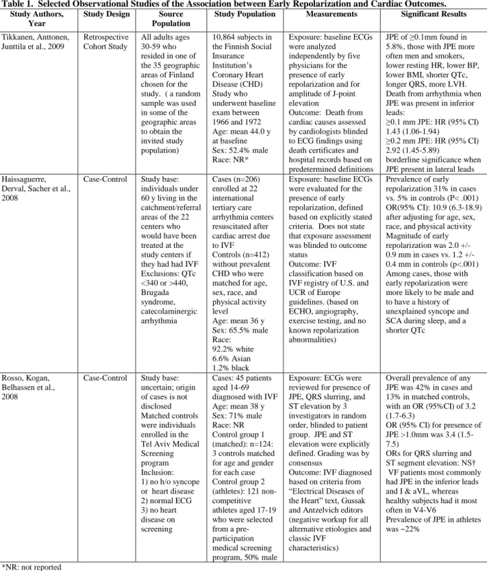
Table 1. Selected Observational Studies of the Association between Early Repolarization and Cardiac Outcomes. Study Authors,
Year
Study Design Source Population
Study Population Measurements Significant Results
Tikkanen, Anttonen, Junttila et al., 2009
Retrospective Cohort Study
All adults ages 30-59 who resided in one of the 35 geographic areas of Finland chosen for the study. ( a random sample was used in some of the geographic areas to obtain the invited study population)
10,864 subjects in the Finnish Social Insurance Institution’s Coronary Heart Disease (CHD) Study who underwent baseline exam between 1966 and 1972 Age: mean 44.0 y at baseline Sex: 52.4% male Race: NR*
Exposure: baseline ECGs were analyzed
independently by five physicians for the presence of early repolarization and for amplitude of J-point elevation
Outcome: Death from cardiac causes assessed by cardiologists blinded to ECG findings using death certificates and hospital records based on predetermined definitions
JPE of ≥0.1mm found in 5.8%, those with JPE more often men and smokers, lower resting HR, lower BP, lower BMI, shorter QTc, longer QRS, more LVH. Death from arrhythmia when JPE was present in inferior leads: ≥0.1 mm JPE: HR (95% CI) 1.43 (1.06-1.94) ≥0.2 mm JPE: HR (95% CI) 2.92 (1.45-5.89) borderline significance when JPE present in lateral leads Haissaguerre,
Derval, Sacher et al., 2008
Case-Control Study base: individuals under 60 y living in the catchment/referral areas of the 22 centers who would have been treated at the study centers if they had had IVF Exclusions: QTc <340 or >440, Brugada syndrome, catecolaminergic arrhythmia
Cases (n=206) enrolled at 22 international tertiary care arrhythmia centers resuscitated after cardiac arrest due to IVF
Controls (n=412) without prevalent CHD who were matched for age, sex, race, and physical activity level
Age: mean 36 y Sex: 65.5% male Race: 92.2% white 6.6% Asian 1.2% black
Exposure: baseline ECGs were evaluated for the presence of early repolarization, defined based on explicitly stated criteria. Does not state that exposure assessment was blinded to outcome status
Outcome: IVF classification based on IVF registry of U.S. and UCR of Europe guidelines. (based on ECHO, angiography, exercise testing, and no known repolarization abnormalities)
Prevalence of early repolarization 31% in cases vs. 5% in controls (P< .001) OR(95% CI): 10.9 (6.3-18.9) after adjusting for age, sex, race, and physical activity Magnitude of early repolarization was 2.0 +/- 0.9 mm in cases vs. 1.2 +/- 0.4 mm in controls (p<.001) Among cases, those with early repolarization were more likely to be male and to have a history of unexplained syncope and SCA during sleep, and a shorter QTc
Rosso, Kogan, Belhassen et al., 2008
Case-Control Study base: uncertain; origin of cases is not disclosed Matched controls were individuals enrolled in the Tel Aviv Medical Screening program Inclusion: 1) no h/o syncope or heart disease 2) normal ECG 3) no heart disease on screening
Cases: 45 patients aged 14-69 diagnosed with IVF Age: mean 38 y Sex: 71% male Race: NR Control group 1 (matched): n=124: 3 controls matched for age and gender for each case Control group 2 (athletes): 121 non-competitive athletes aged 17-19 who were selected from a pre-participation medical screening program, 50% male
Exposure: ECGs were reviewed for presence of JPE, QRS slurring, and ST elevation by 3 investigators in random order, blinded to patient group. JPE and ST elevation were explicitly defined. Grading was by consensus
Outcome: IVF diagnosed based on criteria from “Electrical Diseases of the Heart” text, Gussak and Antzelvich editors (negative workup for all alternative etiologies and classic IVF
characteristics)
Overall prevalence of any JPE was 42% in cases and 13% in matched controls, with an OR (95%CI) of 3.2 (1.7-6.3) OR (95% CI) for presence of JPE >1.0mm was 3.4 (1.5-7.5)
ORs for QRS slurring and
ST segment elevation: NS† VF patients most commonly had JPE in the inferior leads and I & aVL, whereas healthy subjects had it most often in V4-V6 Prevalence of JPE in athletes was ~22%
8
Table 2. Quality Ratings for Studies in Systematic Review.
Study Authors, Year Source population adequately described?* Study population representative of source population?* Potential for selection bias† Potential for measurement bias† Potential for
confounding† Statistical analysis*
Overall internal validity*
Tikkanen, Anttonen, Junttila et al., 2009
good good low low-moderate low good good
Haissaguerre, Derval, Sacher et al., 2008
fair fair high moderate high fair fair
Rosso, Kogan, Belhassen et al., 2008
poor uncertain high moderate high fair poor
*Graded as good, fair, or poor
†Graded as low, moderate, or high potential for bias
Tikkanen Study
The Tikkanen study was a retrospective cohort study that examined the prevalence of early
repolarization and its association with death from cardiac causes, death from all causes, and
death from arrhythmia in Finnish subjects aged 30-59.11 The study population consisted of
10,864 subjects originally recruited for the Finnish Social Insurance Institution’s Coronary Heart
Disease (CHD) Study who underwent baseline exam between 1966 and 1972. Subjects were
followed for a mean of 30±11 years for the purposes of this study. The presence of early
repolarization was determined by visual review of ECGs. Early repolarization patterns were
stratified according to degree of J-point elevation (≥0.1mV or >0.2mV) and according to lead
location as inferior (II, III, aVF) or lateral (I, aVL, V4-V6). Mortality causes and rates were
ascertained using death certificates from Statistics Finland, and death from arrhythmia was
determined by cardiologists using death certificates and hospital records, according to the
Cardiac Arrhythmia Pilot Study definition.
The overall prevalence of early repolarization ≥0.1mV was 5.8%. Adjusted hazard ratios
9
were 1.28 (95% CI 1.04 to 1.59) for JPE ≥0.1 mV, and 2.98 (95% CI 1.85-4.92) for JPE ≥0.2
mV. For all cause mortality adjusted HRs were 1.10 (95%CI .97-1.26) for JPE ≥0.1 mV and
1.54 (95% CI 1.06-2.24) for JPE ≥0.2 mV. For death from arrhythmia, adjusted HRs were 1.43
(95% CI 1.06-1.94) for JPE ≥0.1 mV and 2.92 (95% CI 1.45-5.89) for JPE ≥0.2 mV. In the
lateral leads, statistical significance was borderline for all cause mortality and death from cardiac
causes, and results were not significant for death from arrhythmia.
Study Design
The study’s population-based cohort design is a strength, as is the large sample size. This
is an appropriate research design because cohort studies are useful for identifying exposures that
increase or decrease the risk of disease. For a research question such as this, a randomized
controlled trial is obviously not feasible. The data used in this study are from a prospective
cohort study, which means that ECG data were collected at baseline, and subjects were followed
longitudinally. This clear temporality is another strength of the study. However, we must
remember that this study was indeed a retrospective cohort study, meaning that the data pertinent
to this research question were identified from past records. The design is thus not as strong as
that of a true prospective cohort study, which would have precise and predefined definitions of
cause of death and other variables.
Selection of Study Population
An examination of baseline characteristics shows several baseline differences between
groups. This is to be expected since this is a cohort study and the groups were not randomized.
Those with J point elevation were more often men and smokers, had lower resting resting heart
rates, lower blood pressure, lower BMI, shorter QTc intervals, longer QRS duration, more left
10
less than 2% loss to follow up, and even for these 2%, vital status could be ascertained for the
majority. Initially there was moderate potential for selection bias given the differences in
baseline characteristics, but the authors corrected for many covariates in regression analysis, and
so overall potential for selection bias is low.
Measurements
Turning to measurement, the study has several strengths. ECG review procedures were
standardized: ECGs were read for early repolarization by 5 five physicians, who examined
inferior (II, III, aVF) and lateral (I, aVL, V4-V6) groups. Reading physicians chose not to
examine leads V1-V3 in order to avoid including the highly arrhythmogenic Brugada syndrome.
For outcomes assessment, investigators used death data from Statistics Finland. Finland
maintains an extensive death registry, and the reliability of this registry had been validated by
previous studies. The criteria used by study physicians to ascertain death from arrhythmia were
well explained and based on published guidelines. Lastly, but importantly, outcomes were
assessed by cardiologists blinded to ECG findings.
There were several weaknesses related to measurement. Study authors do not clearly
define their criteria for diagnosis of early repolarization; they state only that they “stratified by
degree of JPE, that was either notched or slurred in 2 consecutive leads.” There is no explicit
mention of what criteria were used to make the initial diagnosis of early repolarization. Thus it is
not clear whether they took ST segment shape or T wave characteristics into account. Authors
also did not specifically define what types of deaths constituted death from cardiac causes.
Finally, there is a limited ability using retrospective analysis of death certificates to determine
whether death is definitively associated with arrhythmia. This is an inherent limitation of the
11
definitions of arrhythmia death. Possible lack of accuracy in death classification could lead to
nondifferential measurement bias; it would affect both the exposed and unexposed groups,
producing bias towards the null. Potential for measurement bias in this study is low to moderate.
Confounding
There are a few possible sources of confounding that deserve mention. The authors state
that they measured CV risk factors, but they do not explicitly list all of the variables measured,
which leads one to wonder whether they included all the pertinent risk factors. For example,
there was no mention of diabetes, claudication, or high density lipoprotein measurement. Since
these are established cardiovascular risk factors, we would hope that they were at least examined
as potential covariates. Similarly, the authors state that the use of medications that affect cardiac
repolarization was rare at the time of baseline exam. Though that may be true, it would be useful
to see data on the prevalence of use of these medications between groups to assure that they
could not have acted as confounders. It is not clear whether use of antiarrhythmics was
measured. Measurement of use of these drugs is important because they affect cardiac
repolarization and are also likely to be associated with death from cardiac causes. Aside from
these factors, other possible sources of confounding appear to be well addressed. Outcomes
assessors were blinded. The study was retrospective, so differential treatment of patients by
physicians would not be an issue. Overall potential for confounding was low after statistical
adjustment.
Statistical Analysis
The Tikkanen study was strong from a statistical standpoint. They had a large number of
events (n=6133 deaths), including 1969 deaths from cardiac causes. Due to the large sample size
12
of type II error. Results were precise, as shown by the generally narrow confidence intervals,
indicating that the results of the study are likely to be reproducible. Authors used Cox
proportional hazards models to examine death outcomes between groups. They controlled well
for confounders, and listed all of the covariates included in the model, though it would have been
helpful to list all of the variables that were initially examined as possible confounders. Another
strong feature of the analysis was the stratification by degree of J point elevation as ≥0.1mV or
>0.2mV. This allows for examination for a dose- response effect, which was present in the
results and strengthens the argument for causality.
Overall Quality
Overall, the Tikkanen study had good internal validity. Apart from a few measurement
issues, the study was sound. The population-based nature of this study enhances its
generalizability. No information on race was provided, but there were likely few non-whites
included in the sample. The study was done in Finland, so there is uncertainty as to whether it
generalizes to the U.S. or to nonwhite racial groups. The prevalence of JPE has been shown to
differ between ethnic groups, and most experts suspect that there is a genetic basis for JPE,
which would lead to differing prevalence in various racial groups. Lastly, it is unclear whether
JPE represents a single pathophysiology or is merely the phenotypic expression of a range of
underlying processes.17 If the latter case proves true, then differential geographic (and ethnic)
distribution of the various underlying processes could mean than the prognosis associated with
JPE varies widely between groups.
One final point on this study is related to clarity of terminology. The authors use the
terms J point elevation and early repolarization interchangeably, even when discussing their
13
repolarization are not synonymous. J point elevation can be seen in a variety of other conditions,
such as Brugada syndrome, pericarditis, acute MI, bundle-branch block, and hyperkalemia.
Since the authors did not list any ECG exclusion criteria with which would have excluded other
potential causes of JPE, referring to these terms interchangeably could be misleading. This study
examines the association of death with early repolarization, not with J-point elevation from any
cause. This lack of clarity is not a threat to the validity of the study, but it is a threat to its
interpretability by readers, especially when published in a journal that is read by a wide
audience, not exclusively by electrocardiologists.
Haissaguerre Study
The Haissaguerre study was a case control study that investigated the prevalence of early
repolarization and its association with idiopathic ventricular fibrillation (IVF).3 Study cases
consisted of 206 individuals enrolled at 22 international tertiary care arrhythmia centers who
were under age 60 and were resuscitated after cardiac arrest due to IVF. Controls (n=412)
without prevalent CHD were matched for age, sex, race, and physical activity level. Early
repolarization was defined as “elevation of the QRS-ST junction of ≥0.1 mV in the inferior (II,
III, aVF) or lateral (I, aVL, V4-V6) leads, manifested as QRS notching or slurring” at the time of
ICD placement. IVF classification was based on guidelines from the Joint Steering Committees
of the Unexplained Cardiac Registry of Europe and IVF registry of United States.
Comparing cases to controls, the prevalence of early repolarization was 31% vs. 5% (p<
.001). After adjustment for age, sex, race, and physical activity, the odds ratio (OR) for the
presence of JPE was 10.9 (95% CI 6.3-18.9). Magnitude of JPE when early repolarization was
14
Study Design
The case control study design does not have quite the same ability as a population-based
cohort study to definitively answer the clinical question. That being said, the role of initial
clinical studies on a topic often is to see whether the hypotheses generated in experimental
studies or observed in case reports is supported by clinical evidence. Case control studies are
often less expensive and logistically easier to do. In fact, it was the results of the Haissaguerre
study that helped generate greater interest in the topic and likely lead to the Tikkanen study
discussed earlier.
Selection of Study Population
In a case-control study, the main issues with selection bias often center on the selection of
controls; controls must reflect the exposure prevalence in the study base for the study to be valid.
In this study, the study base would be people under age 60 living in the catchment/referral areas
of the 22 tertiary care centers who would have been treated at the centers if they had had IVF.
Location of the centers is not reported. The question then is whether the controls chosen for this
study are likely to represent the exposure prevalence in the study base. The control group in this
study was made up of health care professionals. We do not know where they were from or how
they were selected. Therefore it is difficult to say whether this group represents the study base
well, though it is likely that this is not a representative control group. This group of
professionals may differ from the study base on variables such as age and race, which are closely
linked to JPE. If that were the case, the exposure prevalence in this group would not accurately
represent that of the study case. The ideal control group in a case control study is a random
15
It is essential that cases and controls meet the same criteria for inclusion in the study,
otherwise other systematic differences between them could account for observed results. In this
study, controls were required to have a normal echocardiogram, no prevalent heart disease and
no history of syncope. This was a prudent decision by investigators because the cases, in order
to be included as IVF cases, also had to meet similar criteria.
Typically an important part of the assessment of selection bias in case control studies
involves review of the table comparing baseline characteristics of cases to those of controls. The
table given in this study compares only the cases stratified by exposure status; it does not
compare case vs. control baseline characteristics. Therefore, it cannot determine whether the
baseline characteristics between groups show the expected known exposure-disease associations.
Normally the table should include an examination of differences in terms of risk characteristics
between cases and controls. Bearing that in mind, this may be less of an issue in this study,
because for IVF we do not know as much about its risk factors as we do about the risk factors of
many other diseases. On the positive side, no loss to follow-up was reported, and the authors
state no cases were lost to follow-up. This reassures the reader than there is less likely to be
selection bias introduced from differential loss to follow-up.
On the issue of matching, controls were matched for age, sex, race, and physical activity
level using global frequency matching. Matching can be useful because it reduces the
differences between groups on determinants of disease other than the exposure being considered.
However, matching changes the exposure prevalence in the controls if the exposure is related to
the matching variables. In this study matching selects for controls that look more like IVF cases.
This can produce bias toward the null, as it likely leads to higher exposure prevalence than the
16
There is potential for overmatching in this study, which would diminish the ability of the study
to detect an effect. The study did still find significant results, so there is a possibility that the
relationship is even stronger than what is indicated by the results. Overall, there is high potential
for selection bias in this study.
Measurements
Turning now to measurement issues, the Haissaguerre study had several positive traits.
The definition of early repolarization was explicitly defined as quoted above. Thus, it is clear
exactly what criteria ECG readers were looking for in order to identify early repolarization. In
addition, IVF classification was based on international guidelines. These facts are reassuring in
terms of valid and reliable assessment of exposure and outcome.
The authors do not disclose who read the ECGs, how many readers there were, or
whether coding was double-checked. Additionally, they do not indicate that exposure
assessment was blinded to case/control status. This could easily produce bias away from the null
if the unblinded reader believed in a positive association between early repolarization and IVF,
because due to its nature, some subjectivity remains in the interpretation of the early
repolarization pattern. Finally, the authors admit that data collection was not uniform between
centers. This means that measurement may have been unequal between cases at different
centers. Because it is not likely that the controls were distributed equally between the various
centers, it is also likely that measurement was unequal between cases and controls. This would
constitute differential measurement bias, and could produce bias either toward or away from the
null hypothesis depending on the exact nature of measurement differences. Potential for
measurement bias in this study was moderate.
17
Several other possible confounding factors in this study must be discussed. The first is
the possibility of survival bias. Survival bias is possible if early repolarization somehow
provides a survival advantage in IVF patients. In any study, using prevalent cases or using living
cases may introduce bias if exposure is related to duration of disease or prognosis. If survival
bias were operating, it could produce study findings that were the exact opposite of the truth.
This may be a theoretical concern, but since we still do not fully understand the complex
interplay between early repolarization and arrhythmogenesis, survival bias should at the very
least be considered.
A related issue is that of temporality. One must consider the expected temporal
relationship when investigating an association. Ideally, we would look for the presence of early
repolarization on ECGs recorded before IVF occurred. In this study they read ECGs that were
recorded after the event. Therefore, the theoretical possibility exists that the IVF event somehow
caused the finding of early repolarization. When the exposure is influenced by the disease, it is
referred to as protopathic bias. This is by no means a certainty in this situation, but the
possibility needs to be considered. Could the outcome have caused or influenced the exposure?
Several other variables deserved mention in the study to assuage concerns about
confounding. No mention was made of the prevalence of use of antiarrhythmics or other drugs
that might affect cardiac repolarization such as beta-blockers. Similarly, other risk factors for
early repolarization, namely heart rate, were not mentioned. It may be that these variables were
not related to IVF, but they deserved comment.
Statistical Analysis
From an analysis standpoint, the main concern was that were was no discussion about
18
authors used logistic regression to compare the odds of early repolarization between cases and
controls, and controlled for age, race, sex, and physical activity. They do not mention
considering any other possible covariates. This raises the concern that control for confounding
was incomplete, especially in the absence of a table comparing baseline characteristics between
groups.
Statistical analysis was otherwise sound. Investigators used traditional exclusive
sampling for controls. This method can sometimes overestimate the OR because it utilizes the
rare disease assumption. However, it is acceptable in this case because the outcome in question
is rare and incident cases were used. Since the authors used global frequency matching, they did
not need to do a matched analysis. The authors used 2:1 ratio of controls to cases. This is a
good way to increase the power of the study if the number of cases is limited, or if there is a low
prevalence of the exposure among controls, as is true in this study. Finally, the authors excluded
V1-V3 from the analysis to avoid Brugada syndrome, as it is highly arrhythmogenic and could
confound the results.
Overall Quality
Overall, the internal validity of the Haissaguerre study was fair. There were significant
issues with selection bias and measurement. However, some of the concerns are more theoretical
and not necessarily major threats to the study’s validity. In terms of generalizability, the study
contained few athletes or blacks, which are populations with a higher prevalence of early
repolarization, so results may not be generalizable to these populations. The fact that the study
was not population-based also limits its generalizability to some extent.
19
The Rosso study was a case control study that assessed the association between J point elevation
and IVF.9 Cases consisted of 45 patients aged 14-69 who had been diagnosed with IVF after
sudden cardiac arrest. There were 2 control groups. The main control group consisted of 124
controls aged 24-70 years who were matched for age and gender, 3 controls per case. A second
control group included 121 non-competitive athletes aged 17-19 years. JPE was defined as a
positive “humplike deflection” at the J-point. The authors evaluated “QRS slurring” as a
separate entity in this study, and defined it as an R wave gradually slurring into the ST segment
with upright concavity. IVF diagnosis was based on criteria described in a prominent cardiac
electrophysiology text.
Overall prevalence of any JPE was 42% in cases and 13% in matched controls (p=.001).
The OR for the overall presence of any JPE was 3.2 (95%CI 1.7-6.3). Stratified by lead
groupings, ORs for the presence of JPE were 3.2 (95%CI 1.4-7.5) in the inferior leads, 16.9
(95%CI 2.0-140.3) in leads I and aVL, and 0.9 (95%CI 0.2-3.3) in leads V4-V6. Authors found
that the OR for presence of JPE >1.0mm was 3.4 (95%CI 1.5-7.5). The OR for QRS slurring
was 1.3 (95%CI 0.7-2.4), and for ST segment elevation the OR was 1.3 (95%CI 0.7-2.5). Neither
of these measurements added diagnostic value when combined with JPE, and no significant
relationships were found when stratified by lead groupings. IVF patients most commonly had
JPE in the inferior leads and I & aVL, whereas healthy subjects had it most often in leads V4-V6.
The prevalence of JPE in athletes was intermediate, at ~22%.
Selection of Study Population
Similar to the Haissaguerre study discussed above, the Rosso study was a case-control
study. Therefore, one of the main points that must be assessed is whether the choice of controls
20
study base. In this study, the authors do not disclose where the cases came from. It is
impossible to surmise the study base, and therefore difficult to determine whether the controls
used were an accurate representation of the source population. We know that the matched
controls were selected from among 3,500 adults who participated in the Tel Aviv Medical
Screening program in 2007. Controls were individually matched for age and sex. The exact
source of these individuals is not stated, and we do not know how they were recruited for
screening, their reasons for enrolling, or even whether they represent the same geographic area as
the controls. The issue here, as previously stated, is that there is not sufficient information given
to determine whether these controls are an appropriate choice to represent the study base. It is
possible that participants in the Tel Aviv Medical Screening Program do represent the study base
well, but there is no way of knowing. To obtain controls for younger cases, investigators used
“medical personnel and their offspring.” This is a vague, somewhat suspect source of controls.
For the second control group, investigators selected non-competitive athletes aged 17-19 from a
pre-participation medical screening program, 50% of whom were male. No mention is made of
how the participants were chosen from among those who participated in the medical screening
program. As with the previous study, these groups may differ from the study base on variables
that are closely linked to JPE. If this were true, the exposure prevalence in these groups would
not accurately represent that of the study base.
It is also important that cases and controls meet the same criteria for inclusion in the
study, such that if one of the controls had the outcome in question, he or she would have been
included as a case. This means that not only should controls be part of the study base, but that
they also should meet any other inclusion criteria imposed on the cases. A strength of this study
21
syncope or heart disease 2) a normal ECG, and 3) no heart disease uncovered on screening exam.
Again, this is similar to the criteria that sudden cardiac arrest survivors had to meet in order to be
classified as IVF, so this was a good decision by the investigators.
The authors did not include a table of baseline characteristics, so one cannot compare the
cases and controls on a range of characteristics. Typically we want to make sure that cases differ
from controls in ways that we would expect. The failure to provide this information prevents us
from assessing differences between groups and therefore from deciding whether confounding
was adequately addressed in the study design and analysis.
The issues surrounding matching in general were discussed above. In this study, the
matched control subjects were individually matched based on age and sex. This may seem
appropriate, because age and sex are strong risk factors for IVF, and thus could lead to
confounding. However, age and sex are also strongly related to JPE, so matching on these
variables will lead to an artificially high exposure prevalence in the control group and to a
greater chance that cases and controls have a the same exposure history. Again, this would
produce bias towards the null. In contrast, athletes were not age or sex matched. Many
epidemiologists prefer this avoidance of matching, provided that other measures are taken to
account for confounding by these important variables. Matching interferes with the fundamental
goal of having controls approximate the exposure prevalence in the source population.
A strength of this study, and indeed of all case-control studies, is that there no loss to
follow up. Investigators excluded 11 matched controls and 11 athlete controls after selection
mainly due to poor ECG quality. This is not likely to be a large problem because this is not a
22
Still, considering all of the aforementioned issues, there is high potential for selection bias in this
study.
Measurements
From a measurement standpoint, the Rosso study did several things well. JPE was
explicitly defined as a positive “humplike deflection” immediately after the QRS at the onset of
the ST segment. Similarly, QRS slurring was defined when the R wave gradually became the ST
segment with upright concavity, and ST height was measured at most horizontal portion with
electronic calipers. This indicates that exposure measurement is at least somewhat reproducible.
ECGs were reviewed by 3 investigators in random order. Another strength of the study was that
ECG interpretation was blinded to case/control status, thereby helping eliminate the chance of a
bias on the part of ECG readers toward finding more JPE in the case group. Blinding helps
prevent this bias away from the null. Lastly, IVF diagnosis was based on published rigorous
criteria that included negative workup for all alternative etiologies, a compatible history, and all
of the unique IVF characteristics. The only glaring deficiency in terms of measurements was the
fact that there was no discussion of the measurement of possible covariates and other
cardiovascular risk factors. It is unclear whether this was done. The overall potential for
measurement bias is therefore moderate.
Confounding
Apart from issues with selection bias, only one other possible confounding issue stands
out. The authors mention in passing that some of the ECG tracings were old. This brings up an
issue that is related to the use of historical controls in case control studies. Generally, historical
control groups are a poor choice in case control studies because disease diagnosis patterns,
23
over time, thereby introducing many opportunities for confounding if historical controls are used.
Even though in this situation it is some of the cases that are old, not the controls, some of the
same bias could still be operating. However, in this situation the potential for problems with this
issue is less because diagnosis and treatment of IVF have not changed a great deal, and the
occurrence of IVF is not intimately related to deficiencies in treatment or diagnosis. In any case,
we are studying survivors of IVF. This of course also means that survivor bias is again an issue
to consider. Overall potential for confounding is high, largely due to the contribution of
selection bias
Statistical Analysis
In their analysis, the authors compared continuous variables with blocked analysis of
variance, and compared dichotomous variables with conditional logistic regression. Since the
investigators used individual matching to match cases and controls, this must be accounted for
during the statistical analysis. The above measures are appropriate ways in which to account for
matching during analysis. Like the other studies, the Rosso study excluded lead V1-V3 to avoid
events possibly caused by Brugada syndrome. Two patients included as cases were eventually
diagnosed with Brugada syndrome. Authors did a sensitivity analysis excluding the two Brugada
patients that did not change the results.
As discussed earlier, investigators can justify a decision to forgo matching if they account
for covariates by another method. While the control group made up of young athletes was not
matched on any characteristics, it appears that no effort was made to control for any covariates
during analysis. Therefore, differences in prevalence between the athletes and the cases could be
explained by other factors. For example, the mean age of cases was 38 years, whereas the athlete
24
for, or the results are not interpretable. Similarly, in the matched controls it does not seem that
any additional covariates were adjusted for during analysis. Controls were matched on sex and
age, but analysis appeared to be strictly bivariate. The direction of bias produced by this type of
issue is difficult to predict because it depends on a complex interplay between various levels of
different confounders, the baseline levels of which are not available to us.
Overall Quality
The results of the Rosso study results are consistent with other studies, finding a positive
association between JPE and IVF. It is also important to note that while the two previous
studies defined early repolarization as slurring or notching at the J point, the Rosso study
separates notching from slurring. Interestingly, J point notching was found to be significantly
associated with IVF, but QRS slurring was not. Overall, the internal validity of this study was
poor. It is difficult to interpret the results given that there was great potential for selection bias
based on ambiguity in control selection and that little was done to account for selection bias in
the study design and analysis. It is inappropriate to generalize the findings of the study to other
populations because its internal validity is suspect.
DISCUSSION
The Haissaguerre and Rosso studies had difficulty controlling for selection bias. My greatest
concerns were the authors’ failure to fully disclose the source of cases and the comparability of
cases and controls, coupled with inadequate measures to address several potential sources of
confounding. However, these studies laid the foundation for stronger, population-based studies
25
These studies highlight the presence of more general issues with the measurement of
early repolarization. Definitions and descriptions of early repolarization differ between articles,
and varying criteria for diagnosis of early repolarization are used from study to study. This may
lead to questionable reproducibility and comparability of findings. Furthermore, in these studies
the definitions of visually coded ECGs were limited; they used a “broad definition of early
repolarization, with no regard for t-wave amplitude or specification of the shape of the ST
segment.”18
Consensus criteria for the diagnosis of early repolarization on ECG are needed.
The differences in the prevalence of early repolarization between groups were large in the
case control studies, and the effect sizes for the relationship between early repolarization in the
inferior leads and death from arrhythmia were significant and dose-dependent in the cohort
study. The large effects are unlikely to be accounted for completely by the aforementioned
biases. In general, although there were some potential issues with internal validity, they are not
likely to be sufficient to account for the results obtained in these three studies, especially since
some of potential biases actually would have produced bias toward the null hypothesis of no
effect.
Despite various threats to internal validity in these studies, their overall similar findings
increase the likelihood of a true association. Relative consistency of results across several types
of study designs makes it less likely that some unmeasured confounder is operating to cause the
observed association. A trend toward increased risk from early repolarization in the inferior
leads deserves further investigation. All of the studies also had limits in terms of
generalizability. However, the consistency of these findings in a variety of different populations
is meaningful. Though these studies were individually flawed, there is sufficient evidence to
26
arrhythmia in future population-based studies. This research should be actively pursued, as
prevention of sudden cardiac death from ventricular arrhythmia is an important public health
27
REFERENCES
1. Klatsky AL, Oehm R, Cooper RA, Udaltsova N, Armstrong MA. The early repolarization normal variant electrocardiogram: Correlates and consequences. Am J Med. 2003;115(3):171-177.
2. Mehta M, Jain AC, Mehta A. Early repolarization. Clin Cardiol. 1999;22(2):59-65.
3. Haissaguerre M, Derval N, Sacher F, et al. Sudden cardiac arrest associated with early repolarization. N Engl J Med. 2008;358(19):2016-2023.
4. Antzelevitch C, Yan GX. J wave syndromes. Heart Rhythm. 2009.
5. Yan GX, Lankipalli RS, Burke JF, Musco S, Kowey PR. Ventricular repolarization components on the electrocardiogram: Cellular basis and clinical significance. J Am Coll Cardiol. 2003;42(3):401-409.
6. Mehta MC, Jain AC. Early repolarization on scalar electrocardiogram. Am J Med Sci. 1995;309(6):305-311.
7. Goldman MJ. RS-T segment elevation in mid- and left precordial leads as a normal variant. Am Heart J. 1953;46(6):817-820.
8. Wellens HJ. Early repolarization revisited. N Engl J Med. 2008;358(19):2063-2065.
9. Rosso R, Kogan E, Belhassen B, et al. J-point elevation in survivors of primary ventricular fibrillation and matched control subjects: Incidence and clinical significance. J Am Coll Cardiol. 2008;52(15):1231-1238.
10. Nam GB, Kim YH, Antzelevitch C. Augmentation of J waves and electrical storms in patients with early repolarization. N Engl J Med. 2008;358(19):2078-2079.
11. Tikkanen JT, Anttonen O, Junttila MJ, et al. Long-term outcome associated with early repolarization on electrocardiography. N Engl J Med. 2009;361(26):2529-2537.
12. Wellens HJ. Early repolarization revisited. N Engl J Med. 2008;358(19):2063-2065.
13. Gussak I, Antzelevitch C. Early repolarization syndrome: Clinical characteristics and possible cellular and ionic mechanisms. J Electrocardiol. 2000;33(4):299-309.
14. Weaver WD, Peberdy MA. Defibrillators in public places -- one step closer to home. N Engl J Med. 2002;347(16):1223-1224.
28
16. Merchant FM, Noseworthy PA, Weiner RB, Singh SM, Ruskin JN, Reddy VY. Ability of terminal QRS notching to distinguish benign from malignant electrocardiographic forms of early repolarization. Am J Cardiol. 2009;104(10):1402-1406.
17. Myerburg RJ, Castellanos A. Early repolarization and sudden cardiac arrest: Theme or variation on a theme? Nat Clin Pract Cardiovasc Med. 2008;5(12):760-761.
29 Long-Term Prognosis Associated with J-point Elevation in a Large Biracial Cohort:
The ARIC Study
ABSTRACT
BACKGROUND
Sudden cardiac death accounts for greater than half of all cardiovascular disease deaths.
Ventricular arrhythmia is the most common cause of sudden cardiac death. Several studies have demonstrated an association between ventricular fibrillation and early repolarization, an
electrocardiogram finding characterized by elevation of the QRS-ST junction (J-point elevation). A recent population- based study found that early repolarization was associated with increased risk of sudden death from arrhythmia. Further study of this association in more diverse
populations is needed.
METHODS
We assessed the long-term prognosis associated with J-point elevation on electrocardiogram in a biracial cohort of 15,141subjects. These subjects, aged 45-64, were participants in the
Atherosclerosis Risk in Communities (ARIC) study. The primary endpoint was adjudicated sudden cardiac death, and secondary endpoints were fatal/nonfatal coronary heart disease events and all-cause mortality. Mean follow-up time was 17±4 years, and J-point elevation was
considered to be present if J-point amplitude was ≥ 0.1mV in any lead.
RESULTS
J-point elevation was present in 1,866 subjects (12.3%). After adjustment for demographic, clinical, lifestyle, and laboratory data, J-point elevation was not significantly related to
adjudicated SCD in the overall sample (adjusted hazard ratio, 1.23; 95% CI, 0.87-1.75). There was a significant interaction between race and J-point elevation for adjudicated SCD. Whites with J-point elevation had a higher risk of death from adjudicated SCD (adjusted HR, 2.03; 95% CI, 1.28-3.21) than did whites without J-point elevation. However, blacks with J-point elevation did not have an increased risk of death from adjudicated SCD (adjusted HR, 0.82; 95% CI, 0.52-1.30) compared to blacks without the finding. There was also a significant interaction between sex and J-point elevation. J-point elevation significantly increased risk of adjudicated SCD in females (adjusted HR, 2.54; 95% CI, 1.34-4.82). However, the finding of J-point elevation did not similarly increase risk of adjudicated SCD in males (adjusted HR, 1.02; 95% CI, 0.69-1.50).
CONCLUSIONS
30
INTRODUCTION
Sudden cardiac death (SCD) is an important public health problem that accounts for more than
half of all deaths from cardiovascular disease.1-3 The incidence of SCD is estimated to be
between 300,000 and 450,000 annually in the United States (U.S.).1, 4 In most cases, SCD is
thought to be caused by cardiac arrest from ventricular arrhythmia. Despite advances in
treatment in many areas of medicine, outcomes for cardiac arrest remain poor, with overall
survival rates of 5-10%.5 Prompt defibrillation offers the greatest chance of survival in
individuals who have suffered a cardiac arrest. However, approximately 70-80% of cardiac
arrests occur in the home, with no timely access to defibrillation.6, 7
In light of the difficulty of treating cardiac arrest, efforts must focus on prevention. One
preventive strategy is to identify and ameliorate risk factors for SCD. Most individuals who
suffer SCD have underlying coronary heart disease (CHD), and attention must continue to be
paid to major modifiable risk factors for CHD (such as hypertension and hyperlipidemia).
However, 5-10% of those who succumb to SCD do not have CHD. Such patients’ hearts, while
structurally normal , are believed to have an underlying electrical abnormality.1, 8 These patients,
in the absence of evidence of a primary arrhythmogenic disorder, are labeled as having
idiopathic ventricular fibrillation (IVF).1, 2 Identification of people at increased risk of IVF may
allow targeted prevention strategies that could ultimately decrease the burden of SCD.
Although traditionally viewed as benign (especially in young healthy adults), several
case-control studies have suggested that one electrocardiogram (ECG) finding that may be
associated with the development of IVF is the early repolarization pattern. 9-11 Early
repolarization is a common ECG finding that is seen in 1-6% of people. 12-14 It is characterized
31
population-based cohort study showed that early repolarization was associated with increased
risk of death from cardiac causes and with increased risk of death from arrhythmia.14 Though it
is the only population-based study on the topic that we are aware of, its findings may not be
generalizable to other populations. For example, the Finnish cohort likely had few blacks in the
sample, though this was not discussed explicitly. Importantly though, blacks have a higher
prevalence of the early repolarization pattern, and are more likely to experience SCD.1, 9, 12, 15, 16
Research regarding the implications of early repolarization for cardiovascular outcomes in this
high risk population is lacking, and experts have called for investigation into the significance of
these junctional changes in the groups that are at higher risk for them.16
The early repolarization pattern has many morphologic variants.17 A considerable
degree of subjectivity is involved in the diagnosis of early repolarization, and no consensus
criteria currently exist for the diagnosis of early repolarization. Each study on the topic has used
a slightly different definition for the identification of early repolarization, making it difficult to
directly compare results. J-point elevation is not only a central feature of the early repolarization
pattern, but is also a critical component in the diagnosis of other arrhythmogenic ECG patterns
such as Brugada syndrome.9, 16, 18 Like the early repolarization pattern, J point elevation has been
found in some studies to be more common in blacks.19, 20 An examination specifically of J point
elevation has several advantages. It can be determined from computerized ECG coding
programs, and is therefore useful in large epidemiologic studies. J point measurements provide a
quantitative, unambiguous, and objective measurement of ST segment deviation.
The goals of this study were therefore to (1) estimate the prevalence of J-point elevation in a
large biracial cohort of U.S. adults, and (2) examine the association of J-point elevation with risk
32
Specifically, we investigated the association of J-point elevation with sudden cardiac death,
fatal/nonfatal coronary heart disease (CHD) events, and all-cause mortality in a large biracial
cohort.
METHODS
Study Population
The Atherosclerosis Risk in Communities (ARIC) study21 is a prospective, population-based
cohort study designed to investigate the etiology and natural history of cardiovascular disease.
From 1987 to 1989, ARIC investigators used probability sampling to enroll 15,792 men and
women aged 45-64 residing in four U.S. communities: Jackson, Mississippi, Washington
County, Maryland, Forsyth County, North Carolina, and the northwestern suburbs of
Minneapolis, Minnesota.
For the present analysis, we excluded 202 subjects for whom J-point amplitude data were
missing or incomplete. We also excluded 604 subjects with QRS complex duration >120
milliseconds in order to remove cases of bundle branch block, Wolf-Parkinson-White syndrome,
and idioventricular rhythm. Additionally, 48 subjects were excluded for race other than black or
white. Finally, we excluded three individuals who were large positive outliers on the J-point
amplitude variable. After the above exclusions, 15,141 subjects remained for analysis.
Baseline Measurements
At the baseline examination, a standard, resting, supine 12- lead ECG was obtained for
33
locator was used to determine and standardize the positioning of chest electrodes V3-V6. Each
ECG tracing consisted of 10 seconds of each of the 12 leads recorded simultaneously. Tracings
were sent via phone modem to be computer coded at the ARIC ECG Reading Center. Computer
analysis included measurement of the voltage and duration of ECG waves and segments, as well
as ECG classification according to Minnesota Code.22 All records with significant Minnesota
code findings as determined by the computer, as well as a random sample of tracings, were sent
to the ECG coding center to be visually coded. Discrepancies between the computer code and
visual code were adjudicated by a senior coder. A rhythm strip consisting of 2 minutes of lead
V1, II, and V5 was taken immediately following the 12-lead ECG and sent to the ECG coding
center for coding of arrhythmias. Later processing of the ECGs took place at EPICARE
(Epidemiological Cardiology Research Center at Wake Forest University, Winston-Salem, NC),
where the 2001 version of the GE Marquette 12-SL program was used to obtain ST amplitude at
the J-point in relation to the isoelectric line (thus giving both negative and positive values). Each
tracing was checked visually to confirm accuracy of readings.
J-point elevation was defined as a J-point amplitude ≥1.0 mm in any lead. Cornell
Voltage (|S V3| + R in aVL), a measure of ECG-defined left ventricular hypertrophy (LVH), was
also calculated.23 LVH was considered to be present if the Cornell Voltage was >28mm in males
and >22mm in females.23
Anthropometric measurements were taken with subjects wearing scrub suits and no
shoes. Height, measured to the nearest centimeter, and weight measured to the nearest pound
were used to calculate body mass index (BMI) in kg/m2. Three seated blood pressures were
taken using a random-zero sphygmomanometer, and the average of the last two was used.
34
performed on the antecubital vein of seated subjects, and blood samples were analyzed for lipid
levels and chemistry. Diabetes was defined as a fasting glucose level of ≥126 mg/dl, a
nonfasting level of ≥200 mg/dl, self-reported physician diagnosis of diabetes, or pharmacologic
treatment for diabetes. Information regarding race, smoking history, physical activity, family
health history, and educational attainment was obtained through interviews. Education was
categorized as basic (less than high school), intermediate (high school diploma and/or vocational
school), or advanced (some college, graduate school, or professional school). Physical activity
was based on reported level of sport activity using the Baecke physical activity questionnaire.24
Follow-up
Follow-up procedures are described in detail elsewhere.21 The primary outcome was adjudicated
sudden cardiac death. All cases of fatal CHD in ARIC were reviewed by a committee of
physicians. Reviewed cardiovascular deaths were coded as Definite SCD, Possible SCD, or
non-sudden CHD death. For the outcome SCD by 1 hour definition, SCD was defined as non-sudden and
unexpected death within 1 hour of symptom onset. For the outcome SCD by 24 hour definition,
the subject had to have been observed alive with 24 hours of death if the event was unwitnessed.4
Individuals meeting the criteria for SCD by 1 hour definition were also included in the SCD by
24 hour group. Secondary outcomes included fatal/nonfatal CHD events and all-cause mortality.
Fatal/nonfatal CHD event was defined as definite or probable myocardial infarction, or definite
CHD death.21 Myocardial infarction was classified using standardized criteria.21
Events
occurring between the baseline examination and December 31st, 2007 were included in analysis
of all outcomes with the exception of adjudicated sudden cardiac death, for which the end of
35
Statistical Analysis
Continuous variables are presented as means (±SD), and categorical variables are presented as
overall percentages and as percentages for those with and without J point elevation. We
examined the relationships between J point elevation and subject characteristics using 2-sample
t-tests for continuous characteristics and Pearson’s chi square tests for categorical characteristics.
We then examined associations of subject characteristics with sudden cardiac death, again using
2-sample t-tests for continuous variables and Pearson’s chi square tests for categorical variables.
Cox proportional hazards models were used to obtain multivariate-adjusted hazard ratios
for SCD by all three definitions, CHD events, and all-cause mortality for those with versus
without J-point elevation. ECG leads were grouped into anterior (V1-V5), inferior (II, III, aVL),
and lateral (I, aVL,V6) leads, as well as into an overall measure of J-point elevation in any lead.
Results are reported as hazard ratios with 95% confidence intervals. Censoring occurred at the
time of an event, death, loss to follow up, or at the end of follow-up (December 31st, 2007).
Potential covariates included in the initial model were: age, sex, race, BMI, heart rate,
systolic blood pressure, smoking status, high-density lipoprotein level, low-density lipoprotein
level, diabetes, level of physical activity, family history of premature CHD, education level,
Cornell voltage for LVH, electrolyte levels, and presence of major ECG abnormality on
Minnesota Code.22 We also examined data on prevalent CHD (including myocardial infarction
determined by self-report or ECG, self report of heart or arterial surgery, coronary bypass,
balloon angioplasty, coronary artery angioplasty) and on physician-diagnosed stroke, angina, and
36
medications were also studied: digitalis, selective and non-selective beta-blockers, calcium
channel blockers, angiotensin converting enzyme (ACE) inhibitors, and antiarrthymic drugs.
Variables found not to be related to J point amplitude in bivariate analysis were dropped
from the full model in a stepwise fashion, and the resulting reduced models were tested against
the full model using the likelihood ratio test. Tested variables that did not significantly affect
results were dropped from the model. Using this method, we created a set of reduced models fit
to J-point elevation in any lead as the main exposure and adjudicated sudden cardiac death as the
outcome. This same set of covariates was then employed across all exposures and all outcomes
in order to enhance comparability and reproducibility. Given the uncertain nature of the
relationship between LVH and J-point elevation, we conducted a sensitivity analysis by
removing Cornell voltage from the models, which produced no meaningful change in the results.
Model 1 adjusts for demographic factors. Model 2 adjusts for demographic and clinical data.
Model 3 adjusts for demographic, clinical, lifestyle, and laboratory data.
The proportional hazards assumption was tested for each model using the test of
Schoenfeld residuals. We used stratified Cox models to stratify on any variables that violated the
proportional hazards assumption, and this produced no meaningful change in the results.
Violations of the proportional hazards assumption by J-point elevation were checked graphically
and judged to be trivial. We also undertook pre-specified subgroup analyses by race, sex, heart
rate, and prevalent CHD in order to assess whether effect modification was present. We tested
for the presence of these interactions individually using the likelihood ratio test. When a
significant interaction was present, we calculated hazard ratios for the subgroups individually
37
For the lead groupings in which significant results were consistently observed, we
examined the individual component leads, both as continuous measures of J-point amplitude and
as a dichotomized measure of J-point elevation. For continuous variables, the linearity
assumption was tested using a test for linear trend.
Statistical analyses were performed using STATA 11.0 (Stata Corp., College Station,
Texas). All reported p values are 2-sided, with a p-value of less than 0.05 considered to indicate
statistical significance.
This secondary analysis was exempted from full review by the Office of Human
Research Ethics of the University of North Carolina at Chapel Hill.
RESULTS
Sample Characteristics
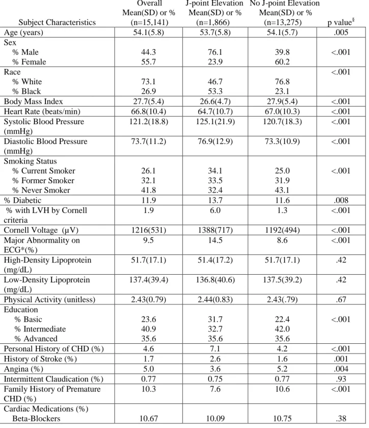
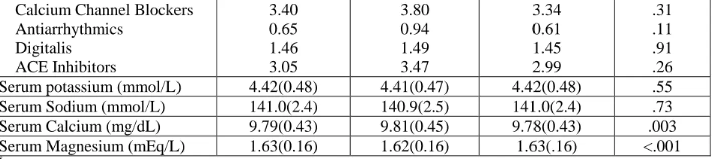
Baseline characteristics of the overall sample and of subjects with and without J-point elevation
are shown in Table 1. Overall, the sample was 44.3% male and 26.9% black. However, among
subjects with J-point elevation, 76.1% were male and 53.3% were black. Subjects with J-point
elevation also had lower BMI, lower heart rate, and higher blood pressure. They were more
likely to be smokers (34.1% vs. 25.0%, p<.001), to have LVH (6.0% vs 1.3%, p<0.001), and
were more likely to have a major Minnesota Code abnormality on ECG (14.5 vs. 8.6%,
p<0.001). Finally, those with J-point elevation were more likely to have a history of CHD or
other vascular disease, but were less likely to have a family history of premature CHD.
38
J-point elevation was present in 1,866 of 15,141 subjects (12.3%) (Table 2). Elevation in the
anterior leads (V1-V5) was found in 1,767 subjects (11.7%). Elevation in the inferior leads (II,
III, aVL) was found in 119 subjects (0.8%), and 92 subjects (0.6%) had elevation in the lateral
leads (I, aVL,V6). Males were more likely than females to have J-point elevation in at least one
lead (21.2% vs. 5.3%, p<0.001). Among blacks, there was a 24.4% prevalence of J-point
elevation; prevalence among whites was 7.9%. J-point elevation was significantly more
common among younger subjects (13.1%) than among older subjects (11.5%).
Overall Risk of Death and Cardiac Outcomes
After a mean follow-up of 17±4years, 3,555 subjects (23.5%) died. Of these deaths, 237 were
due to adjudicated SCD, representing 6.6% of all deaths. Using the 1 hour definition of SCD,
244 SCDs occurred, representing 6.9% of all deaths. When this group is expanded to include
SCD by 24 hour definition in addition to SCD by 1 hour definition, 482 deaths are attributed to
SCD. This figure represents 13.6% of all deaths during follow-up. During follow-up, 1,764
subjects experienced confirmed fatal or nonfatal CHD events. Of these events, 339 were fatal
CHD, 105 were fatal MI, and 1,320 were nonfatal MI.
In unadjusted analysis, those with J-point elevation in any lead were approximately 2.3
times more likely to suffer adjudicated SCD (adjusted HR, 2.28; 95%CI, 1.69-3.07, Table 3).
Statistically significant relationships were also present between J-point elevation and all other
outcomes in unadjusted analysis. After adjustment for race, sex, and age, J-point elevation was
no longer significantly related to adjudicated SCD (adjusted HR, 1.31; 95% CI, 0.94-1.82), and
39
lifestyle, and laboratory data in addition to demographic factors, these relationships remained
nonsignificant. Hazard ratios for leads V1-V5 individually are shown in supplemental table 1.
Among subjects with J-point elevation in any lead, there was a significant interaction
between race and J-point elevation for the primary outcome adjudicated SCD (Table 4). Whites
with J-point elevation had a higher risk of death from adjudicated SCD (adjusted HR, 2.03; 95%
CI, 1.28-3.21) than did whites without J-point elevation. However, blacks with J-point elevation
did not have an increased risk of death from adjudicated SCD (adjusted HR, 0.82; 95% CI,
0.52-1.30) compared to blacks without the finding. The ratio of hazard ratios for whites compared to
blacks was 2.46, indicating a large difference between these groups in the risk associated with
J-point elevation.
A significant interaction was also present between sex and J-point elevation. J-point
elevation significantly increased risk of adjudicated SCD in females (adjusted HR, 2.54; 95% CI,
1.34-4.82). However, the finding of J-point elevation did not similarly increase risk of
adjudicated SCD in males (adjusted HR, 1.02; 95% CI, 0.69-1.50). The ratio of hazard ratios for
females compared to males for adjudicated RSCD was 2.50, indicating that J-point elevation
connotes a much greater risk of SCD in females than it does in males. The ratios of hazard ratios
are nearly universally above one, indicating that the trend of increased risk associated with
J-point elevation in whites and females is consistent across outcomes.
DISCUSSION
Our study suggests that the finding of J-point elevation in white individuals is associated with an
increased risk of SCD. This association was not shown to be present among black individuals.